. 2022 Jul 5;8(1):9.
doi: 10.1038/s41514-022-00090-1.

Age-dependent cognitive impairment, hydrocephalus and leukocyte infiltration in transgenic mice with endothelial expression of human EPHX2

Affiliations

Age-dependent cognitive impairment, hydrocephalus and leukocyte infiltration in transgenic mice with endothelial expression of human EPHX2

Catherine M Davis et al. NPJ Aging. .

Abstract

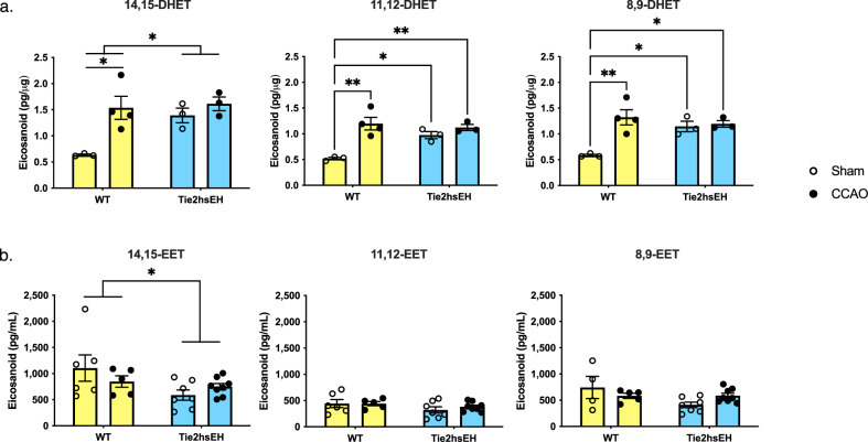
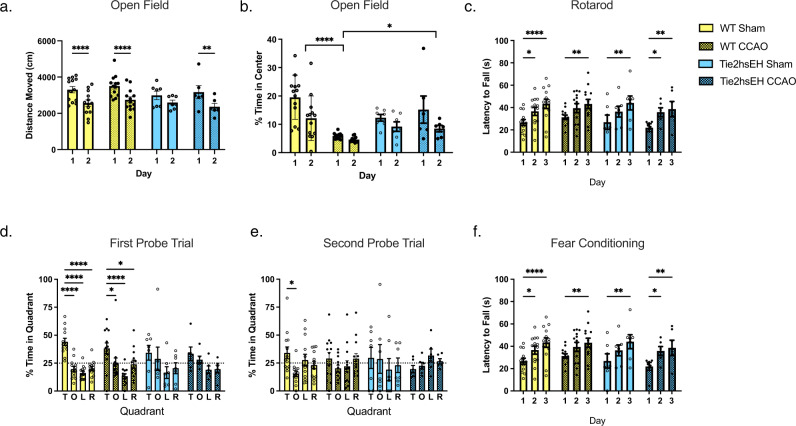
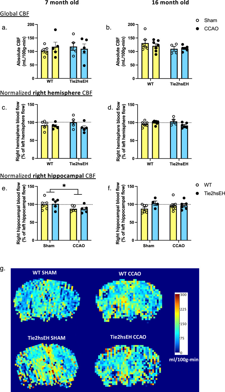
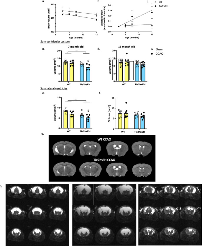
Soluble epoxide hydrolase (sEH) is upregulated in microvascular endothelium of human brain with vascular cognitive impairment (VCI). Transgenic endothelial expression of human sEH in mice (Tie2hsEH) induces endothelial dysfunction (ED), a pathogenetic mechanism of VCI. We sought to determine if endothelial upregulation of sEH is sufficient to cause cognitive impairment, and if cognitive impairment due to chronic hypoperfusion induced by unilateral common carotid artery occlusion (CCAO) is exacerbated in Tie2hsEH mice. Behavioral performance was assessed by the open field, rotarod, novel object, Morris water maze and fear conditioning tests. Cerebral blood flow and brain morphology were evaluated by MRI, and inflammatory changes investigated using immunohistochemistry and flow cytometry. We demonstrate that transgenic endothelial expression of sEH is sufficient to induce cognitive impairment, associated with leukocyte infiltration, brain atrophy and accelerated, age-dependent ventriculomegaly, identifying ED and sEH upregulation as potential underlying mechanisms and therapeutic targets for VCI.


Conflict of interest statement

Financial competing interests are as follows: N.J.A. is co-inventor of technologies related to sEH and EETs signaling that have been licensed by OHSU and JHU to Precision Molecular and Vasocardea. This potential conflict of interest has been reviewed and managed by OHSU. All other authors declare no competing interests.

Figures

Fig. 1
Fig. 1. Endothelial DHETs are increased in Tie2hsEH mice and by CCAO, plasma EETs are decreased.
Four months following surgery DHETs were assessed in acutely isolated cerebral endothelial cells (a) and EETs in plasma (b) by LC–MS/MS. a Endothelial 14,15-DHETs were increased in Tie2hEH mice compared to WT, 11,12- and 8,9-DHETs are also increased in Tie2hsEH sham endothelium compared to WT sham; 14,15-, 11,12- and 8,9-DHET were all increased by CCAO surgery in WT but not Tie2hsEH mice (n = 3–4/group). b Plasma 14,15-EETs were reduced in Tie2hsEH mice compared to WT; there was no effect of surgery. *p < 0.05, **p < 0.01, 2-way ANOVA, n = 4–8/group. Bar graphs display mean ± SEM.
Fig. 2
Fig. 2. Tie2hsEH genotype and CCAO lead to impaired spatial memory retention.
Three months after surgery behavioral performance was assessed. Locomotor activity and anxiety- related behavior were assessed by the open field test. a Locomotor activity was unaffected by genotype or surgery. WT sham, WT CCAO and Tie2hsEH UCCAO groups all exhibited reduced distance moved on day 2 of testing. **p < 0.01, ****p < 0.0001 vs. day 1 of the same group, 2-way ANOVA with Sidak’s multiple comparisons test, n = 6–13/group. b Anxiety-related behavior was determed by time spent in the more anxiety-provoking center of the open field; time spent in the center was reduced by CCAO compared to sham in WT. Time in center was unaffected by genotype in sham, or by CCAO in Tie2hsEH, compared to their sham. *p < 0.05, ****p < 0.0001, 2-way ANOVA with Tukey’s multiple comparisons test, n = 7–13/group. c Locomotor performance was assessed by rotarod test. All groups, regardless of surgery or genotype improved performance over the 3 days of testing; no differences were detected between groups. *p < 0.05, **p < 0.01, ****p < 0.0001 vs. trial 1 of the same group, 2-way ANOVA with Tukey’s multiple comparison test. Spatial learning and memory were assessed in the Morris water maze test. d In the first probe trial, WT sham and CCAO mice spend more time in the target quadrant than the other quadrants. Tie2hsEH mice, both sham and CCAO, did not show preference for a particular quadrant. e In the second probe trial, WT sham mice spend more time in the target compared to the opposite quadrant, however the WT CCAO group did not. In the second probe trial, neither Tie2hsEH sham nor CCAO showed preference for a quadrant. T target, O opposite, L left, R right. *p < 0.05, ****p < 0.0001, 2-way ANOVA with Tukey’s multiple comparison test, n = 6–13/treatment. f No differences between groups were observed in contextual fear memory tasks, 2-way ANOVA, n = 6–13 mice/genotype/treatment. Bar graphs display mean ± SEM.
Fig. 3
Fig. 3. Unilateral hippocampal decrease in CBF in CCAO mice.
CBF was measured using ASL-MRI perfusion in 7- and 16-month-old mice, 4 months following CCAO surgery. Global CBF, and right hemispheric CBF, were unaffected by surgery in either genotype (ad). Hippocampal blood flow in the ipsilateral hemisphere was reduced in both WT and Tie2hsEH 7-month-old, but not 16-month-old mice (e, f). g Representative CBF maps from 7-month-old cohort. *p < 0.05, 2-way ANOVA, n = 4–6/group, data are represented as mean ± SEM.
Fig. 4
Fig. 4. Age-dependent atrophy and ventriculomegaly in Tie2hsEH mice.
Ventricular and total brain volume were measured by T2-weighted MRI. Longitudinal T2-weighted MRI shows that Tie2hsEH mice have a smaller brain volume than WT at 7 and 8 months of age (a), *p < 0.05, **p < 0.01, 2-way ANOVA with Sidak’s multiple comparison test, n = 4–18/group. Ventricle size, normalized to brain volume, increases with age in Tie2hsEH mice, but not WT (b); linear regression analysis shows that the slope of Tie2hsEH is steeper than that of WT (0.06 vs. 0.006, respectively, p = 0.0038), with increased ventricle/brain ratio in Tie2hsEH mice at 9 and 12 months of age, *p < 0.05, 2-way ANOVA with Sidak’s multiple comparison test, n = 3–14/group. Ventricular volume is reduced in Tie2hsEH mice compared to WT, in 7-month-old mice (c), but not in 16-month-old-mice (d); CCAO surgery does not alter ventricle volume in either age group. Lateral ventricles are also reduced in volume in the 7- but not 16-month cohort (e, f); a reduction in volume is also observed by CCAO surgery in the 7- but not 16-month cohort. *p < 0.05, ***p < 0.001, #p < 0.05 WT sham vs. Tie2hsEH sham, $p < 0.05 WT CCAO vs. Tie2hsEH CCAO, 2-way ANOVA with Sidak’s multiple comparison test, n = 5–7/group. Representative sequential T2-weighted image slices are shown of both genotypes at 7 months of age (g). Severe hydrocephalus was observed in 3 out of 18 naïve12-month-old Tie2hsEH mice, but not WT, by ASL-MRI perfusion (n/s Fisher’s exact test); MRI images of the 3 hydrocephalic Tie2hsEH brains (h). Data are represented as mean ± SEM.
Fig. 5
Fig. 5. Increased leukocyte infiltration in Tie2hsEH mice.
CD45, Iba1 and GFAP were assessed by immunohistochemistry, leukocytes and neutrophils by flow cytometry, in 7-month-old mice. Iba1 immunoreactivity is increased in fornix by CCAO in Tie2hsEH mice, but not in WT (a). GFAP levels are unaffected in fornix by either genotype or surgery (b); representative images of GFAP (red) and Iba1 (green) immunolabelling (c). CD45-positive cells within brain parenchyma are increased in Tie2hsEH vs. WT (d); expression is unaffected by CCAO surgery, e representative image of CD45-positive cells by immunolabeling. Genotype-specific increase in leukocytes is also observed in naïve mice by flow cytometry; infiltrating leukocytes were determined by high expression of CD45, utilizing CD11b staining to improve resolution of leukocytes from microglia (f, g). Further analysis reveals that this increase is characterized by selective recruitment of neutrophils, defined as CD45hiCD11b + CD11c-Ly6cmidSSChi (h, i). *p < 0.05, 2-way ANOVA with Sidak’s multiple comparisons test (histology), two-tailed t-test (flow cytometry), n = 4–6/group, data are represented as mean ± SEM.

Similar articles

Cited by

References

    1. Iadecola C. Vascular and metabolic factors in Alzheimer’s disease and related dementias: introduction. Cell. Mol. Neurobiol. 2016;36:151–154. doi: 10.1007/s10571-015-0319-y. - DOI - PMC - PubMed
    1. Snowdon DA, et al. Brain infarction and the clinical expression of Alzheimer disease. The Nun Study. JAMA. 1997;277:813–817. doi: 10.1001/jama.1997.03540340047031. - DOI - PubMed
    1. Wang F, et al. Dysfunction of cerebrovascular endothelial cells: prelude to vascular dementia. Front. Aging Neurosci. 2018;10:376. doi: 10.3389/fnagi.2018.00376. - DOI - PMC - PubMed
    1. Davis CM, Liu X, Alkayed NJ. Cytochrome P450 eicosanoids in cerebrovascular function and disease. Pharmacol. Ther. 2017;179:31–46. doi: 10.1016/j.pharmthera.2017.05.004. - DOI - PMC - PubMed
    1. Zuloaga KL, et al. High fat diet-induced diabetes in mice exacerbates cognitive deficit due to chronic hypoperfusion. J. Cereb. Blood Flow Metab. 2016;36:1257–1270. doi: 10.1177/0271678X15616400. - DOI - PMC - PubMed