Neuro-oncological Ventral Antigen 2 Regulates Splicing of Vascular Endothelial Growth Factor Receptor 1 and Is Required for Endothelial Function
- PMID: 35927413
- PMCID: PMC9988812
- DOI: 10.1007/s43032-022-01044-4
Neuro-oncological Ventral Antigen 2 Regulates Splicing of Vascular Endothelial Growth Factor Receptor 1 and Is Required for Endothelial Function
Abstract
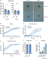
Pre-eclampsia (PE) affects 2-8% of pregnancies and is responsible for significant morbidity and mortality. The maternal clinical syndrome (defined by hypertension, proteinuria, and organ dysfunction) is the result of endothelial dysfunction. The endothelial response to increased levels of soluble FMS-like Tyrosine Kinase 1 (sFLT1) is thought to play a central role. sFLT1 is released from multiple tissues and binds VEGF with high affinity and antagonizes VEGF. Expression of soluble variants of sFLT1 is a result of alternative splicing; however, the mechanism is incompletely understood. We hypothesize that neuro-oncological ventral antigen 2 (NOVA2) contributes to this. NOVA2 was inhibited in human umbilical vein endothelial cells (HUVECs) and multiple cellular functions were assessed. NOVA2 and FLT1 expression in the placenta of PE, pregnancy-induced hypertension, and normotensive controls was measured by RT-qPCR. Loss of NOVA2 in HUVECs resulted in significantly increased levels of sFLT1, but did not affect expression of membrane-bound FLT1. NOVA2 protein was shown to directly interact with FLT1 mRNA. Loss of NOVA2 was also accompanied by impaired endothelial functions such as sprouting. We were able to restore sprouting capacity by exogenous VEGF. We did not observe statistically significant regulation of NOVA2 or sFLT1 in the placenta. However, we observed a negative correlation between sFLT1 and NOVA2 expression levels. In conclusion, NOVA2 was found to regulate FLT1 splicing in the endothelium. Loss of NOVA2 resulted in impaired endothelial function, at least partially dependent on VEGF. In PE patients, we observed a negative correlation between NOVA2 and sFLT1.
Keywords: Alternative Splicing; Endothelium; FLT1; Placenta; Pre-eclampsia.
© 2022. The Author(s).
Conflict of interest statement
The authors declare no competing interests.
Figures


Similar articles
-
Soluble FLT1 sensitizes endothelial cells to inflammatory cytokines by antagonizing VEGF receptor-mediated signalling.Cardiovasc Res. 2011 Feb 15;89(3):671-9. doi: 10.1093/cvr/cvq346. Epub 2010 Dec 7. Cardiovasc Res. 2011. PMID: 21139021 Free PMC article.
-
Low-dose aspirin reduces hypoxia-induced sFlt1 release via the JNK/AP-1 pathway in human trophoblast and endothelial cells.J Cell Physiol. 2019 Aug;234(10):18928-18941. doi: 10.1002/jcp.28533. Epub 2019 Apr 19. J Cell Physiol. 2019. PMID: 31004367
-
A novel human-specific soluble vascular endothelial growth factor receptor 1: cell-type-specific splicing and implications to vascular endothelial growth factor homeostasis and preeclampsia.Circ Res. 2008 Jun 20;102(12):1566-74. doi: 10.1161/CIRCRESAHA.108.171504. Epub 2008 May 30. Circ Res. 2008. PMID: 18515749
-
Placental-specific sFLT-1: role in pre-eclamptic pathophysiology and its translational possibilities for clinical prediction and diagnosis.Mol Hum Reprod. 2017 Feb 10;23(2):69-78. doi: 10.1093/molehr/gaw077. Mol Hum Reprod. 2017. PMID: 27986932 Review.
-
Soluble fms-like tyrosine kinase 1 and soluble endoglin are elevated circulating anti-angiogenic factors in pre-eclampsia.Pregnancy Hypertens. 2012 Oct;2(4):358-67. doi: 10.1016/j.preghy.2012.06.003. Epub 2012 Jul 6. Pregnancy Hypertens. 2012. PMID: 26105603 Review.
Cited by
-
Alternative Splicing Changes Promoted by NOVA2 Upregulation in Endothelial Cells and Relevance for Gastric Cancer.Int J Mol Sci. 2023 Apr 30;24(9):8102. doi: 10.3390/ijms24098102. Int J Mol Sci. 2023. PMID: 37175811 Free PMC article.
References
-
- Palmer KR, Tong S, Kaitu'u-Lino TJ. Placental-specific sFLT-1: role in pre-eclamptic pathophysiology and its translational possibilities for clinical prediction and diagnosis. Mol Hum Repr. 2016;23(2):69–78. - PubMed
Publication types
MeSH terms
Substances
LinkOut - more resources
Full Text Sources
Medical
Molecular Biology Databases
Miscellaneous

