. 2022 Jul 18;8(1):10.
doi: 10.1038/s41514-022-00091-0.

Paracrine-mediated rejuvenation of aged mesenchymal stem cells is associated with downregulation of the autophagy-lysosomal pathway

Affiliations

Paracrine-mediated rejuvenation of aged mesenchymal stem cells is associated with downregulation of the autophagy-lysosomal pathway

George Hung et al. NPJ Aging. .

Abstract

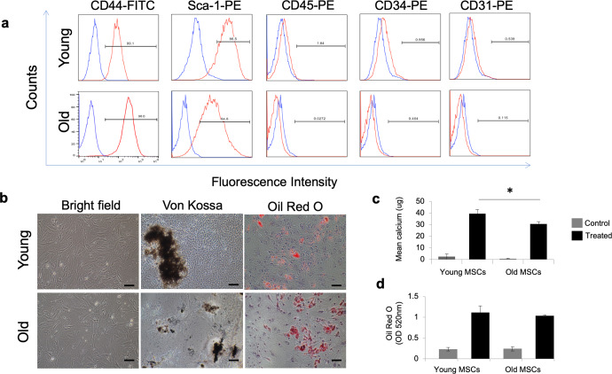
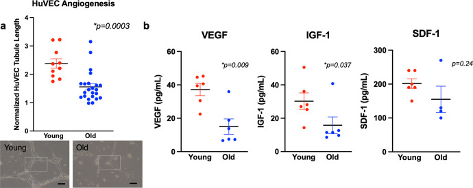
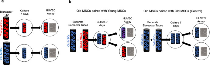
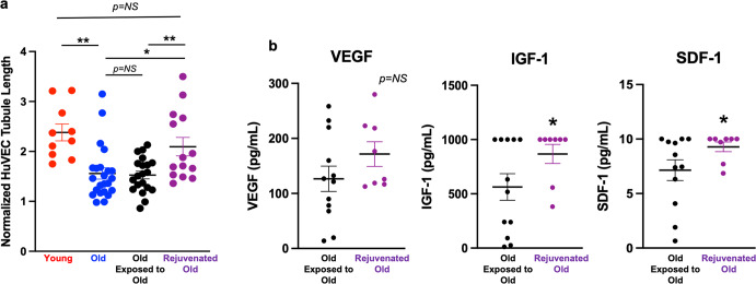
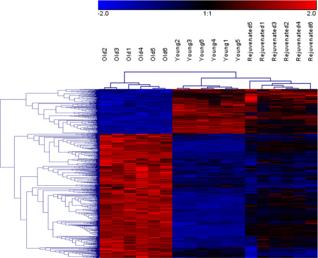
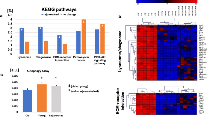
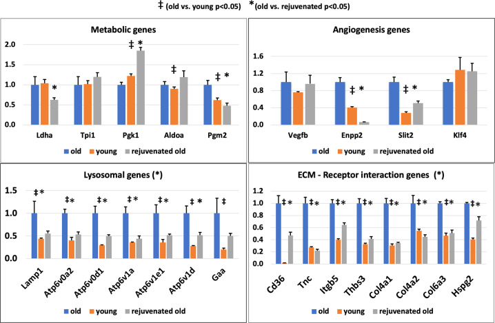
Age-related differences in stem-cell potency contribute to variable outcomes in clinical stem cell trials. To help understand the effect of age on stem cell potency, bone marrow-derived mesenchymal stem cells (MSCs) were isolated from young (6 weeks) and old (18-24 months) mice. HUVEC tubule formation (TF) induced by the old and young MSCs and ELISA of conditioned media were compared to one another, and to old MSCs after 7 d in indirect co-culture with young MSCs. Old MSCs induced less TF than did young (1.56 ± 0.11 vs 2.38 ± 0.17, p = 0.0003) and released lower amounts of VEGF (p = 0.009) and IGF1 (p = 0.037). After 7 d in co-culture with young MSCs, TF by the old MSCs significantly improved (to 2.09 ± 0.18 from 1.56 ± 0.11; p = 0.013), and was no longer different compared to TF from young MSCs (2.09 ± 0.18 vs 2.38 ± 0.17; p = 0.27). RNA seq of old MSCs, young MSCs, and old MSCs following co-culture with young MSCs revealed that the age-related differences were broadly modified by co-culture, with the most significant changes associated with lysosomal pathways. These results indicate that the age-associated decreased paracrine-mediated effects of old MSCs are improved following indirect co-culture with young MSC. The observed effect is associated with broad transcriptional modification, suggesting potential targets to both assess and improve the therapeutic potency of stem cells from older patients.


Conflict of interest statement

The authors declare no competing interests.

Figures

Fig. 1
Fig. 1. MSC characterization and in vitro differentiation capacity.
a Flow cytometry results show that MSCs from both young and aged donor mice were uniformly positive for MSCs markers CD44 and Sca-1 and negative for hematopoietic stem cells markers CD34 and CD45 and endothelial marker CD31 (n = 6). b MSCs from young and aged donor mice display fibroblast-like morphology on bright field microscopy (left panels; scale bar = 20 μm). Following exposure to differentiation media MSCs from both young and aged mice showed osteogenic (middle panels) and adipogenic (right panels) differentiation as detected by Von Kossa and oil red O staining, respectively. c Quantitative assessment of calcium deposition showed significantly less calcium deposition in aged MSCs compared to young following exposure to differentiation media (p ≤ 0.05; t test; n = 3 each group). d There was no significant difference in lipid content between groups (t test; n = 3 each). MSCs cultured in control medium served as negative control for both conditions. Error bars show s.d.
Fig. 2
Fig. 2. HUVEC tubule formation and relevant PF release: young v old.
a Young MSCs housed in bioreactor tubes (n = 10) stimulated significantly greater TF by HUVECs over 18 h than did the same number of old MSCs (n = 23; p < 0.001; permutation test; scale bar = 20 μm). b Conditioned media from young MSCs produced significantly greater amounts of VEGF (p = 0.009; Mann–Whitney; n = 6 for each group) and IGF-1 (p = 0.047; Mann–Whitney; n = 6 for each group) than did conditioned media from old MSCs; there was no significant difference in SDF-1 production (Mann–Whitney; n = 6 for young, n = 4 for old). Error bars show s.e.m.
Fig. 3
Fig. 3. Workflow for human umbilical vascular endothelial cell (HUVEC) angiogenesis experiments.
a The same number of MSCs obtained from old or young C57BL mice were placed in separate bioreactor tubes and grown at standard culture conditions. After 7 d the bioreactor tubes were placed separately in plates containing HUVECs in media without any supplement. Tubule formation (TF) was assessed after 18 h. b For the rejuvenation experiments, two separate bioreactor tubes were placed in the same well for 7 d. Pairings included old MSCs grown with separate old MSCs (control) and old MSCs grown separately with young MSCs (rejuvenation). After 7 d the bioreactor tubes were removed and used separately to stimulate HUVEC angiogenesis with TF assessed after 18 h.
Fig. 4
Fig. 4. Rejuvenating effect of exposure of old MSCs to young MSCs on HUVEC tubule formation and PF release.
a Co-culture of old MSCs with young MSCs housed in a separate bioreactor tube for 7 d (“Old × Young”; n = 14 in each group) “rejuvenated” the old MSCs. TF stimulated by the rejuvenated old MSCs was significantly greater than that of unexposed old MSCs and that of old MSCs grown in co-culture for 7 d with other old MSCs (“Old × Old”; n = 12) and was not significantly different from that of the young MSCs. b The rejuvenated old MSCs also produced significantly greater amounts of IGF-1 (p < 0.05; Mann–Whitney) and SDF-1 (p < 0.05; Mann–Whitney) than did old MSCs paired with other old MSCs; there was no significant difference in the release of VEGF (p = NS; Mann–Whitney). For figures: *p < 0.05, **p ≤ 0.01. Error bars show s.e.m.
Fig. 5
Fig. 5. Principal component analysis (PCA) of > 20,000 analyzed genes.
PCA of young, old, and rejuvenated old MSCs (6 replicates for each condition) reveals three distinct gene expression patterns.
Fig. 6
Fig. 6. Differentially expressed genes (DEGs) with more “youthful” expression in rejuvenated old MSCs.
Of the 2831 observed DEGs between the old and the rejuvenated old MSCs, 831 of those in the rejuvenated MSCs had more “youthful” expression, i.e. expression of DEGs that was more like that of the young MSCs, than that of the old MSCs. Gene expression levels are color-coded with blue and red representing low and high expression levels, respectively.
Fig. 7
Fig. 7. KEGG pathway analysis shows more youthful expression of lysosomal pathways in rejuvenated old MSCs.
a Over-represented functional categories based on KEGG pathways of the 831 transcripts associated with a more youthful expression profile (* indicates false discovery rate FDR < 5%). b Hierarchical clustering of genes of the lysosome/phagosome (upper panel) and ECM–receptor interaction (bottom panel). Gene expression levels are color-coded with blue and red representing low and high expression levels, respectively. Expression levels of the rejuvenated old cells cluster with those of the young MSCs rather than those with the old cells. c Results from a functional autophagy assay comparing old, young, and rejuvenated old MSCs showed significant differences between old and young MSCs (p < 0.05; t test) and old vs rejuvenated old MSCs (*p < 0.05; t test). [a.u.] indicates arbitrary units. Error bars show s.e.m.
Fig. 8
Fig. 8. RT-PCR of selected transcripts confirms more youthful expression of rejuvenated MSCs.
rtPCR of selected transcripts identified as having more youthful expression in RNA sequencing analysis showed broad agreement, with 20/24 factors showing more youthful expression in rejuvenated MSCs compared to old MSCs when normalized to housekeeping gene 18S. Selected transcripts include those associated with metabolism, ECM–Receptor interaction, autophagy / lysosome, and angiogenesis. p < 0.05 for old vs. young (t test). *p < 0.05 for old vs. rejuvenated (t test). Error bars show s.e.m.

References

    1. Spyridopoulos I, et al. Telomere gap between granulocytes and lymphocytes is a determinant for hematopoetic progenitor cell impairment in patients with previous myocardial infarction. Arterioscler. Thromb. Vasc. Biol. 2008;28:968–974. doi: 10.1161/ATVBAHA.107.160846. - DOI - PubMed
    1. Fan M, et al. The effect of age on the efficacy of human mesenchymal stem cell transplantation after a myocardial infarction. Rejuvenation Res. 2010;13:429–438. doi: 10.1089/rej.2009.0986. - DOI - PubMed
    1. Perin EC, et al. A randomized study of transendocardial injection of autologous bone marrow mononuclear cells and cell function analysis in ischemic heart failure (FOCUS-HF) Am. Heart J. 2011;161:1078–1087.e1073. doi: 10.1016/j.ahj.2011.01.028. - DOI - PubMed
    1. Bhatnagar A, et al. Bone marrow cell characteristics associated with patient profile and cardiac performance outcomes in the LateTIME-Cardiovascular Cell Therapy Research Network (CCTRN) trial. Am. Heart J. 2016;179:142–150. doi: 10.1016/j.ahj.2016.06.018. - DOI - PMC - PubMed
    1. Contreras A, et al. Identification of cardiovascular risk factors associated with bone marrow cell subsets in patients with STEMI: a biorepository evaluation from the CCTRN TIME and LateTIME clinical trials. Basic Res. Cardiol. 2017;112:3. doi: 10.1007/s00395-016-0592-z. - DOI - PMC - PubMed