

Glycan shield of the ebolavirus envelope glycoprotein GP
- PMID: 35927436
- PMCID: PMC9352669
- DOI: 10.1038/s42003-022-03767-1
Glycan shield of the ebolavirus envelope glycoprotein GP
Abstract
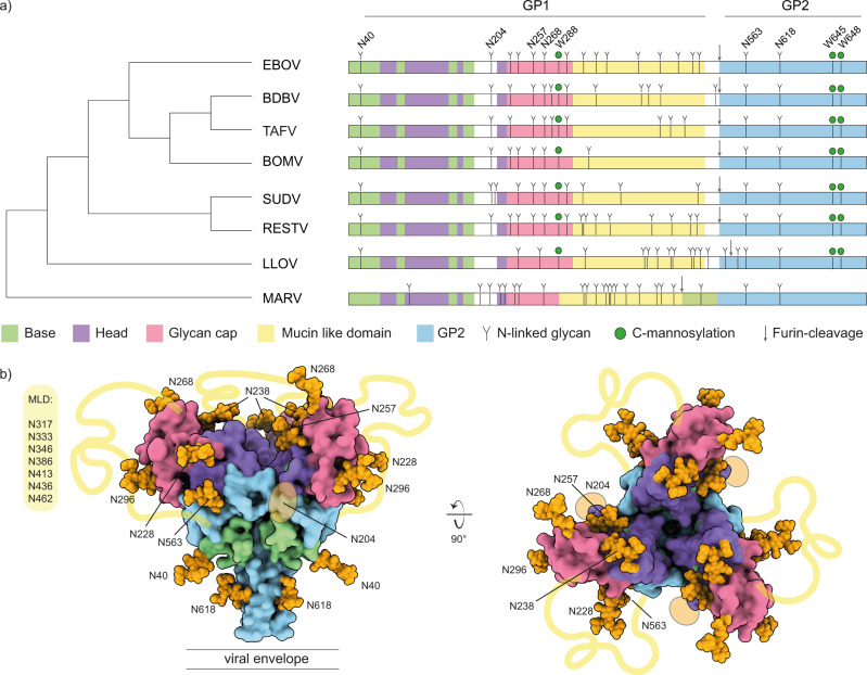
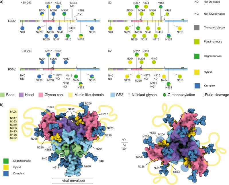
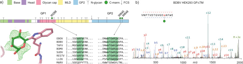
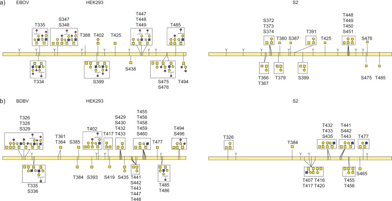
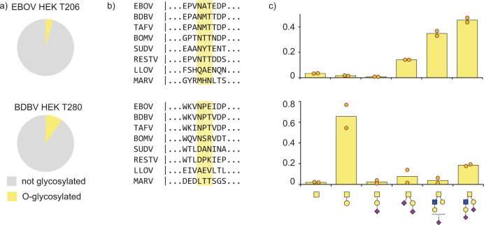
The envelope glycoprotein GP of the ebolaviruses is essential for host cell entry and the primary target of the host antibody response. GP is heavily glycosylated with up to 17 N-linked sites, numerous O-linked glycans in its disordered mucin-like domain (MLD), and three predicted C-linked mannosylation sites. Glycosylation is important for host cell attachment, GP stability and fusion activity, and shielding from neutralization by serum antibodies. Here, we use glycoproteomics to profile the site-specific glycosylation patterns of ebolavirus GP. We detect up to 16 unique O-linked glycosylation sites in the MLD, and two O-linked sites in the receptor-binding GP1 subunit. Multiple O-linked glycans are observed within N-linked glycosylation sequons, suggesting crosstalk between the two types of modifications. We confirmed C-mannosylation of W288 in full-length trimeric GP. We find complex glycosylation at the majority of N-linked sites, while the conserved sites N257 and especially N563 are enriched in unprocessed glycans, suggesting a role in host-cell attachment via DC-SIGN/L-SIGN. Our findings illustrate how N-, O-, and C-linked glycans together build the heterogeneous glycan shield of GP, guiding future immunological studies and functional interpretation of ebolavirus GP-antibody interactions.
© 2022. The Author(s).
Conflict of interest statement
The authors declare no competing interests.
Figures
Similar articles
-
Targeting host O-linked glycan biosynthesis affects Ebola virus replication efficiency and reveals differential GalNAc-T acceptor site preferences on the Ebola virus glycoprotein.J Virol. 2024 Jun 13;98(6):e0052424. doi: 10.1128/jvi.00524-24. Epub 2024 May 17. J Virol. 2024. PMID: 38757972 Free PMC article.
-
Comprehensive functional analysis of N-linked glycans on Ebola virus GP1.mBio. 2014 Jan 28;5(1):e00862-13. doi: 10.1128/mBio.00862-13. mBio. 2014. PMID: 24473128 Free PMC article.
-
Comparison of N- and O-linked glycosylation patterns of ebolavirus glycoproteins.Virology. 2017 Feb;502:39-47. doi: 10.1016/j.virol.2016.12.010. Epub 2016 Dec 13. Virology. 2017. PMID: 27984785
-
Structure and Role of O-Linked Glycans in Viral Envelope Proteins.Annu Rev Virol. 2023 Sep 29;10(1):283-304. doi: 10.1146/annurev-virology-111821-121007. Epub 2023 Jul 6. Annu Rev Virol. 2023. PMID: 37285578 Review.
-
Glycan Shielding and Modulation of Hepatitis C Virus Neutralizing Antibodies.Front Immunol. 2018 Apr 27;9:910. doi: 10.3389/fimmu.2018.00910. eCollection 2018. Front Immunol. 2018. PMID: 29755477 Free PMC article. Review.
Cited by
-
Ebola Virus Activates IRE1α-Dependent XBP1u Splicing.Viruses. 2022 Dec 30;15(1):122. doi: 10.3390/v15010122. Viruses. 2022. PMID: 36680162 Free PMC article.
-
Targeting host O-linked glycan biosynthesis affects Ebola virus replication efficiency and reveals differential GalNAc-T acceptor site preferences on the Ebola virus glycoprotein.J Virol. 2024 Jun 13;98(6):e0052424. doi: 10.1128/jvi.00524-24. Epub 2024 May 17. J Virol. 2024. PMID: 38757972 Free PMC article.
-
Quantification and Site-Specific Analysis of Co-occupied N- and O-Glycopeptides.J Proteome Res. 2024 Dec 6;23(12):5449-5461. doi: 10.1021/acs.jproteome.4c00574. Epub 2024 Nov 5. J Proteome Res. 2024. PMID: 39498894 Free PMC article.
-
The GATE glycoprotein complex enhances human cytomegalovirus entry in endothelial cells.Nat Microbiol. 2025 Jul;10(7):1605-1616. doi: 10.1038/s41564-025-02025-4. Epub 2025 Jun 30. Nat Microbiol. 2025. PMID: 40588592 Free PMC article.
-
Divergent antibody recognition profiles are generated by protective mRNA vaccines against Marburg and Ravn viruses.Res Sq [Preprint]. 2024 Mar 26:rs.3.rs-4087897. doi: 10.21203/rs.3.rs-4087897/v1. Res Sq. 2024. Update in: Nat Commun. 2025 Jul 1;16(1):5702. doi: 10.1038/s41467-025-60057-0. PMID: 38585993 Free PMC article. Updated. Preprint.
References
Publication types
MeSH terms
Substances
Grants and funding
LinkOut - more resources
Full Text Sources
Medical
Molecular Biology Databases
