

Molecular profile and response to energy deficit of leptin-receptor neurons in the lateral hypothalamus
- PMID: 35927440
- PMCID: PMC9352899
- DOI: 10.1038/s41598-022-16492-w
Molecular profile and response to energy deficit of leptin-receptor neurons in the lateral hypothalamus
Abstract
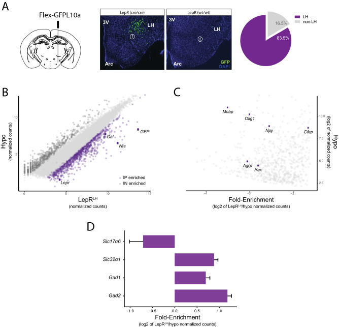
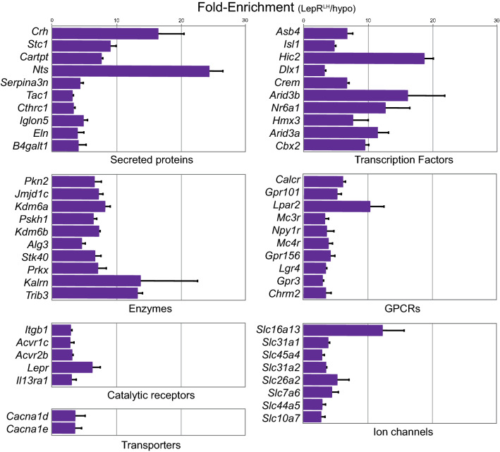
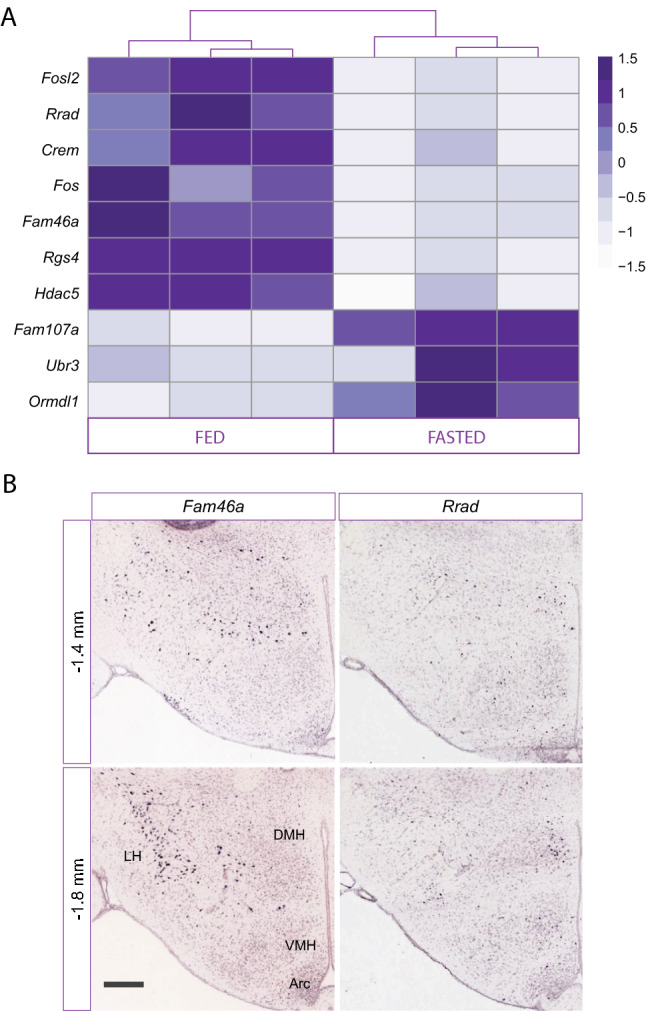
Leptin exerts its effects on energy balance by inhibiting food intake and increasing energy expenditure via leptin receptors in the hypothalamus. While LepR neurons in the arcuate nucleus of the hypothalamus, the primary target of leptin, have been extensively studied, LepR neurons in other hypothalamic nuclei remain understudied. LepR neurons in the lateral hypothalamus contribute to leptin's effects on food intake and reward, but due to the low abundance of this population it has been difficult to study their molecular profile and responses to energy deficit. We here explore the transcriptome of LepR neurons in the LH and their response to energy deficit. Male LepR-Cre mice were injected in the LH with an AAV carrying Cre-dependent L10:GFP. Few weeks later the hypothalami from fed and food-restricted (24-h) mice were dissected and the TRAP protocol was performed, for the isolation of translating mRNAs from LepR cells in the LH, followed by RNA sequencing. After mapping and normalization, differential expression analysis was performed with DESeq2. We confirm that the isolated mRNA is enriched in LepR transcripts and other known neuropeptide markers of LepRLH neurons, of which we investigate the localization patterns in the LH. We identified novel markers of LepRLH neurons with association to energy balance and metabolic disease, such as Acvr1c, Npy1r, Itgb1, and genes that are differentially regulated by food deprivation, such as Fam46a and Rrad. Our dataset provides a reliable and extensive resource of the molecular makeup of LH LepR neurons and their response to food deprivation.
© 2022. The Author(s).
Conflict of interest statement
The authors declare no competing interests.
Figures

Similar articles
-
Disrupted Leptin Signaling in the Lateral Hypothalamus and Ventral Premammillary Nucleus Alters Insulin and Glucagon Secretion and Protects Against Diet-Induced Obesity.Endocrinology. 2016 Jul;157(7):2671-85. doi: 10.1210/en.2015-1998. Epub 2016 May 16. Endocrinology. 2016. PMID: 27183315
-
Effects of GABA and Leptin Receptor-Expressing Neurons in the Lateral Hypothalamus on Feeding, Locomotion, and Thermogenesis.Obesity (Silver Spring). 2019 Jul;27(7):1123-1132. doi: 10.1002/oby.22495. Epub 2019 May 14. Obesity (Silver Spring). 2019. PMID: 31087767 Free PMC article.
-
Altered function of arcuate leptin receptor expressing neuropeptide Y neurons depending on energy balance.Mol Metab. 2023 Oct;76:101790. doi: 10.1016/j.molmet.2023.101790. Epub 2023 Aug 9. Mol Metab. 2023. PMID: 37562743 Free PMC article.
-
Genetic dissection of neuronal pathways controlling energy homeostasis.Obesity (Silver Spring). 2006 Aug;14 Suppl 5:222S-227S. doi: 10.1038/oby.2006.313. Obesity (Silver Spring). 2006. PMID: 17021371 Review.
-
The growing complexity of the control of the hypothalamic pituitary thyroid axis and brown adipose tissue by leptin.Vitam Horm. 2025;127:305-362. doi: 10.1016/bs.vh.2024.07.005. Epub 2024 Aug 21. Vitam Horm. 2025. PMID: 39864945 Review.
Cited by
-
The Integrated Function of the Lateral Hypothalamus in Energy Homeostasis.Cells. 2025 Jul 8;14(14):1042. doi: 10.3390/cells14141042. Cells. 2025. PMID: 40710295 Free PMC article. Review.
-
Control of energy homeostasis by the lateral hypothalamic area.Trends Neurosci. 2023 Sep;46(9):738-749. doi: 10.1016/j.tins.2023.05.010. Epub 2023 Jun 22. Trends Neurosci. 2023. PMID: 37353461 Free PMC article. Review.
-
Lateral hypothalamus and eating: cell types, molecular identity, anatomy, temporal dynamics and functional roles.Exp Mol Med. 2025 May;57(5):925-937. doi: 10.1038/s12276-025-01451-y. Epub 2025 May 1. Exp Mol Med. 2025. PMID: 40307571 Free PMC article. Review.
References
-
- Rezai-Zadeh K, Yu S, Jiang Y, Laque A, Schwartzenburg C, Morrison CD, Derbenev AV, Zsombok A, Münzberg H. Leptin receptor neurons in the dorsomedial hypothalamus are key regulators of energy expenditure and body weight, but not food intake. Mol. Metab. 2014;3(7):681–693. doi: 10.1016/j.molmet.2014.07.008. - DOI - PMC - PubMed
Publication types
MeSH terms
Substances
LinkOut - more resources
Full Text Sources
Molecular Biology Databases
Miscellaneous
