

Melatonin enhances autologous adipose-derived stem cells to improve mouse ovarian function in relation to the SIRT6/NF-κB pathway
- PMID: 35927704
- PMCID: PMC9351187
- DOI: 10.1186/s13287-022-03060-2
Melatonin enhances autologous adipose-derived stem cells to improve mouse ovarian function in relation to the SIRT6/NF-κB pathway
Abstract
Background: Premature ovarian insufficiency (POI) is the main cause of female infertility. Adipose-derived stem cells (ADSCs) are ideal candidates for the treatment of POI. However, some deficient biological characteristics of ADSCs limit their utility. This study investigated whether melatonin (MLT)-pretreated autologous ADSCs were superior to ADSCs alone in the treatment of the POI mouse model.
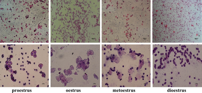
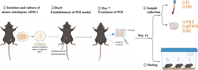
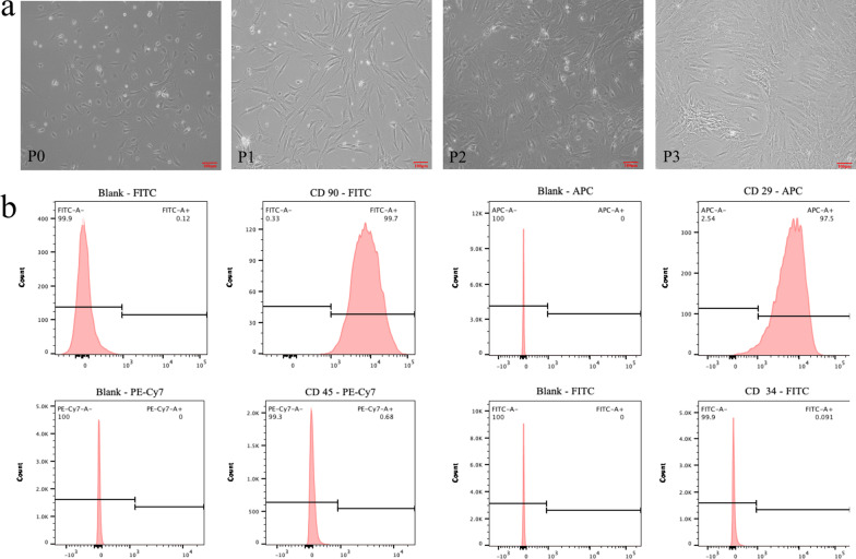
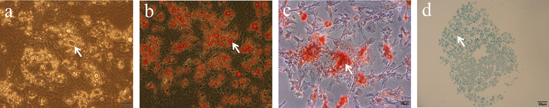
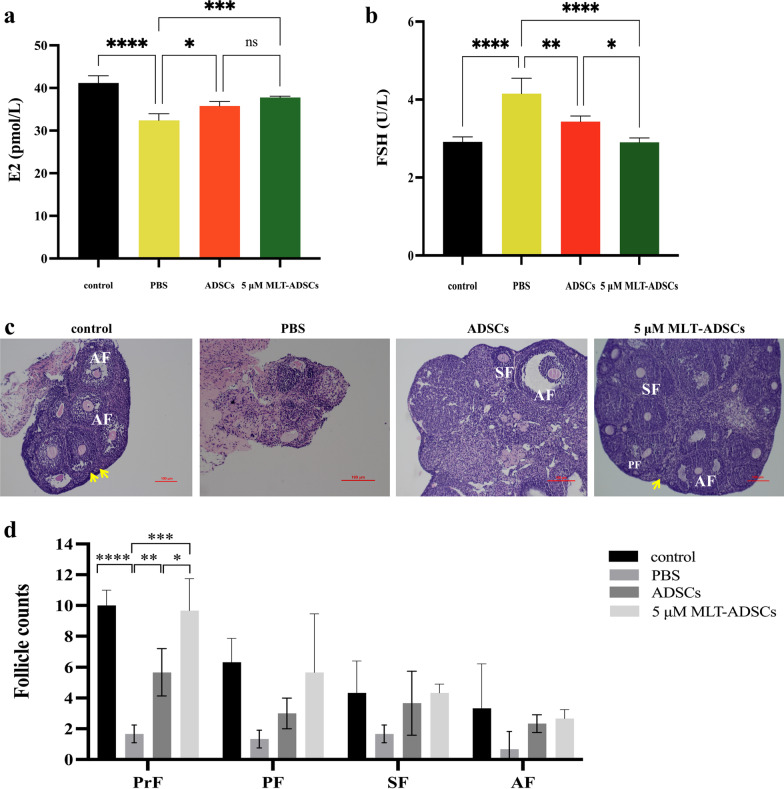
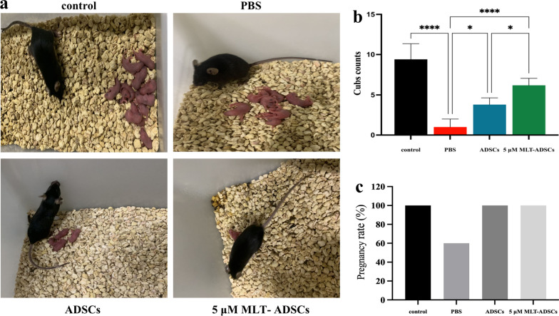
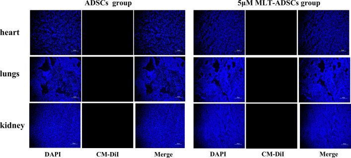
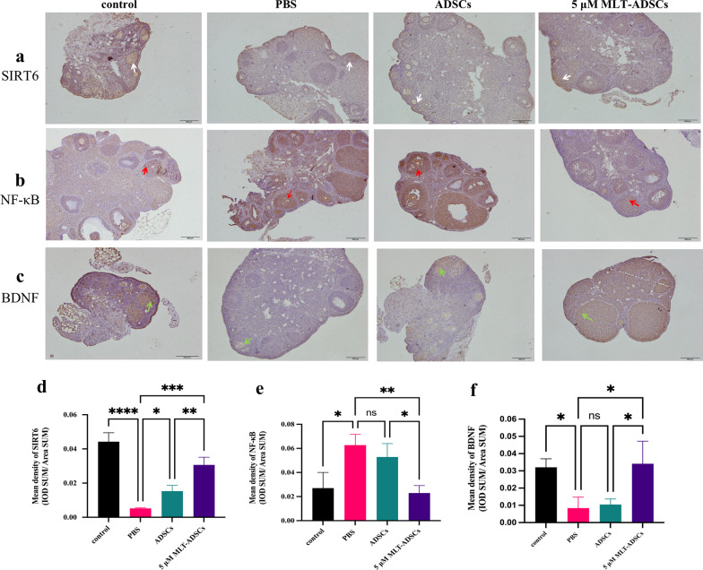
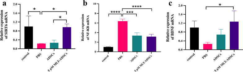
Methods: Autologous ADSCs were isolated and cultured in MLT-containing medium. Surface markers of ADSCs were detected by flow cytometry. To determine the effect of MLT on ADSCs, CCK-8 assay was used to detect ADSCs proliferation and enzyme-linked immunosorbent assay (ELISA) was used to detect the secretion of cytokines. The POI model was established by intraperitoneal injection of cyclophosphamide and busulfan. Then, MLT-pretreated autologous ADSCs were transplanted into mice by intraovarian injection. After 7 days of treatment, ovarian morphology, follicle counts, and sex hormones levels were evaluated by hematoxylin and eosin (H&E) staining and ELISA, and the recovery of fertility was also observed. The expressions of SIRT6 and NF-κB were detected by immunohistochemical (IHC) staining and quantitative real-time polymerase chain reaction (qRT-PCR).
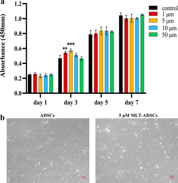
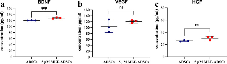
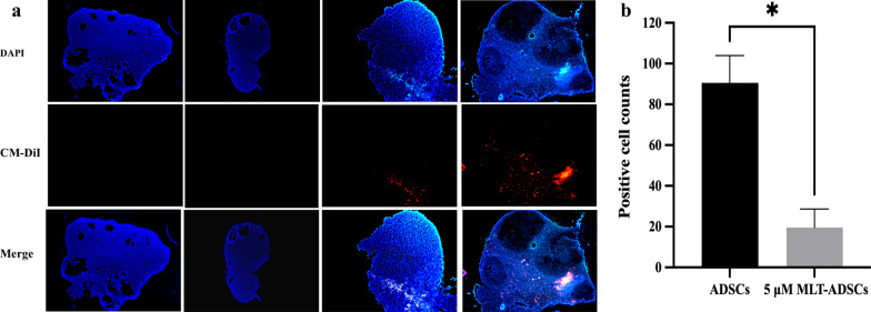
Results: Flow cytometry showed that autologous ADSCs expressed CD90 (99.7%) and CD29 (97.5%). MLT can not only promote the proliferation of ADSCs but also boost their secretory function, especially when ADSCs were pretreated with 5 µM MLT for 3 days, improving the interference effect. After transplantation of autologous ADSCs pretreated with 5 µM MLT, the serum hormone levels and reproductive function were significantly recovered, and the mean counts of primordial follicle increased. At the same time, the expression of SIRT6 was remarkably increased and the expression of NF-κB was significantly decreased in this group.
Conclusions: MLT enhances several effects of ADSCs in restoring hormone levels, mean primordial follicle counts, and reproductive capacity in POI mice. Meanwhile, our results suggest that the SIRT6/NF-κB signal pathway may be the potential therapeutic mechanism for ADSCs to treat POI.
Keywords: Autologous adipose-derived stem cells; Melatonin; Premature ovarian insufficiency; SIRT6/NF-κB pathway.
© 2022. The Author(s).
Conflict of interest statement
There is no conflict of interest to declare.
Figures
Similar articles
-
Transplantation of adipose-derived stem cells combined with collagen scaffolds restores ovarian function in a rat model of premature ovarian insufficiency.Hum Reprod. 2016 May;31(5):1075-86. doi: 10.1093/humrep/dew041. Epub 2016 Mar 9. Hum Reprod. 2016. PMID: 26965432
-
Drug-free in vitro activation combined with 3D-bioprinted adipose-derived stem cells restores ovarian function of rats with premature ovarian insufficiency.Stem Cell Res Ther. 2022 Jul 26;13(1):347. doi: 10.1186/s13287-022-03035-3. Stem Cell Res Ther. 2022. PMID: 35883196 Free PMC article.
-
Exosomes derived from human adipose mesenchymal stem cells improve ovary function of premature ovarian insufficiency by targeting SMAD.Stem Cell Res Ther. 2018 Aug 9;9(1):216. doi: 10.1186/s13287-018-0953-7. Stem Cell Res Ther. 2018. PMID: 30092819 Free PMC article.
-
The Evaluation of Ovarian Function Recovery Following Treatment of Primary Ovarian Insufficiency: A Systematic Review.Front Endocrinol (Lausanne). 2022 Apr 28;13:855992. doi: 10.3389/fendo.2022.855992. eCollection 2022. Front Endocrinol (Lausanne). 2022. PMID: 35573993 Free PMC article.
-
The Potency of Adipose-Derived Mesenchymal Stem Cells to Increase Ovarian Function in Primary Ovarian Insufficiency: A Systematic Review of in vivo Studies.Gynecol Obstet Invest. 2025 Feb 18:1-11. doi: 10.1159/000544721. Online ahead of print. Gynecol Obstet Invest. 2025. PMID: 39965550
Cited by
-
3D hUC-MSC spheroids exhibit superior resistance to autophagy and apoptosis of granulosa cells in POF rat model.Reproduction. 2024 Jul 13;168(2):e230496. doi: 10.1530/REP-23-0496. Print 2024 Aug 1. Reproduction. 2024. PMID: 38912966 Free PMC article.
-
Melatonin Receptors and Serotonin: Age-Related Changes in the Ovaries.J Pers Med. 2024 Sep 22;14(9):1009. doi: 10.3390/jpm14091009. J Pers Med. 2024. PMID: 39338263 Free PMC article.
-
Sirtuins in mitophagy: key gatekeepers of mitochondrial quality.Mol Cell Biochem. 2025 Jul 24. doi: 10.1007/s11010-025-05358-0. Online ahead of print. Mol Cell Biochem. 2025. PMID: 40705152 Review.
-
Advances in mesenchymal stem cell and exosome-based therapies for aging and age-related diseases.Stem Cell Res Ther. 2025 Jul 26;16(1):401. doi: 10.1186/s13287-025-04318-1. Stem Cell Res Ther. 2025. PMID: 40713733 Free PMC article. Review.
-
Autologous Human Mesenchymal Stem Cell-Based Therapy in Infertility: New Strategies and Future Perspectives.Biology (Basel). 2023 Jan 10;12(1):108. doi: 10.3390/biology12010108. Biology (Basel). 2023. PMID: 36671799 Free PMC article. Review.
References
Publication types
MeSH terms
Substances
LinkOut - more resources
Full Text Sources
Medical
