. 2022 Jul 19:13:951009.
doi: 10.3389/fmicb.2022.951009. eCollection 2022.

YWHAG inhibits influenza a virus replication by suppressing the release of viral M2 protein

Affiliations

YWHAG inhibits influenza a virus replication by suppressing the release of viral M2 protein

Haiying Mao et al. Front Microbiol. .

Abstract

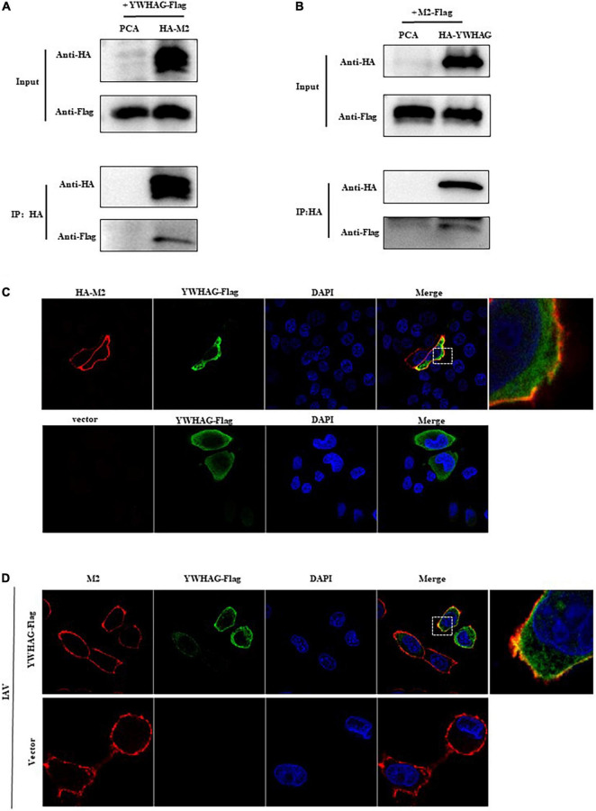
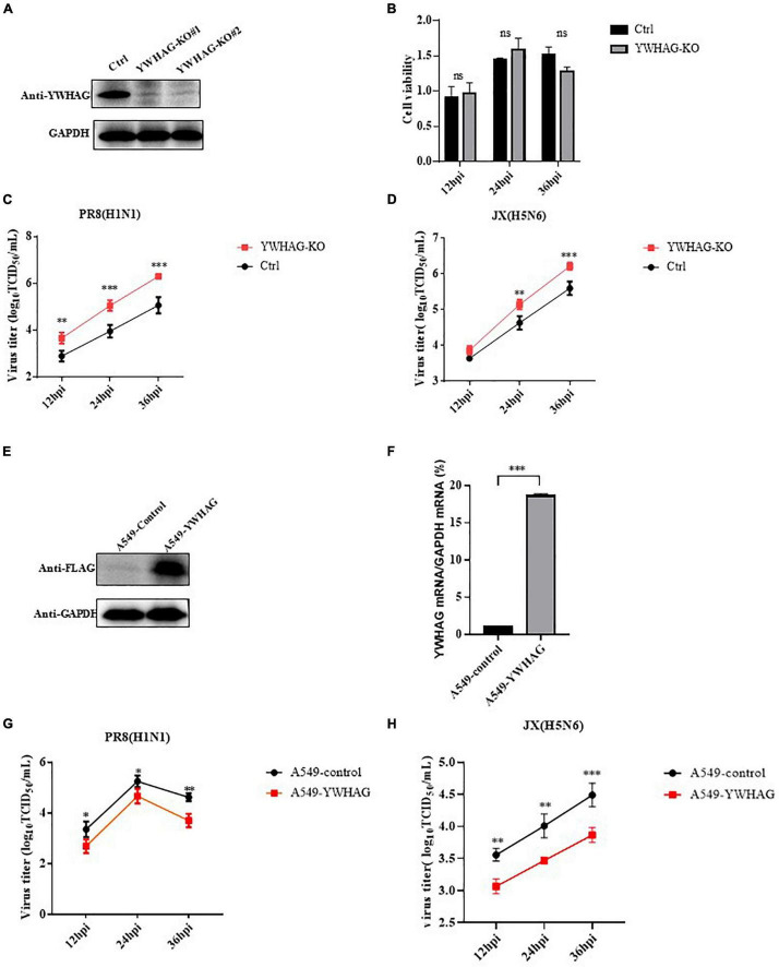
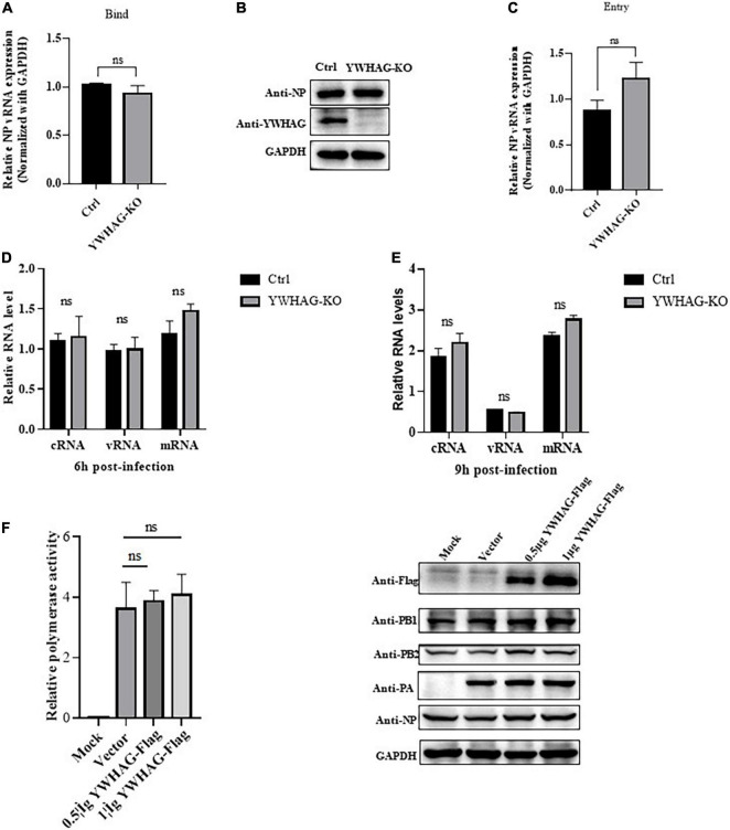
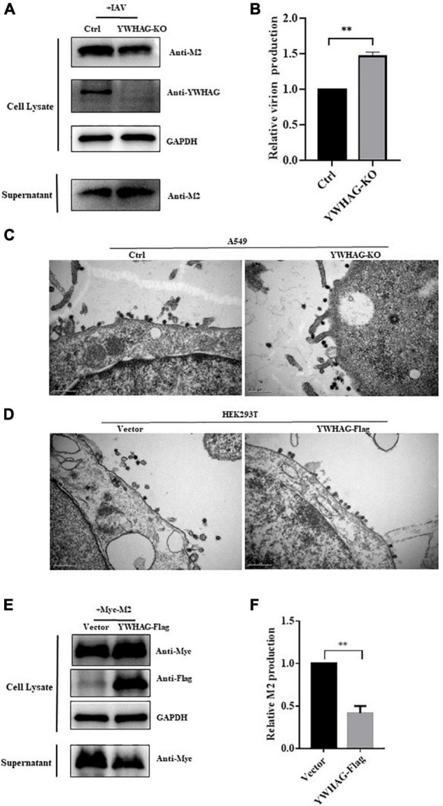
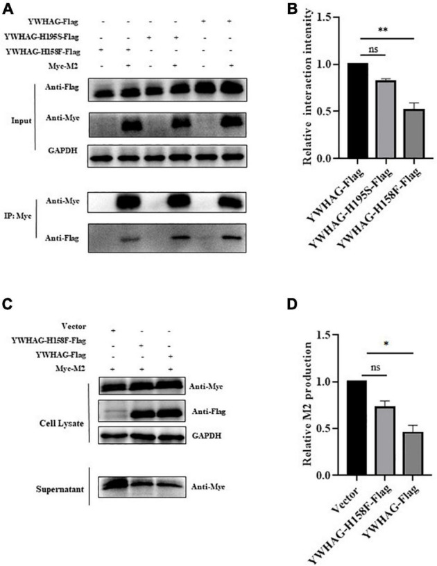
Influenza A virus (IAV) poses a serious threat to human life and property. The IAV matrix protein 2 (M2) is significant in viral budding. Increasing studies have proven the important roles of host factors in IAV replication. In this study, immunoprecipitation combined with mass spectrometry revealed that the host protein tyrosine 3-monooxygenase/tryptophan 5-monooxygenase activation protein gamma (YWHAG), which belongs to the 14-3-3 protein scaffold family, interacts with M2. Their interactions were further confirmed by co-immunoprecipitation (Co-IP), immunofluorescence, and confocal microscopy of virus-infected HeLa cells. Moreover, we constructed YWHAG-KO and YWHAG-overexpressing cells and found that YWHAG knockout significantly increased viral production, whereas its overexpression reduced the titer of virus progeny. Therefore, YWHAG is a negative regulatory factor during IAV infection. Further, YWHAG knockout or overexpression had no effect on the binding, entry, or viral RNA replication in the early stages of the virus life cycle. On the contrary, it impaired the release of virions at the plasma membrane as determined using transmission electron microscopy and suppressed the M2-mediated budding of the influenza virus. Importantly, the H158F mutation of YWHAG was found to affect interaction with M2 and its budding. Collectively, our work demonstrates that YWHAG is a novel cellular regulator that targets and mediates the interaction and release of M2.

Keywords: M2 protein; YWHAG; influenza A virus; protein–protein interaction; viral budding.


Conflict of interest statement

The authors declare that the research was conducted in the absence of any commercial or financial relationships that could be construed as a potential conflict of interest.

Figures

FIGURE 1
FIGURE 1
The host factor YWHAG interacts with M2. (A,B) Flag-tagged YWHAG-transfected with empty vector or HA-tagged M2 in HEK293T cells. (A) Flag-tagged M2 co-transfected with empty vector or HA-tagged YWHAG in HEK293T cells. (B) Cell lysates were incubated with anti-HA beads for 2 h at 25°C or at 4°C overnight. The beads were washed with PBST 4–5 times and IP eluates were subjected to western blotting analysis. (C) HeLa cells were co-transfected HA-tagged M2 and Flag-tagged YWHAG or empty vector, the cells were fixed and stained with mouse anti-Flag mAb and rabbit anti-HA pAb, and then incubated with Alexa Fluor 488 anti-mouse IgG (H + L) (green) and Alexa Fluor 594 goat anti-Rabbit IgG (H + L) (red). The nuclei were stained with DAPI. (D) Flag-tagged YWHAG or empty vector was transfected into HeLa cells and infected with PR8 at MOI = 1. The anti-M2 rabbit red fluorescent antibody and anti-Flag mouse green fluorescent antibody were used for staining. The interaction part enclosed by the white box was enlarged, as shown in the right.
FIGURE 2
FIGURE 2
YWHAG inhibits influenza A virus (IAV) replication. (A) Endogenous YWHAG in A549 cells was knocked out using the CRISPR/Cas9 system. YWHAG expression was determined by western blotting. (B) Viability of YWHAG-KO cells was measured using a CCK8 assay. The data are presented as mean ± standard deviation (SD). (C,D) Control (Ctrl) or YWHAG-knockout (YWHAG-KO) A549 cells were infected with PR8 (H1N1) (C) or JX (H5N6). (D) The supernatants were harvested and viral titers were determined at the indicated times. Stable overexpression of Flag-tagged YWHAG was confirmed by western blotting using a mouse anti-Flag mAb (E) and reverse-transcription quantitative PCR (RT-qPCR) (F) compared with that in control cells transduced with an empty retrovirus. (G,H) YWHAG-overexpressing cells or control cells were infected with PR8 (H1N1) (G), JX (H5N6) (H) at MOI = 0.01. Supernatants were collected at different times, and virus titers were determined using a TCID50 assay on MDCK cells. The data are presented as the means ± SD by t-test (*p < 0.05; **p < 0.01; ***p < 0.001).
FIGURE 3
FIGURE 3
YWHAG does not affect the binding, entry, replication, and transcription of IAV in the early stages of infection. (A,B) YWHAG-KO or control cells were incubated with PR8 at MOI = 5, at 4°C for 1 h, the viral copy number of bound viruses in the cell lysates was assessed using reverse-transcription quantitative PCR (RT-qPCR) (A) and western blotting (B). (C) YWHAG-KO and control cells were incubated with PR8 at an MOI = 5 at 4°C for 1 h and then allowed to internalize bound IAV by incubation at 37°C for 30 min, followed by the addition of exogenous NA to remove the cell-surface virions and incubation for another 30 min. Viral entry was measured using reverse-transcription quantitative PCR (RT-qPCR). (D,E) YWHAG-KO or control cells were infected with PR8 at MOI = 5. Total RNA was harvested at 6 h (D) and 9 h (E) post-infection, and the nucleoprotein (NP) in vRNA, cRNA, and mRNA was analyzed using reverse-transcription quantitative PCR (RT-qPCR). (F) HEK293T cells were co-transfected with pPolI-Luc and expression plasmids containing viral PB1, PB2, PA, or NP of H1N1 together with an empty vector or Flag-tagged YWHAG. At 24 h post-transfection, western blotting and a dual-luciferase assay were performed wherein the relative firefly luciferase activity was normalized to that of the internal control.
FIGURE 4
FIGURE 4
YWHAG suppresses M2-mediated budding of IAV. (A,B) YWHAG-KO or control cells were infected with PR8 at MOI = 5 for 10 h and the supernatants were collected by ultracentrifugation. Cell lysates and virions were determined by western blotting (A). Relative virions production was calculated based on the M2 protein band intensities and expressed as the ratios of viral lysate/cell lysate (B). The level of virions from control cells was set as 100%. (C,D) YWHAG-KO or control cells (C) and HEK293T cells transfected with Flag-tagged YWHGA or empty vector (D) were infected with PR8 at MOI = 5 for 10 h; thin sections were prepared and analyzed by electron microscopy. Scale bars indicate 500 nm. (E,F) HEK293T cells were transfected with Myc-tagged M2 and Flag-tagged YWHAG. The culture supernatants were harvested by ultracentrifugation. Cell lysates and centrifuged supernatants samples were examined by western blotting (E). The intensities of relative M2 production were determined using Image J (F). The data are presented as the means ± SD by t-test (**p < 0.01).
FIGURE 5
FIGURE 5
YWHAG with H158F mutation inhibits M2 budding. (A) HEK293T cells were transfected to produce Flag-tagged YWHAG protein variants together with the indicated Myc-tagged M2. Protein interactions were detected using co-immunoprecipitation. (B) Relative interaction intensity was calculated based on protein band intensities and expressed as the ratios of IP (Flag/Myc). (C,D) HEK293T cells were transfected with Myc-tagged M2 and Flag-tagged YWHAG variants. Culture supernatants were harvested by ultracentrifugation. Cell lysates and supernatant samples were examined using western blotting (C) and the relative M2 production was analyzed (D). The data are presented as the means ± SD by t-test (*p < 0.5; **p < 0.01).

Similar articles

Cited by

References

    1. Aghazadeh Y., Papadopoulos V. (2016). The role of the 14-3-3 protein family in health, disease, and drug development. Drug Discov. Today 21 278–287. 10.1016/j.drudis.2015.09.012 - DOI - PubMed
    1. Borkenhagen L. K., Allen M. W., Runstadler J. A. (2021). Influenza virus genotype to phenotype predictions through machine learning: a systematic review. Emerg. Microbes Infect. 10 1896–1907. 10.1080/22221751.2021.1978824 - DOI - PMC - PubMed
    1. Bustad H. J., Skjaerven L., Ying M., Halskau Ø, Baumann A., Rodriguez-Larrea D. (2012). The peripheral binding of 14-3-3γ to membranes involves isoform-specific histidine residues. PLoS One 7:e49671. 10.1371/journal.pone.0049671 - DOI - PMC - PubMed
    1. Cau Y., Valensin D., Mori M., Draghi S., Botta M. (2018). Structure, Function, Involvement in Diseases and Targeting of 14-3-3 Proteins: An Update. Curr. Med. Chem. 25 5–21. 10.2174/0929867324666170426095015 - DOI - PubMed
    1. Chauhan R. P., Gordon M. L. (2022). An overview of influenza A virus genes, protein functions, and replication cycle highlighting important updates. Virus Genes [Epub ahead of print]. 10.1007/s11262-022-01904-w - DOI - PubMed