

Role of immunocytochemistry in cervical cancer screening
- PMID: 35928527
- PMCID: PMC9345115
- DOI: 10.25259/CMAS_03_17_2022
Role of immunocytochemistry in cervical cancer screening
Abstract
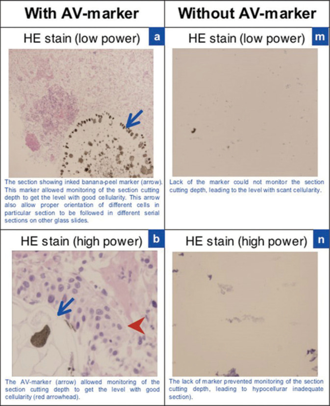
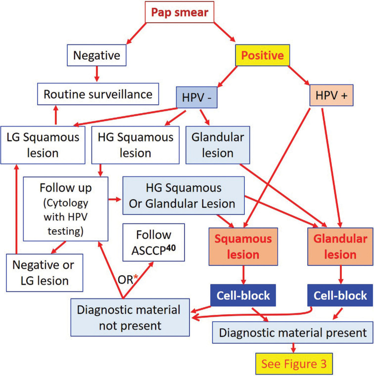
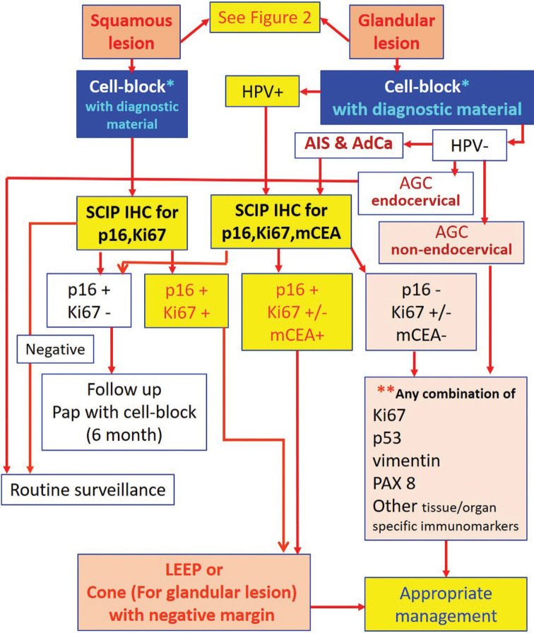
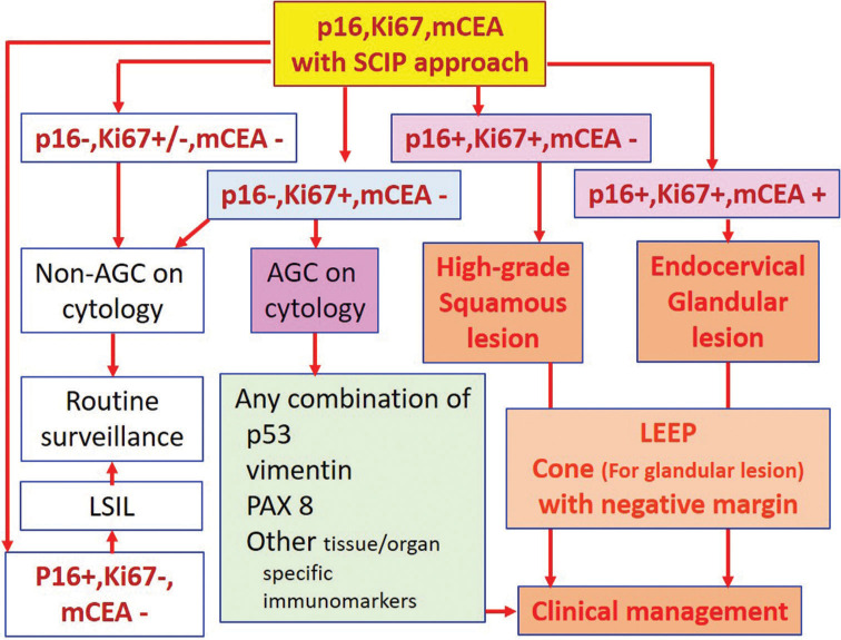
The cervical cancer screening has been based conventionally on cytologic analysis. With advances in understanding the role of human papillomavirus, cotesting has been applied. But most of the patients subjected to colposcopy did not benefit, except in cases with HSIL [high-grade squamous intraepithelial lesion] cytology. Because of this, a step to increase the sensitivity to detect cancers and pre-cancers but with higher specificity with minimal overdiagnosis leading to prevention of unindicated cervical biopsies is highly desired. Such triaging step in cases with abnormal screening results is expected to minimize invasive interventions because of low false positivity. With availability of methodologies leading to quantitatively and qualitatively enhanced cell-blocks from residual liquid based cytology specimens, immunostaining can be performed for multiple immunomarkers with potential objectivity to triage initial screening test results. This is enhanced further with inclusion of AV marker in the cell-blocks and application of SCIP (subtractive coordinate immunoreactivity pattern) approach. The cell-blocks are also resource for performing other ancillary studies including molecular pathology and proteomics/metabolomics as potential tests in future. This review explores application of residual liquid based cytology specimen for cell-blocking with application of ancillary studies in algorithmic manner as adjunct to ASCCP management guidelines for improved patient care.
Keywords: Cell-block; Cervical cancer; Cervical cytology; HPV; Immunocytochemistry; Immunohistochemistry; Ki-67; LBC; Liquid based cytology; PAP test; Papanicolaou smear; p16.
© 2022 Cytopathology Foundation Inc, Published by Scientific Scholar.
Figures
Similar articles
-
Role of Cervical Cancer Biomarkers p16 and Ki67 in Abnormal Cervical Cytological Smear.J Obstet Gynaecol India. 2021 Feb;71(1):72-77. doi: 10.1007/s13224-020-01380-y. Epub 2020 Nov 18. J Obstet Gynaecol India. 2021. PMID: 33814802 Free PMC article.
-
p16 immunocytochemistry on cell blocks as an adjunct to cervical cytology: Potential reflex testing on specially prepared cell blocks from residual liquid-based cytology specimens.Cytojournal. 2011 Jan 31;8:1. doi: 10.4103/1742-6413.76379. Cytojournal. 2011. PMID: 21369522 Free PMC article.
-
Evaluating the Performance of p16INK4a Immunocytochemistry in Cervical Cancer Screening.Cancer Manag Res. 2020 Sep 25;12:9067-9075. doi: 10.2147/CMAR.S273079. eCollection 2020. Cancer Manag Res. 2020. PMID: 33061601 Free PMC article.
-
Evidence regarding human papillomavirus testing in secondary prevention of cervical cancer.Vaccine. 2012 Nov 20;30 Suppl 5:F88-99. doi: 10.1016/j.vaccine.2012.06.095. Vaccine. 2012. PMID: 23199969 Review.
-
Immunocytochemistry of effusion fluids: Introduction to SCIP approach.Cytojournal. 2022 Jan 31;19:3. doi: 10.25259/CMAS_02_05_2021. eCollection 2022. Cytojournal. 2022. PMID: 35541032 Free PMC article. Review.
Cited by
-
Field-Applicable Loop-Mediated Isothermal Amplification for the Detection of Seven Common Human Papillomavirus Subtypes.Trop Med Infect Dis. 2024 Oct 12;9(10):240. doi: 10.3390/tropicalmed9100240. Trop Med Infect Dis. 2024. PMID: 39453267 Free PMC article.
-
Spectrum of cervicovaginal Pap smears in newly established tertiary care medical institute.Cytojournal. 2023 Aug 29;20:20. doi: 10.25259/Cytojournal_8_2023. eCollection 2023. Cytojournal. 2023. PMID: 37681076 Free PMC article.
-
Clinical utility of HPV typing and quantification combined with PAX1/ZNF582 methylation detection in accurate cervical cancer screening.Cytojournal. 2023 Sep 1;20:26. doi: 10.25259/Cytojournal_46_2022. eCollection 2023. Cytojournal. 2023. PMID: 37681081 Free PMC article.
-
Deep Learning-Based Screening of Urothelial Carcinoma in Whole Slide Images of Liquid-Based Cytology Urine Specimens.Cancers (Basel). 2022 Dec 30;15(1):226. doi: 10.3390/cancers15010226. Cancers (Basel). 2022. PMID: 36612222 Free PMC article.
References
-
- Shidham V, D'Amore K, Varsegi G. Objective and definitive subcategorization of LSIL with p16INK4a immunocytochemistry on cell block sections of cervical cytology specimens. Cancer Cytopathol. 2009;117:349–450.
-
- Shidham VB, Mehrotra R, Varsegi G, D'Amore KL, Hunt B, Narayan R. p16 INK4a immunocytochemistry on cell blocks as an adjunct to cervical cytology: Potential reflex testing on specially prepared cell blocks from residual liquid-based cytology specimens. CytoJournal. 2011;8:1. doi: 10.4103/1742-6413.76379. - DOI - PMC - PubMed
-
- NextGen CelBlokingTM kits. AV BioInnovation. Available from: https://www.avbioinnovation.com [Last accessed on 2022 Apr 26]
Publication types
LinkOut - more resources
Full Text Sources
Research Materials