

Joubert-Plus syndrome with an atretic cephalocele: a case report
- PMID: 35928591
- PMCID: PMC9343393
- DOI: 10.1016/j.radcr.2022.07.038
Joubert-Plus syndrome with an atretic cephalocele: a case report
Abstract
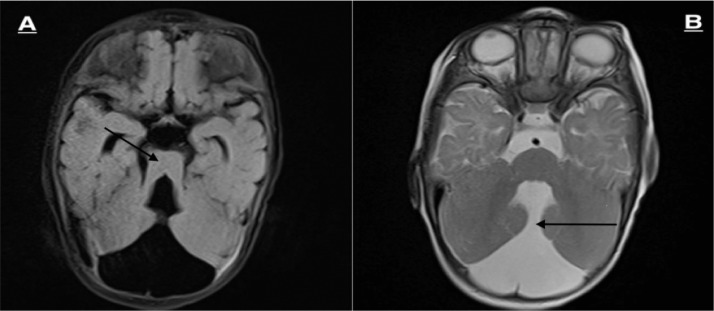
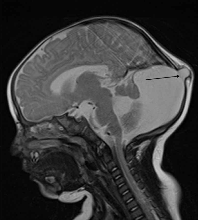
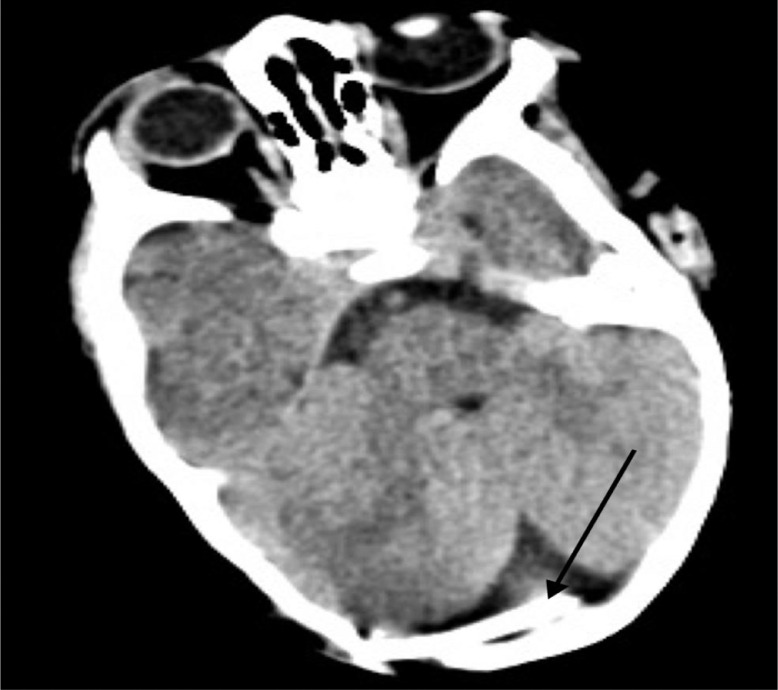
Joubert syndrome is a rare heterogeneous disease affecting the cerebellum. It usually presents with hypotonia, abnormal breathing pattern, with distinctive cerebellar and brain stem malformation called the molar tooth sign. It may present with different organ involvement or with other neurological alterations such as Dandy-Walker syndrome. Joubert syndrome with dandy walker syndrome is called Joubert-Plus syndrome, an exceedingly rare entity. Dandy-Walker syndrome is defined by hypoplasia and upward rotation of the cerebellar vermis and cystic dilation of the fourth ventricle. Atretic cephalocele is another rare diagnosis which is characterized by a herniation of intracranial contents through a skull defect. Herein, we present a case of a 6-month-old patient who presented with floppiness and a scalp nodule. After further evaluation, he was diagnosed with Joubert-Plus syndrome with an atretic cephalocele.
Keywords: Atretic cephalocele; Case report; Dandy-Walker; Joubert; Joubert-Plus.
© 2022 The Authors. Published by Elsevier Inc. on behalf of University of Washington.
Figures
Similar articles
-
Joubert Plus syndrome in a child with Dandy-Walker malformation and occipital cephalocele: A case report.Radiol Case Rep. 2025 Jun 27;20(9):4701-4705. doi: 10.1016/j.radcr.2025.05.103. eCollection 2025 Sep. Radiol Case Rep. 2025. PMID: 40677886 Free PMC article.
-
Dandy-Walker malformation masking the molar tooth sign: an illustrative case with magnetic resonance imaging follow-up.J Child Neurol. 2010 Nov;25(11):1419-22. doi: 10.1177/0883073810370477. Epub 2010 Sep 7. J Child Neurol. 2010. PMID: 20823032
-
Cerebello-oculo-renal syndromes including Arima, Senior-Löken and COACH syndromes: more than just variants of Joubert syndrome.Am J Med Genet. 1999 Oct 29;86(5):459-69. Am J Med Genet. 1999. PMID: 10508989 Review.
-
Clinical nosologic and genetic aspects of Joubert and related syndromes.J Child Neurol. 1999 Oct;14(10):660-6; discussion 669-72. doi: 10.1177/088307389901401007. J Child Neurol. 1999. PMID: 10511339 Review.
-
Quantitative assessment of brainstem development in Joubert syndrome and Dandy-Walker syndrome.J Child Neurol. 2001 Oct;16(10):751-8. doi: 10.1177/088307380101601008. J Child Neurol. 2001. PMID: 11669349
Cited by
-
Joubert Plus syndrome in a child with Dandy-Walker malformation and occipital cephalocele: A case report.Radiol Case Rep. 2025 Jun 27;20(9):4701-4705. doi: 10.1016/j.radcr.2025.05.103. eCollection 2025 Sep. Radiol Case Rep. 2025. PMID: 40677886 Free PMC article.
References
Publication types
LinkOut - more resources
Full Text Sources
