

The Dynamic Effect of Anterior Cruciate Ligament Deficiency on Patellar Height
- PMID: 35928660
- PMCID: PMC9283625
- DOI: 10.1007/s43465-022-00632-5
The Dynamic Effect of Anterior Cruciate Ligament Deficiency on Patellar Height
Abstract
Background: The anterior tibial translation (ATT) in case of Anterior Cruciate Ligament (ACL) tear can lead to dynamic alterations of the extensor apparatus biomechanics. The aim of this study is to evaluate the dynamic effect of isolated ACL deficiency on patellar height. The hypothesis is that the ATT of ACL-insufficient knees dynamically reduces patellar height.
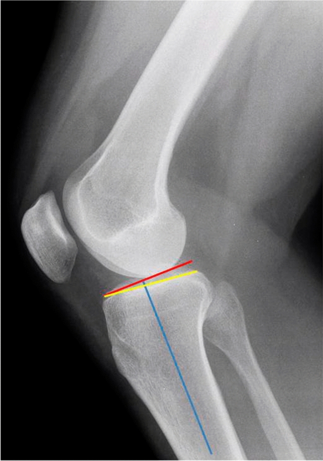
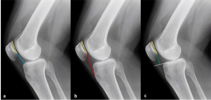
Methods: Skeletally mature patients who underwent ACL reconstruction using hamstring graft between January and December 2018 were included in this study. The Posterior Tibial Slope (PTS), Caton-Deschamps (CDI), modified Insall-Salvati (MISI), and Blackburne-Peel (BPI) indices were calculated in standard lateral and TELOS X-rays. The mean of the measurements calculated between two observers was used to compare these parameters.
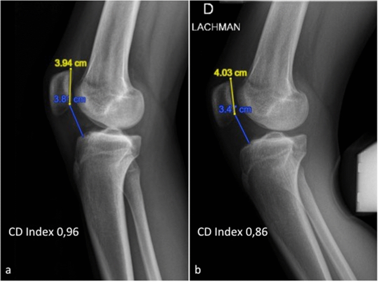
Results: 95 patients (M: 57; F: 38; 95 knees) were included in the study with a mean age of 31.8 years (16-56 years old). Significant patellar height reduction (CDI: 0.11 [- 0.32; 0.31]; MISI: 0.09 [- 0.66; 0.30]) was reported in TELOS compared with standard lateral knee radiography (p < 0.001). 20.0% of the study knees reported an abnormal CDI and 84.2% (16/19 knees) of them reduced this index to within normal limits in TELOS. 20.0% of the knees with mild patella alta reduced CDI in TELOS but always remained above 1.2.
Conclusions: The abnormal ATT in case of ACL-deficient knees results in a lowering effect of the patella in TELOS X-rays. In patients with ACL tear and anterior pain the reconstructive ligament surgery should be performed to avoid also chronic anterior knee pain.
Level of evidence: Basic Science Study (Case Series).
Clinical relevance: The decrease in patellar height in stress-X-rays compared with standard lateral knee radiography in ACL deficient knees, should be considered as a possible contributing cause of anterior pain in these patients.
Keywords: Anterior Cruciate Ligament; Anterior Knee Pain; Anterior Tibial Translation; Patellar Height.
© Indian Orthopaedics Association 2022.
Conflict of interest statement
Conflict of InterestFL has nothing to disclose. MB has nothing to disclose. CB has nothing to disclose. PSR reports personal fees from Arthrex, personal fees from Depuy (Johnson&Johnson), outside the submitted work. SL reports personal fees from Lepine, personal fees from Depuy (Johnson&Johnson), personal fees from Heraeus, personal fees from Smith & Nephew, personal fees from Stryker, personal fees from Medacta, other from Corin, other from Amplitude, outside the submitted work. ES reports personal fees from Smith & Nephew, grants from Corin, outside the submitted work.
Figures
Similar articles
-
How Does Isolated Medial Patellofemoral Ligament Reconstruction Influence Patellar Height?Am J Sports Med. 2020 Mar;48(4):895-900. doi: 10.1177/0363546520902132. Epub 2020 Feb 14. Am J Sports Med. 2020. PMID: 32058795
-
Effect of Infratuberosity Anterior Closing Wedge Osteotomy for Posterior Tibial Slope Correction on Patellar Height in Patients Undergoing Revision ACL Reconstruction.Am J Sports Med. 2025 Apr;53(5):1061-1067. doi: 10.1177/03635465251323623. Epub 2025 Mar 14. Am J Sports Med. 2025. PMID: 40087814
-
The Influence of Tibial Tubercle-Sparing Slope-Reducing Osteotomy on Patellar Height in Patients Undergoing Revision ACL Reconstruction.Am J Sports Med. 2024 Mar;52(4):919-927. doi: 10.1177/03635465241228264. Epub 2024 Feb 22. Am J Sports Med. 2024. PMID: 38385201
-
Radiographic Investigation of Coronal Plane and Patellar Height and Changes Following Tibial Deflection Osteotomy for Correction of Tibial Slope in Combination With ACL Reconstruction.Am J Sports Med. 2024 Mar;52(3):691-697. doi: 10.1177/03635465231222643. Epub 2024 Jan 29. Am J Sports Med. 2024. PMID: 38284182
-
Passive anterior tibia translation in anterior cruciate ligament-injured, anterior cruciate ligament-reconstructed and healthy knees: a systematic review.Musculoskelet Surg. 2019 Aug;103(2):121-130. doi: 10.1007/s12306-018-0572-6. Epub 2018 Oct 16. Musculoskelet Surg. 2019. PMID: 30328030 Free PMC article.
Cited by
-
Infratubercle Anterior Closing Wedge Osteotomy Corrects Sagittal Alignment without Affecting Coronal Alignment or Patellar Height.J Clin Med. 2024 Aug 11;13(16):4715. doi: 10.3390/jcm13164715. J Clin Med. 2024. PMID: 39200857 Free PMC article.
References
-
- Hernández LM, Micheo WF, Amy E. Rehabilitation update for the anterior cruciate ligament injuredpatient: current concepts. Boletín de la Asociación Médica de Puerto Rico. 2006;98(1):62–72. - PubMed
-
- Micheo, W., Hernández, L., Seda, C. (2010). Evaluation, management, rehabilitation, and prevention of anterior cruciate ligament injury: Current concepts. PM and R, 2(10), 935–944. 10.1016/j.pmrj.2010.06.014 - PubMed
-
- Li, G., DeFrate, L.E., Sun, H., Gill, T.J. (2004). In vivo elongation of the anterior cruciate ligament and posterior cruciate ligament during knee flexion. American Journal of Sports Medicine, 32(6), 1415–1420. 10.1177/0363546503262175 - PubMed
-
- Smith, B.A., Livesay, G.A., Woo, S.L.Y. (1993). Biology and biomechanics of the anterior cruciate ligament. Clinics in Sports Medicine, 12(4), 637–670 - PubMed
-
- Kessler, M.A., Behrend, H., Henz, S., Stutz, G., Rukavina, A., Kuster, M.S. (2008). Function, osteoarthritis and activity after ACL-rupture: 11 Years follow-up results of conservative versus reconstructive treatment. Knee Surgery, Sports Traumatology, Arthroscopy, 16(5), 442–448. 10.1007/s00167-008-0498-x - PubMed
LinkOut - more resources
Full Text Sources
Research Materials