

Establishment and evaluation of retroperitoneal liposarcoma patient-derived xenograft models: an ideal model for preclinical study
- PMID: 35928724
- PMCID: PMC9346387
- DOI: 10.7150/ijms.70706
Establishment and evaluation of retroperitoneal liposarcoma patient-derived xenograft models: an ideal model for preclinical study
Abstract
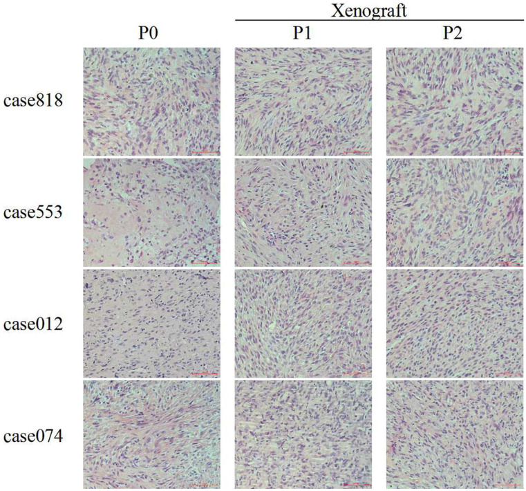
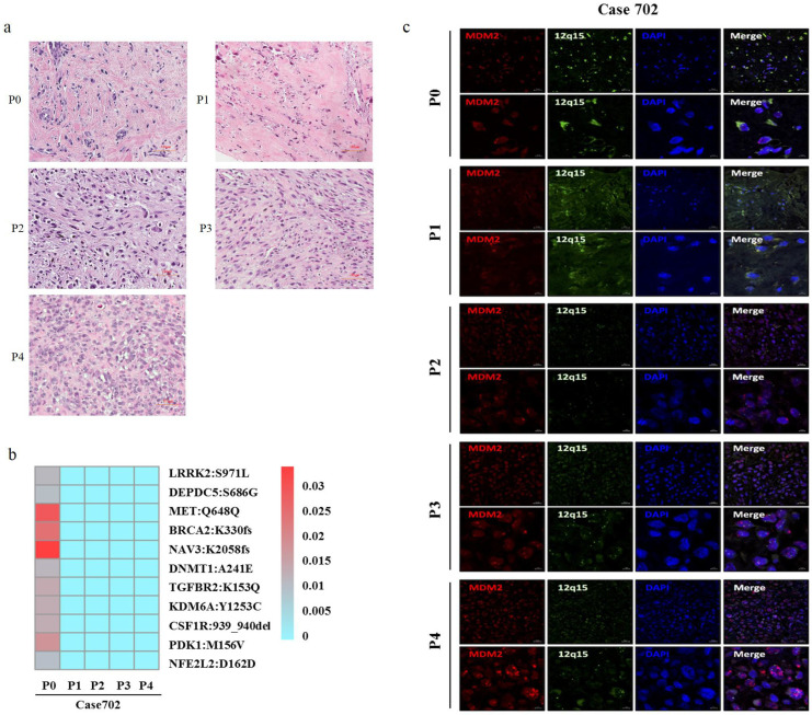
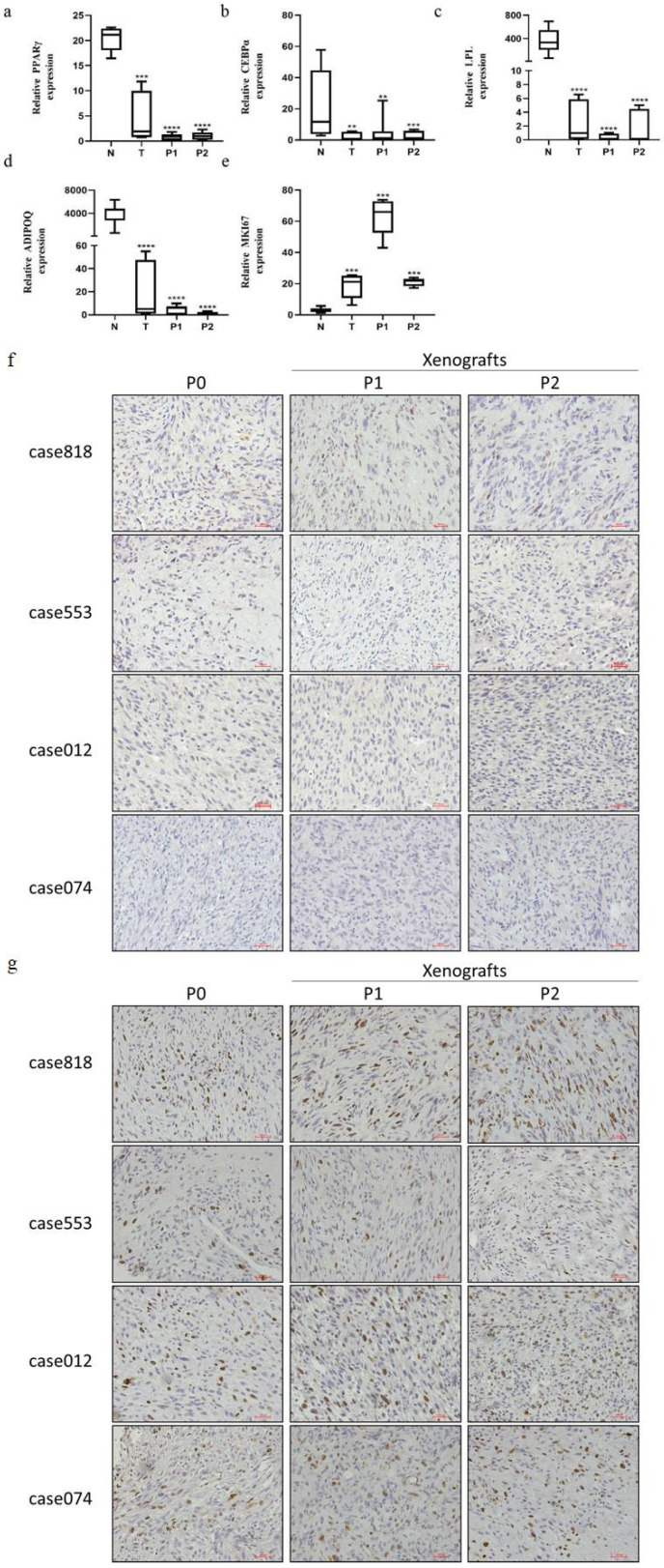
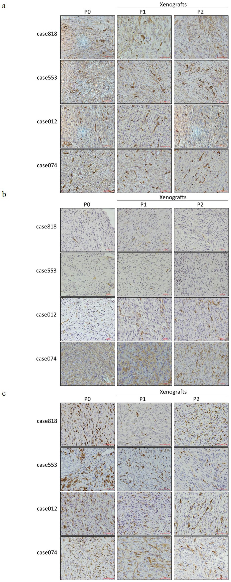
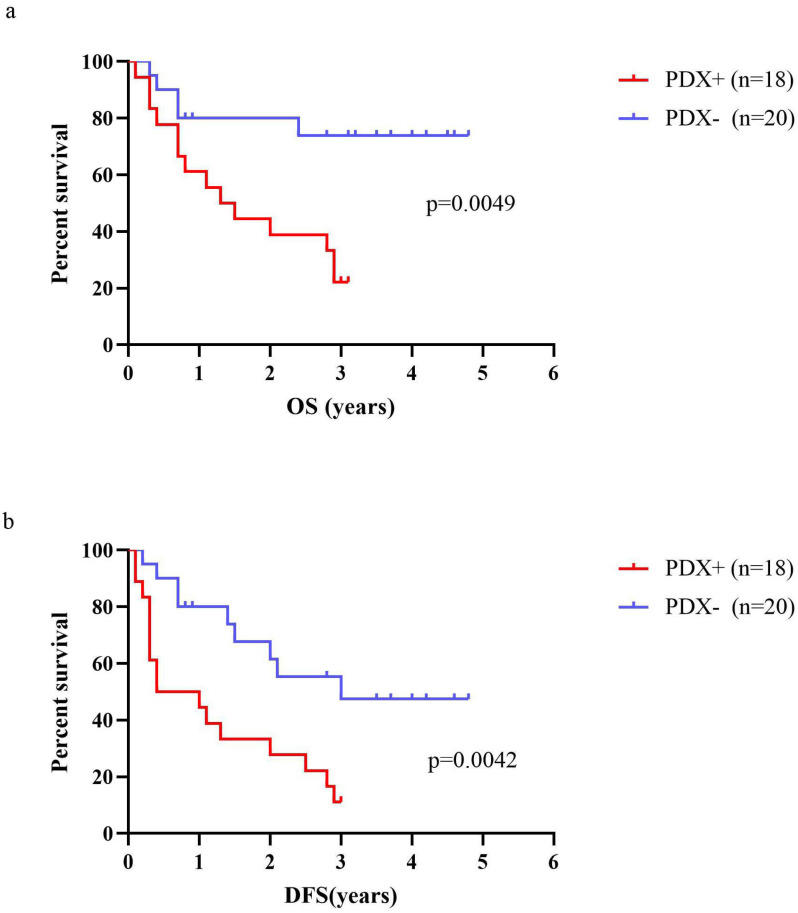
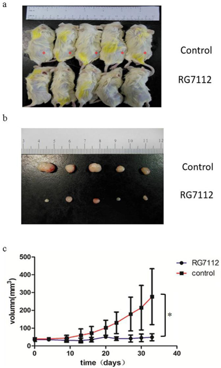
Retroperitoneal liposarcoma (RLPS) is one of the most common subtypes of retroperitoneal soft tissue sarcomas. It is characterized by poor sensitivity to radiotherapy and chemotherapy and a low success rate of complete surgical resection. However, there are few reliable preclinical RLPS models for target discovery and therapy research. In this study, we aimed to establish RLPS patient-derived xenograft (PDX) models that are useful for biological research and preclinical drug trials. A total of 56 freshly resected RLPS tissues were subcutaneously transplanted into non-obese diabetic-severe combined immune deficient (NOD-SCID) mice, with subsequent xenotransplantation into second-generation mice. The tumor engraftment rate of first generation PDXs was 44.64%, and higher success rates were obtained from implantations of dedifferentiated, myxous, pleomorphic, high-grade liposarcomas and those with retroperitoneal organ infiltration. The first- and second- generation PDX models preserved the histopathological morphology, gene mutation profiles and MDM2 amplification of the primary tissues. PDX models can also provide the benefit of retaining original tumor biology and microenvironment characteristics, such as abnormal adipose differentiation, elevated Ki67 levels, high microvessel density, cancer-associated fibroblast presence, and tumor-associated macrophage infiltration. Overall survival (OS) and disease-free survival (DFS) of patients with successful first-generation PDX engraftment were significantly poorer than those with failed engraftment. Treatment with MDM2 inhibitor RG7112 significantly suppressed tumor growth of DDLPS PDX in mice. In conclusion, we successfully established RLPS PDX models that were histologically, genetically, and molecularly consistent with the original tissues. These models might provide opportunities for advancing RLPS tumor biology research, facilitating the development of novel drugs, particularly those targeting MDM2 amplification, adipose differentiation process, angiogenesis, cancer-associated fibroblasts, and so on.
Keywords: Retroperitoneal liposarcoma; patient-derived xenograft (PDX); prognosis; treatment evaluation; tumor biology research.
© The author(s).
Conflict of interest statement
Competing Interests: The authors have declared that no competing interest exists.
Figures
Similar articles
-
Selinexor versus doxorubicin in dedifferentiated liposarcoma PDXs: evidence of greater activity and apoptotic response dependent on p53 nuclear accumulation and survivin down-regulation.J Exp Clin Cancer Res. 2021 Mar 1;40(1):83. doi: 10.1186/s13046-021-01886-x. J Exp Clin Cancer Res. 2021. PMID: 33648535 Free PMC article.
-
Establishment and genetically characterization of patient-derived xenograft models of cervical cancer.BMC Med Genomics. 2022 Sep 8;15(1):191. doi: 10.1186/s12920-022-01342-5. BMC Med Genomics. 2022. PMID: 36076209 Free PMC article.
-
Establishment and Characterization of Histologically and Molecularly Stable Soft-tissue Sarcoma Xenograft Models for Biological Studies and Preclinical Drug Testing.Mol Cancer Ther. 2019 Jun;18(6):1168-1178. doi: 10.1158/1535-7163.MCT-18-1045. Epub 2019 Apr 8. Mol Cancer Ther. 2019. PMID: 30962320
-
Application of Highly Immunocompromised Mice for the Establishment of Patient-Derived Xenograft (PDX) Models.Cells. 2019 Aug 13;8(8):889. doi: 10.3390/cells8080889. Cells. 2019. PMID: 31412684 Free PMC article. Review.
-
Patient-Derived Xenograft Models in Cervical Cancer: A Systematic Review.Int J Mol Sci. 2021 Aug 29;22(17):9369. doi: 10.3390/ijms22179369. Int J Mol Sci. 2021. PMID: 34502278 Free PMC article.
Cited by
-
The Pivotal Role of Preclinical Animal Models in Anti-Cancer Drug Discovery and Personalized Cancer Therapy Strategies.Pharmaceuticals (Basel). 2024 Aug 9;17(8):1048. doi: 10.3390/ph17081048. Pharmaceuticals (Basel). 2024. PMID: 39204153 Free PMC article. Review.
-
Xenografting Human Musculoskeletal Sarcomas in Mice, Chick Embryo, and Zebrafish: How to Boost Translational Research.Biomedicines. 2024 Aug 21;12(8):1921. doi: 10.3390/biomedicines12081921. Biomedicines. 2024. PMID: 39200384 Free PMC article. Review.
-
Modeling the Tumor Microenvironment and Cancer Immunotherapy in Next-Generation Humanized Mice.Cancers (Basel). 2023 May 30;15(11):2989. doi: 10.3390/cancers15112989. Cancers (Basel). 2023. PMID: 37296949 Free PMC article. Review.
References
-
- Dumitra S, Gronchi A. The Diagnosis and Management of Retroperitoneal Sarcoma. Oncology (Williston Park, N.Y.) 2018;32(9):464–9. - PubMed
MeSH terms
Supplementary concepts
LinkOut - more resources
Full Text Sources
Research Materials
