. 2022 Aug 2;11(8):e1408.
doi: 10.1002/cti2.1408. eCollection 2022.

Anti-C1q autoantibodies from systemic lupus erythematosus patients enhance CD40-CD154-mediated inflammation in peripheral blood mononuclear cells in vitro

Affiliations

Anti-C1q autoantibodies from systemic lupus erythematosus patients enhance CD40-CD154-mediated inflammation in peripheral blood mononuclear cells in vitro

Pascal Alexander Rabatscher et al. Clin Transl Immunology. .

Abstract

Objectives: Systemic lupus erythematosus (SLE) is a clinically heterogeneous autoimmune disease with complex pathogenic mechanisms. Complement C1q has been shown to play a major role in SLE, and autoantibodies against C1q (anti-C1q) are strongly associated with SLE disease activity and severe lupus nephritis suggesting a pathogenic role for anti-C1q. Whereas C1q alone has anti-inflammatory effects on human monocytes and macrophages, C1q/anti-C1q complexes favor a pro-inflammatory phenotype. This study aimed to elucidate the inflammatory effects of anti-C1q on peripheral blood mononuclear cells (PBMCs).

Methods: Isolated monocytes, isolated T cells and bulk PBMCs of healthy donors with or without concomitant T cell activation were exposed to C1q or complexes of C1q and SLE patient-derived anti-C1q (C1q/anti-C1q). Functional consequences of C1q/anti-C1q on cells were assessed by determining cytokine secretion, monocyte surface marker expression, T cell activation and proliferation.

Results: Exposure of isolated T cells to C1q or C1q/anti-C1q did not affect their activation and proliferation. However, unspecific T cell activation in PBMCs in the presence of C1q/anti-C1q resulted in increased TNF, IFN-γ and IL-10 secretion compared with C1q alone. Co-culture and inhibition experiments showed that the inflammatory effect of C1q/anti-C1q on PBMCs was due to a direct CD40-CD154 interaction between activated T cells and C1q/anti-C1q-primed monocytes. The CD40-mediated inflammatory reaction of monocytes involves TRAF6 and JAK3-STAT5 signalling.

Conclusion: In conclusion, C1q/anti-C1q have a pro-inflammatory effect on monocytes that depends on T cell activation and CD40-CD154 signalling. This signalling pathway could serve as a therapeutic target for anti-C1q-mediated inflammation.

Keywords: C1q; CD40; T cells; anti‐C1q autoantibodies; monocytes; systemic lupus erythematosus.


Conflict of interest statement

The authors declare no conflict of interest.

Figures
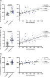
Figure 1
Figure 1
Increase in TNF, IFN‐γ and IL‐10 secretion in peripheral blood mononuclear cells (PBMCs) after T cell activation correlates with anti‐C1q levels. PBMCs activated by tetrameric anti‐CD3/CD28 complexes were cultured on C1q preincubated with the serum of 20 systemic lupus erythematosus patients (C1q/anti‐C1q) or with the serum of 20 healthy donors (C1q/NHS) for 24 h. Cell culture supernatants were analysed for (a) TNF, (b) IFN‐γ and (c) IL‐10 secretion by ELISA. Data points represent the median cytokine concentration obtained from PBMCs of four unrelated healthy donors from independent experiments exposed to one single serum sample. (Left) Horizontal lines with error bars show median with IQR. Mann–Whitney U‐test, ***P < 0.001, ****P < 0.0001. (Right) Solid line represents linear regression with dotted lines indicating the 95% confidence bands. Spearman's rank correlation of cytokine secretion and anti‐C1q levels, ***P < 0.001, ****P < 0.0001.
Figure 2
Figure 2
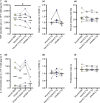
T cell proliferation, activation and IL‐10 secretion in activated T cells are not affected by bound C1q, bound C1q/anti‐C1q and soluble C1q, respectively, whereas TNF secretion is decreased in the presence of soluble C1q. T cells were isolated from peripheral blood mononuclear cells (PBMCs) of healthy donors and cultured on bound HSA, bound C1q, bound C1q preincubated with anti‐C1q‐positive systemic lupus erythematosus serum (bound C1q/anti‐C1q), or together with soluble C1q without coating. T cells were activated by tetrameric anti‐CD3/CD28. Cytokines (a) TNF and (b) IL‐10 as well as activation markers (c) CD25 and (d) CD69 were analysed after 24 h by ELISA and flow cytometry, respectively. For proliferation assessment, cells were stained with CFSE prior to the experiment and (e) per cent dividing cells and (f) proliferation index were analysed by flow cytometry after 96 h. Data points represent six different healthy donors used to obtain PBMCs analysed in independent experiments with connecting lines linking data points of a single individual. Median values are shown as solid horizontal lines. The Friedman test with Dunn's posttest correction (all vs bound HSA), *P < 0.05. (c, d) Relative intensity is calculated by normalising MFI of bound C1q, bound C1q/anti‐C1q and soluble C1q to bound HSA. The horizontal dashed line marks the relative change in intensity of 1.0 (Supplementary figure 4a depicts the gating strategy).
Figure 3
Figure 3
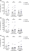
Presence of CD14+ cells is essential for the increased cytokine secretion in the presence of C1q/anti‐C1q complexes. Peripheral blood mononuclear cells (PBMCs), monocytes and T cells (co‐culture 1:5 ratio) and CD14‐depleted PBMCs (89–95% efficacy) were cultured on C1q preincubated with anti‐C1q‐negative NHS (C1q/NHS) or anti‐C1q‐positive systemic lupus erythematosus serum (C1q/anti‐C1q) and activated by tetrameric anti‐CD3/CD28 complexes for 24 h. Cell culture supernatants were analysed by ELISA for (a) TNF, (b) IFN‐γ and (c) IL‐10 secretion. Median cytokine concentrations are shown as horizontal lines, and data points represent independent experiments analysing six different healthy donors used to obtain PBMCs with connecting lines linking data points of a single individual. The Wilcoxon matched‐rank test, *P < 0.05; ns, not significant.
Figure 4
Figure 4
Increase in TNF secretion after exposure to C1q/anti‐C1q complexes requires cell–cell contact between monocytes and T cells and involves CD40 downregulation in CD11+ cells. (a) Peripheral blood mononuclear cells (PBMCs) or monocytes and T cells (1:5 ratio) co‐cultured either together or separated by 0.4 μm pore polyester membrane inserts (monocytes in the receiver plate, T cells in the permeable support system) were exposed to bound C1q, which was preincubated with anti‐C1q‐negative NHS (C1q/NHS) or anti‐C1q‐positive systemic lupus erythematosus serum (C1q/anti‐C1q). Cells were activated with tetrameric anti‐CD3/CD28 complexes for 24 h. Cell culture supernatants were analysed for TNF secretion by ELISA. Median cytokine concentrations are shown as horizontal lines, and data points represent seven different healthy donors analysed in independent experiments. (b) Analyses of CD40, CD80 and CD86 in CD11c+ cells were performed by flow cytometry after 24 h of cell culture. Median MFIs are shown as horizontal lines, and data points represent six different healthy donors analysed in independent experiments. Connecting lines link data points of a single donor used to obtain cells. The Wilcoxon matched‐rank test, *P < 0.05; ns, not significant. Flow cytometry histograms show one donor representative for six healthy donors (Supplementary figure 4b depicts the gating strategy).
Figure 5
Figure 5
CD40 signalling to monocytes is essential for increased TNF secretion after exposure to C1q/anti‐C1q complexes. (a) Peripheral blood mononuclear cells (PBMCs) from healthy donors were cultured in the presence of C1q, which was preincubated with anti‐C1q‐negative NHS (C1q/NHS) or anti‐C1q‐positive systemic lupus erythematosus (SLE) serum (C1q/SLE) for 24 h. The addition of a blocking mouse anti‐CD154 IgG antibody (5.0 μg mL−1) showed a decrease in TNF secretion compared with the isotype control (5 μg mL−1). (b) CD40–CD154 signalling to monocytes was confirmed by co‐culturing IFN‐γ‐primed (500 U mL−1, 18 h) monocytes to CD154‐expressing RD cells in the presence (C1q/anti‐C1q) or absence (C1q/NHS) of C1q/anti‐C1q complexes for 24 h. Cell culture supernatants were analysed for TNF secretion by ELISA. Median cytokine concentrations of TNF are shown as horizontal lines. Data points represent (a) six and (b) eight different healthy donors analysed in independent experiments. Connecting lines link data points of a single donor used to obtain PBMCs. The Wilcoxon matched‐signed test, *P < 0.05, **P < 0.01; ns, not significant.
Figure 6
Figure 6
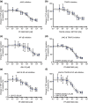
TNF secretion occurs via partially redundant intracellular CD40 signalling pathways JAK3‐STAT5 and TRAF6. Monocytes were isolated from Peripheral blood mononuclear cells (PBMCs) of healthy donors and cultured in the presence of C1q/anti‐C1q. Cells were preincubated with 500 U mL−1 IFN‐γ for 18 h. Prior to the addition of CD154‐expressing RD cells, monocytes were treated with (a) PF‐06651600 0–10 μm (JAK3 inhibitor), (b) TRAF6 inhibitor 6877002 0–20 μm, (c) JSH‐23 0–30 μm (NF‐κB inhibitor), (d) PF‐06651600 0–10 μm plus TRAF6 inhibitor 6877002 20 μm, (e) PF‐06651600 0–10 μm plus JSH 30 μm and (f) PF‐06651600 0–10 μm plus TRAF6 inhibitor 6877002 20 μm and JSH‐23 30 μm for 4 h. Cell culture supernatants were analysed for TNF secretion by ELISA after 24 h of monocyte/RD cell co‐culture. Data points represent mean inhibition of TNF secretion, normalised to the secretion without the addition of any inhibitor, of (a–e) six and (f) four different healthy donors. Error bars show standard deviations, solid lines show a four‐parametric nonlinear regression, and dashed lines show the 95% confidence bands.

Similar articles

Cited by

References

    1. Mok CC, Lau CS. Pathogenesis of systemic lupus erythematosus. J Clin Pathol 2003; 56: 481–490. - PMC - PubMed
    1. Yaniv G, Twig G, Shor DB‐A et al. A volcanic explosion of autoantibodies in systemic lupus erythematosus: a diversity of 180 different antibodies found in SLE patients. Autoimmun Rev 2015; 14: 75–79. - PubMed
    1. Tas SW, Quartier P, Botto M, Fossati‐Jimack L. Macrophages from patients with SLE and rheumatoid arthritis have defective adhesion in vitro, while only SLE macrophages have impaired uptake of apoptotic cells. Ann Rheum Dis 2006; 65: 216–221. - PMC - PubMed
    1. Herrmann M, Voll RE, Zoller OM, Hagenhofer M, Ponner BB, Kalden JR. Impaired phagocytosis of apoptotic cell material by monocyte‐derived macrophages from patients with systemic lupus erythematosus. Arthritis Rheum 1998; 41: 1241–1250. - PubMed
    1. Walport MJ. Complement. Second of two parts. N Engl J Med 2001; 344: 1140–1144. - PubMed