. 2022 Jul 1;24(2):290.
doi: 10.3892/ol.2022.13410. eCollection 2022 Aug.

HS-1793 inhibits cell proliferation in lung cancer by interfering with the interaction between p53 and MDM2

Affiliations

HS-1793 inhibits cell proliferation in lung cancer by interfering with the interaction between p53 and MDM2

Chungun Lim et al. Oncol Lett. .

Abstract

The transcription factor or tumor suppressor protein p53 regulates numerous cellular functions, including cell proliferation, invasion, migration, senescence and apoptosis, in various types of cancer. HS-1793 is an analog of resveratrol, which exhibits anti-cancer effects on various types of cancer, including breast, prostate, colon and renal cancer, and multiple myeloma. However, to the best of our knowledge, the role of HS-1793 in lung cancer remains to be examined. The present study aimed to investigate the anti-cancer effect of HS-1793 on lung cancer and to determine its association with p53. The results revealed that HS-1793 reduced cell proliferation in lung cancer and increased p53 stability, thereby elevating the expression levels of the target genes p21 and mouse double minute 2 homolog (MDM2). When the levels of MDM2, a negative regulator of p53, are increased under normal conditions, MDM2 binds and degrades p53; however, HS-1793 inhibited this binding, confirming that p53 protein stability was increased. In conclusion, the findings of the present study provide new evidence that HS-1793 may inhibit lung cancer proliferation by disrupting the p53-MDM2 interaction.

Keywords: HS-1793; MDM2; anti-cancer; lung cancer; p53.


Conflict of interest statement

The authors declare that they have no competing interests.

Figures
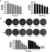
Figure 1.
Figure 1.
HS-1793 diminishes the viability of A549 and H460 cells. (A) A549 and H460 cells were treated with HS-1793 at different concentrations (0, 1, 2.5, 5, 7.5, and 10 µM) for 24 h, and absorbance was measured at 450 nm after 1 h incubation, using a CCK-8 reagent to confirm cell viability. Results are expressed as fold change. Data indicate the mean ± standard deviation (SD) of three independent experiments. *P<0.05, **P<0.01 vs. control. (B) The clonogenic assay was performed using the same cells as above. After treatment with HS-1793 in a dose-dependent manner, the media was replaced after 24 h. The media was then changed once every 2–3 days; the cells were fixed after 7 days of observation and counted by staining with crystal violet. Results are expressed as fold change. Data indicate the mean ± SD of three independent experiments. *P<0.05, **P<0.01 vs. control. CCK-8, cell counting kit-8.
Figure 2.
Figure 2.
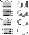
HS-1793 increases p53 protein expression. The protein expression of p53 in (A) A549 and (B) H460 cells was measured by western blot analysis. GAPDH was used as a loading control at each concentration (0, 2.5, 5, 7.5, and 10 µM) for 24 h. The experiment was independently performed twice, quantified through the ImageJ program, normalized with GAPDH, and displayed as a graph. The expression of p53 in (C) A549 and (D) H460 cells was confirmed in a time-dependent manner (0, 3, 6, 9, 12, and 24 h) by selecting 5 µM concentration of HS-1793, which showed a significant increase in the data above. Results are expressed as fold change. Data indicate the mean ± standard deviation of two independent experiments. *P<0.05, **P<0.01 vs. control. GAPDH, glyceraldehyde-3-phosphate dehydrogenase.
Figure 3.
Figure 3.
Increased p53 expression by HS-1793 upregulates its target genes p21 and MDM2. (A and B) A549 and H460 cells were treated with HS-1793 (A) in a dose-dependent manner for 24 h and (B) in a time-dependent manner (5 µM). The cells were subjected to western blotting using the indicated antibodies. This experiment was independently performed twice, protein expression was quantified through the ImageJ program, normalized to tubulin expression, and displayed as a graph. Results are expressed as fold change. Data indicate the mean ± standard deviation of two independent experiments. *P<0.05, **P<0.01 vs. control. MDM2, mouse double minute 2 homolog.
Figure 4.
Figure 4.
HS-1793 increases protein stability but not p53 transcription. (A) A549 and (B) H460 cells were pre-treated with HS-1793 for 6 h; incubated with CHX (20 µg/ml) for 0, 1, 3, 6, and 9 h; harvested; and analyzed using western blot. Band intensity was quantified by normalizing p53 with GAPDH as a control using the ImageJ program. (C and D) A549 and H460 cells were treated with HS-1793 (C) in a dose-dependent manner for 24 h and (D) in a time-dependent manner (5 µM). mRNA expression of the cells was measured using a quantitative polymerase chain reaction. B2M was used as a control and normalized to obtain a relative quantification graph. Results are expressed as fold change. Data indicate the mean ± standard deviation of independent experiments performed twice. CHX, cycloheximide; GAPDH, glyceraldehyde-3-phosphate dehydrogenase; B2M, beta-2-microglobulin.
Figure 5.
Figure 5.
HS-1793 blocks the interaction between p53 and MDM2. The interaction between p53 and MDM2 was confirmed by immunoprecipitation with p53 and MDM2 antibodies. (A) After 9 h treatment with HS-1793 in A549, cells were harvested, and the cell are lysed in lysis buffer A. The lysates (1 mg) was incubated with primary antibodies (2 µg; anti-p53 or 1 µg anti-MDM2) overnight at 4°C. Subsequently, the supernatant was incubated with 50 µl protein A/G sepharose beads for 4 h at 4°C, and the beads were washed with lysis buffer A. A western blot experiment was then performed with the lysate to confirm the interaction. (B) In H460, HS-1793 was treated for 18 h, and the experiment was conducted under the same conditions as above (A). MDM2, mouse double minute 2 homolog.

Similar articles

Cited by

References

    1. Mangiameli G, Cioffi U, Alloisio M, Testori A. Lung metastases: Current surgical indications and new perspectives. Front Surg. 2022;9:884915. doi: 10.3389/fsurg.2022.884915. - DOI - PMC - PubMed
    1. J Saller J, Boyle TA. Molecular pathology of lung cancer. Cold Spring Harb Perspect Med. 2022;12:a037812. doi: 10.1101/cshperspect.a037812. - DOI - PMC - PubMed
    1. Isaacs J, Stinchcombe TE. Neoadjuvant and adjuvant systemic therapy for early-stage non-small-cell lung cancer. Drugs. 2022;82:855–863. doi: 10.1007/s40265-022-01721-3. - DOI - PubMed
    1. Tabbò F, DE Filippis M, Jacobs F, Novello S. Strengths and pitfalls of brigatinib in non-small cell lung cancer patients' management. Minerva Med. 2022;113:315–332. doi: 10.23736/S0026-4806.21.07693-X. - DOI - PubMed
    1. Thomas AF, Kelly GL, Strasser A. Of the many cellular responses activated by TP53, which ones are critical for tumour suppression? Cell Death Differ. 2022;29:961–971. doi: 10.1038/s41418-022-00996-z. - DOI - PMC - PubMed