Dapagliflozin Improves Diabetic Cardiomyopathy by Modulating the Akt/mTOR Signaling Pathway
- PMID: 35928916
- PMCID: PMC9345717
- DOI: 10.1155/2022/9687345
Dapagliflozin Improves Diabetic Cardiomyopathy by Modulating the Akt/mTOR Signaling Pathway
Retraction in
-
Retracted: Dapagliflozin Improves Diabetic Cardiomyopathy by Modulating the Akt/mTOR Signaling Pathway.Biomed Res Int. 2023 Jun 21;2023:9765065. doi: 10.1155/2023/9765065. eCollection 2023. Biomed Res Int. 2023. PMID: 37388370 Free PMC article.
Abstract
Background: Dapagliflozin can significantly improve heart failure, and Cx43 is one of the molecular mechanisms of heart failure. This study investigated the effect of dapagliflozin on Cx43 and Akt/mTOR signaling pathway in ventricular myocytes.
Methods: A rat model of type 2 diabetes mellitus was established by high-fat diet combined with streptozotocin, and the animals were treated randomly with dapagliflozin. The morphological changes of the myocardium were observed by hematoxylin eosin staining, and the expression and distribution of Cx43 in ventricular myocytes were detected by immunohistochemistry. And Western blot determined the expressions of Cx43, Akt, mTOR, p62, and LC3 proteins in rat myocardium.
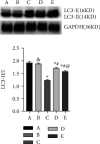
Results: Compared with the normal control group, the heart rate of diabetic rats decreased significantly (p < 0.05), QRS wave of ECG widened, and QT interval prolonged (p < 0.05). Dapagliflozin treatment in diabetic rats resulted in improvements in these ECG indexes (p < 0.05) with early administration group obtaining greater efficacy than the late administration group (p < 0.05). In the normal control group, the cardiomyocytes were arranged orderly, and the expression of Cx43 was dense, uniform, and regular, which was higher than that in the intercalated disc. In the diabetic control model group, the cardiomyocytes were enlarged and presented disorderly with detection of Cx43 in the cytoplasm. Early use of dapagliflozin better improved these myocardial tissue lesions. Of note, as diabetic rats exhibited decreased expression of Cx43, Akt, and mTOR (p < 0.05), increased p62 expression (p < 0.05), and decreased LC3-II/I ratio (p < 0.05), administration of dapagliflozin partially reversed the expression of the above proteins (p < 0.05) with greater improvement in the early administration group compared with the late administration group (p < 0.05).
Conclusions: Dapagliflozin increases the expression of Cx43 in cardiomyocytes of diabetic rats and thereby alleviates heart failure partly through regulating the Akt/mTOR signaling pathway.
Copyright © 2022 Mengxiang Ren et al.
Conflict of interest statement
The authors have no relevant financial or nonfinancial interests to disclose.
Figures


Similar articles
-
Farrerol Alleviates Diabetic Cardiomyopathy by Regulating AMPK-Mediated Cardiac Lipid Metabolic Pathways in Type 2 Diabetic Rats.Cell Biochem Biophys. 2024 Sep;82(3):2427-2437. doi: 10.1007/s12013-024-01353-2. Epub 2024 Jun 15. Cell Biochem Biophys. 2024. PMID: 38878100
-
Oral treatment with a zinc complex of acetylsalicylic acid prevents diabetic cardiomyopathy in a rat model of type-2 diabetes: activation of the Akt pathway.Cardiovasc Diabetol. 2016 May 6;15:75. doi: 10.1186/s12933-016-0383-8. Cardiovasc Diabetol. 2016. PMID: 27153943 Free PMC article.
-
Dapagliflozin attenuates diabetic cardiomyopathy through erythropoietin up-regulation of AKT/JAK/MAPK pathways in streptozotocin-induced diabetic rats.Chem Biol Interact. 2021 Sep 25;347:109617. doi: 10.1016/j.cbi.2021.109617. Epub 2021 Aug 12. Chem Biol Interact. 2021. PMID: 34391751
-
Dapagliflozin and Ticagrelor Have Additive Effects on the Attenuation of the Activation of the NLRP3 Inflammasome and the Progression of Diabetic Cardiomyopathy: an AMPK-mTOR Interplay.Cardiovasc Drugs Ther. 2020 Aug;34(4):443-461. doi: 10.1007/s10557-020-06978-y. Cardiovasc Drugs Ther. 2020. PMID: 32335797
-
Dapagliflozin Ameliorates STZ-Induced Cardiac Hypertrophy in Type 2 Diabetic Rats by Inhibiting the Calpain-1 Expression and Nuclear Transfer of NF-κB.Comput Math Methods Med. 2022 Jan 20;2022:3293054. doi: 10.1155/2022/3293054. eCollection 2022. Comput Math Methods Med. 2022. Retraction in: Comput Math Methods Med. 2023 Dec 6;2023:9864038. doi: 10.1155/2023/9864038. PMID: 35096128 Free PMC article. Retracted.
Cited by
-
The New Role of SGLT2 Inhibitors in the Management of Heart Failure: Current Evidence and Future Perspective.Pharmaceutics. 2022 Aug 18;14(8):1730. doi: 10.3390/pharmaceutics14081730. Pharmaceutics. 2022. PMID: 36015359 Free PMC article. Review.
-
Research Hotspots and Frontier Trends of Autophagy in Diabetic Cardiomyopathy From 2014 to 2024: A Bibliometric Analysis.J Multidiscip Healthc. 2025 Feb 13;18:837-860. doi: 10.2147/JMDH.S507217. eCollection 2025. J Multidiscip Healthc. 2025. PMID: 39963325 Free PMC article.
-
Retracted: Dapagliflozin Improves Diabetic Cardiomyopathy by Modulating the Akt/mTOR Signaling Pathway.Biomed Res Int. 2023 Jun 21;2023:9765065. doi: 10.1155/2023/9765065. eCollection 2023. Biomed Res Int. 2023. PMID: 37388370 Free PMC article.
References
-
- Petrie M. C., Verma S., Docherty K. F., et al. Effect of dapagliflozin on worsening heart failure and cardiovascular death in patients with heart failure with and without diabetes. Journal of the American Medical Association . 2020;323(14):1353–1368. doi: 10.1001/jama.2020.1906. - DOI - PMC - PubMed
Publication types
MeSH terms
Substances
LinkOut - more resources
Full Text Sources
Medical
Miscellaneous

