. 2023 Jan 25;51(2):501-516.
doi: 10.1093/nar/gkac633.

Construction of a cross-species cell landscape at single-cell level

Affiliations

Construction of a cross-species cell landscape at single-cell level

Renying Wang et al. Nucleic Acids Res. .

Abstract

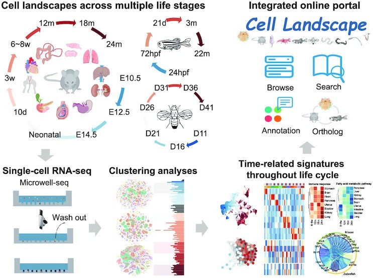
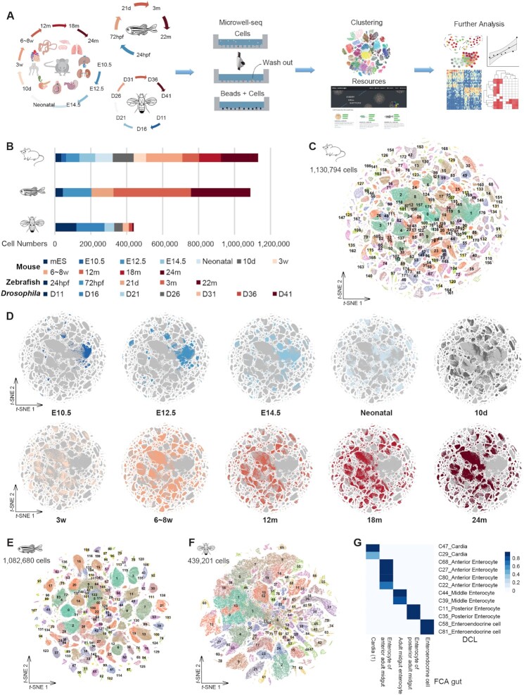
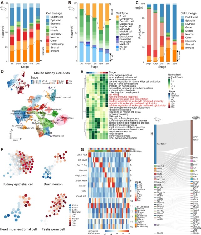
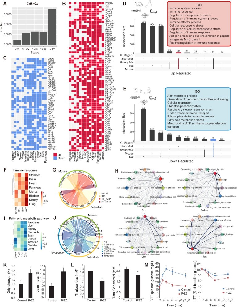
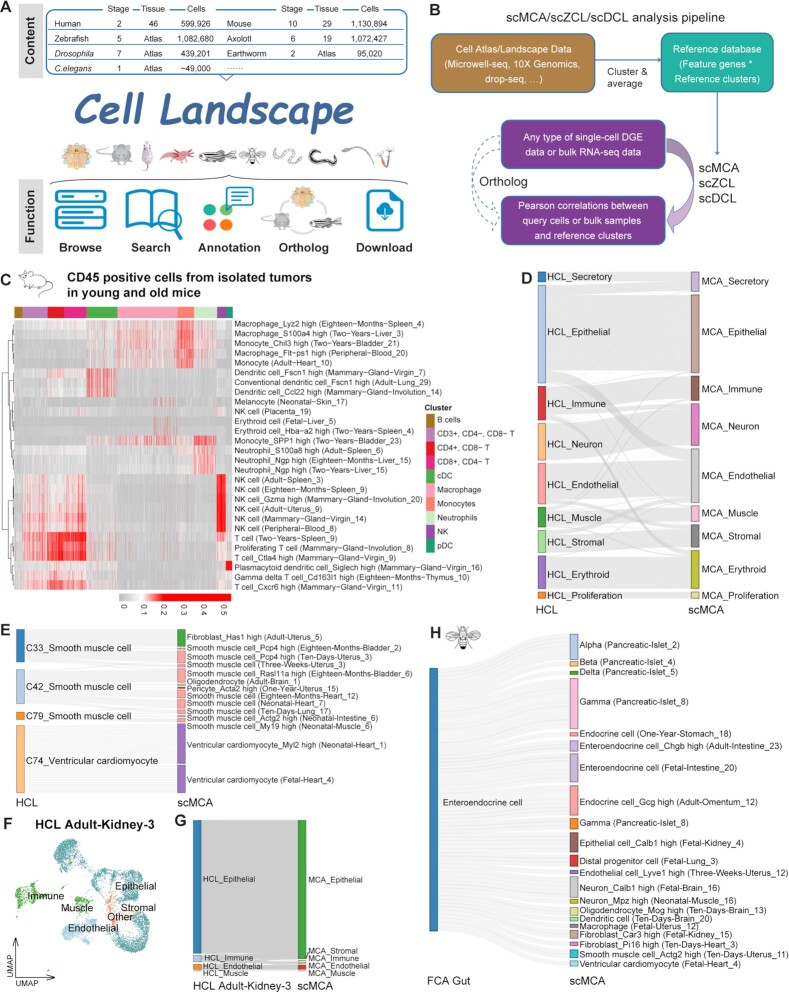
Individual cells are basic units of life. Despite extensive efforts to characterize the cellular heterogeneity of different organisms, cross-species comparisons of landscape dynamics have not been achieved. Here, we applied single-cell RNA sequencing (scRNA-seq) to map organism-level cell landscapes at multiple life stages for mice, zebrafish and Drosophila. By integrating the comprehensive dataset of > 2.6 million single cells, we constructed a cross-species cell landscape and identified signatures and common pathways that changed throughout the life span. We identified structural inflammation and mitochondrial dysfunction as the most common hallmarks of organism aging, and found that pharmacological activation of mitochondrial metabolism alleviated aging phenotypes in mice. The cross-species cell landscape with other published datasets were stored in an integrated online portal-Cell Landscape. Our work provides a valuable resource for studying lineage development, maturation and aging.

Plain language summary

How many cell types are there in nature? How do they change during the life cycle? These are two fundamental questions that researchers have been trying to understand in the area of biology. In this study, single-cell mRNA sequencing data were used to profile over 2.6 million individual cells from mice, zebrafish and Drosophila at different life stages, 1.3 million of which were newly collected. The comprehensive datasets allow investigators to construct a cross-species cell landscape that helps to reveal the conservation and diversity of cell taxonomies at genetic and regulatory levels. The resources in this study are assembled into a publicly available website at http://bis.zju.edu.cn/cellatlas/.


Figures

Graphical Abstract
Graphical Abstract
Construction of a cross-species cell landscape at the single-cell level and revealing signatures and common pathways that changed throughout the life cycle.
Figure 1.
Figure 1.
The cross-species cell landscape was constructed using Microwell-seq. (A) Overview of the experimental and bioinformatics workflow. (B) Bar plot showing the number of analyzed cells per stage by Microwell-seq from each species. (C) t-SNE visualization of 1 130 794 single cells from all tissues across all stages of mice, colored by cluster identity. (D) t-SNE visualization of the MCDAA, colored by stage. (E) t-SNE visualization of 1 082 680 single cells from all tissues across all stages of zebrafish, colored by cluster identity. (F) t-SNE visualization of 439 201 single cells from all tissues across all stages of Drosophila, colored by cluster identity. (G) Heatmap showing the correspondence between cell types in DCL gut (this study, row) and FCA gut (tissue-wide study from Li et al., 2022, column). Blue refers to a mean area under the receiver operating characterstic curve > 0.9.
Figure 2.
Figure 2.
Time-related signatures across mice, zebrafish and Drosophila. (A) Bar plot showing the fraction of cell lineages for five life stages of mouse. (B) Bar plot showing the fraction of immune cell types for five life stages of mouse. (C) Bar plot showing the fraction of cell lineages for five life stages of zebrafish. (D) UMAP visualization of the Mouse Kidney Cell Atlas across all stages of mouse kidney, colored by stage and cell type (PT, proximal tubule cell; NPC, nephronic progenitor cell; DLOH, descending loop of Henle; ALOH, ascending loop of Henle; PC, distal collecting duct principal cell; IC, collecting duct intercalated cell; DCT, distal convoluted tubule cell; EC, endothelial cell.) (E) Heatmap showing normalized GO AUCell Scores of the Mouse Kidney Cell Atlas across all stages of mouse kidney. (F) Relatedness network for the main cell types of mouse kidney, brain, heart and testis based on similarity of regulon activities. Each dot represents the aggregated cell type within each stage (see Supplementary Methods) and colored by stage. Only nodes with > 50 cells are selected. The edges between nodes represent the Spearman correlation coefficient calculated based on the aggregated regulon activity scores and filtered with Spearman correlation coefficient > 0.9. (G) Heatmap showing the aggregated module activities of TFs clustered by fuzzy c-means from mice. (H) Sankey plot showing the homologous relationships among vertebrate developmental-related TFs.
Figure 3.
Figure 3.
Aging-related signatures across multiple species. (A) Bar plot showing the fractions of cells expressing Cdkn2a at different mouse life stages. (B) Heatmap showing consensus up-regulated genes in 14 tissues of mice. The red cell represents up-regulated genes in the aging process. (C) Heatmap showing consensus down-regulated genes in 14 tissues of mice. The blue cell represents down-regulated genes in the aging process. (D) UpSet plot showing GO terms for up-regulated genes in multiple species. Representative GO terms for up-regulated genes in mice, rats and zebrafish are shown in the red box. (E) UpSet plot showing GO terms for down-regulated genes in multiple species. Representative GO terms for down-regulated genes in five species are shown in the blue box. (F) Heatmap showing normalized AUCell score of Immune response in eight mice tissues during four aging stages. (G) A circle plot showing the up-regulated homologous TFs between mice and zebrafish, colored by TF family. (H) Ligand and receptor analysis of 3w, 6–8w, 12m and 18m kidneys using CellPhoneDB. Line thickness indicates the degree of association between cell types. (I) Heatmap showing normalized AUCell score of the Fatty acid metabolic pathway in eight mice tissues during four aging stages. (J) Circular plot showing the down-regulated homologous TFs among the three species, colored by TF family. (K) Bar plot showing that PGZ reverses aging-induced frailty and sarcopenia in mice by increasing grip strength and lean mass in aging mice (n = 5). (L) Bar plot showing that PGZ reverses aging-induced hyperlipidemia in mice by decreasing triglycerides and total cholesterol in aging mice (n = 5). (M) Line chart showing that PGZ reverses aging-induced insulin resistance (n = 5).
Figure 4.
Figure 4.
The main function of Cell Landscape. (A) Overview of the Cell Landscape website construction. (B) Diagram showing the pipeline for scMCA, scZCL, scDCL and their cross-species analysis. (C) scMCA results for isolated tumors in young (6–8w) and old (20–22m) mice (n = 9966 cells). Each row represents one cell type in our reference. Each column represents data from a single cell. Pearson correlation coefficient was used to evaluate cell type gene expression similarity. Red indicates a high correlation; gray indicates a low correlation. (D) The Sankey plot showing the highest correlation coefficient pairs in each cell type between HCL and scMCA, merged by cell lineage. (E) The Sankey plot showing the highest correlation coefficient pairs in each cell type between muscle cells in HCL and corresponding cell types in scMCA. (F) t-SNE visualization of Adult-Kidney-3 in HCL, colored by cell lineage. (G) The Sankey plot showing the highest correlation coefficient pairs in each cell type between Adult-Kidney-3 in HCL and scMCA, merged by cell lineage. (H) The Sankey plot showing the highest correlation coefficient pairs in each cell type between EEs in FCA gut and corresponding cell types in scMCA.

References

    1. Tang F., Barbacioru C., Wang Y., Nordman E., Lee C., Xu N., Wang X., Bodeau J., Tuch B.B., Siddiqui A.et al. .. mRNA-Seq whole-transcriptome analysis of a single cell. Nat. Methods. 2009; 6:377–382. - PubMed
    1. Hashimshony T., Wagner F., Sher N., Yanai I.. CEL-Seq: single-cell RNA-Seq by multiplexed linear amplification. Cell Rep. 2012; 2:666–673. - PubMed
    1. Ramskold D., Luo S., Wang Y.C., Li R., Deng Q., Faridani O.R., Daniels G.A., Khrebtukova I., Loring J.F., Laurent L.C.et al. .. Full-length mRNA-Seq from single-cell levels of RNA and individual circulating tumor cells. Nat. Biotechnol. 2012; 30:777–782. - PMC - PubMed
    1. Shalek A.K., Satija R., Adiconis X., Gertner R.S., Gaublomme J.T., Raychowdhury R., Schwartz S., Yosef N., Malboeuf C., Lu D.et al. .. Single-cell transcriptomics reveals bimodality in expression and splicing in immune cells. Nature. 2013; 498:236–240. - PMC - PubMed
    1. Treutlein B., Brownfield D.G., Wu A.R., Neff N.F., Mantalas G.L., Espinoza F.H., Desai T.J., Krasnow M.A., Quake S.R.. Reconstructing lineage hierarchies of the distal lung epithelium using single-cell RNA-seq. Nature. 2014; 509:371–375. - PMC - PubMed

Publication types