. 2022 Aug 16;11(16):e024581.
doi: 10.1161/JAHA.121.024581. Epub 2022 Aug 5.

von Willebrand Factor: A Central Regulator of Arteriovenous Fistula Maturation Through Smooth Muscle Cell Proliferation and Outward Remodeling

Affiliations

von Willebrand Factor: A Central Regulator of Arteriovenous Fistula Maturation Through Smooth Muscle Cell Proliferation and Outward Remodeling

Suzanne L Laboyrie et al. J Am Heart Assoc. .

Abstract

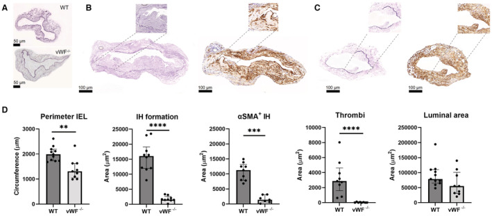
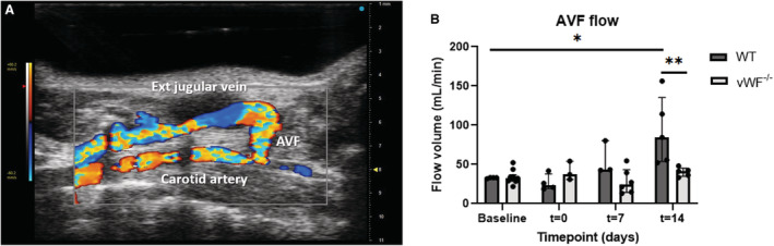
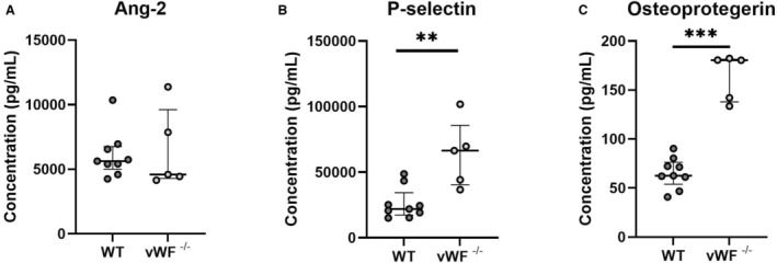
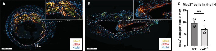
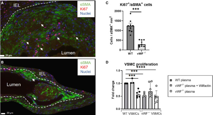
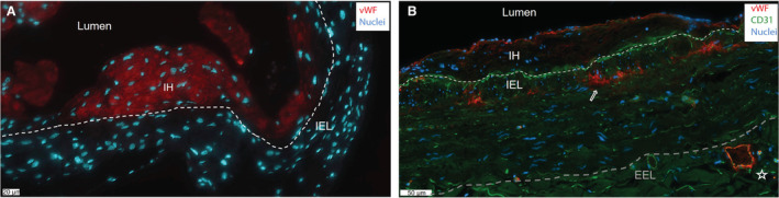
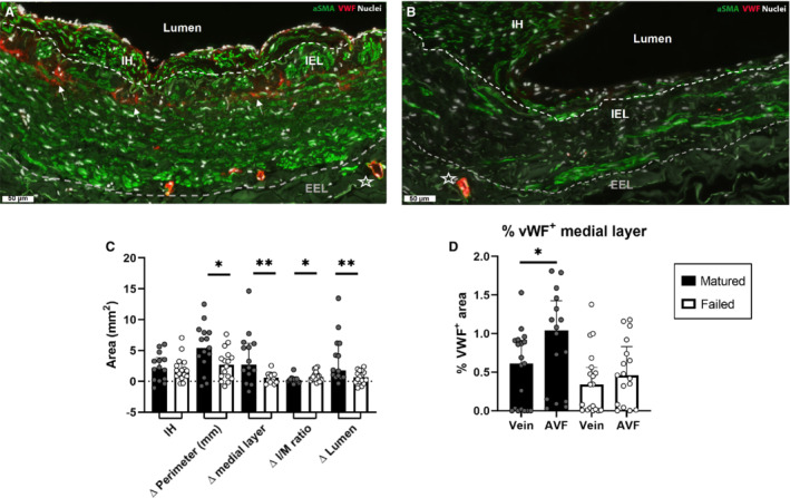
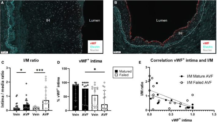
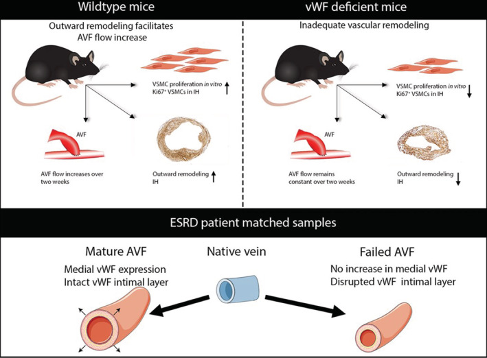
Background Arteriovenous fistula (AVF) maturation failure is a main limitation of vascular access. Maturation is determined by the intricate balance between outward remodeling and intimal hyperplasia, whereby endothelial cell dysfunction, platelet aggregation, and vascular smooth muscle cell (VSMC) proliferation play a crucial role. von Willebrand Factor (vWF) is an endothelial cell-derived protein involved in platelet aggregation and VSMC proliferation. We investigated AVF vascular remodeling in vWF-deficient mice and vWF expression in failed and matured human AVFs. Methods and Results Jugular-carotid AVFs were created in wild-type and vWF-/- mice. AVF flow was determined longitudinally using ultrasonography, whereupon AVFs were harvested 14 days after surgery. VSMCs were isolated from vena cavae to study the effect of vWF on VSMC proliferation. Patient-matched samples of the basilic vein were obtained before brachio-basilic AVF construction and during superficialization or salvage procedure 6 weeks after AVF creation. vWF deficiency reduced VSMC proliferation and macrophage infiltration in the intimal hyperplasia. vWF-/- mice showed reduced outward remodeling (1.5-fold, P=0.002) and intimal hyperplasia (10.2-fold, P<0.0001). AVF flow in wild-type mice was incremental over 2 weeks, whereas flow in vWF-/- mice did not increase, resulting in a two-fold lower flow at 14 days compared with wild-type mice (P=0.016). Outward remodeling in matured patient AVFs coincided with increased local vWF expression in the media of the venous outflow tract. Absence of vWF in the intimal layer correlated with an increase in the intima-media ratio. Conclusions vWF enhances AVF maturation because its positive effect on outward remodeling outweighs its stimulating effect on intimal hyperplasia.

Keywords: AVF maturation; VSMC; intimal hyperplasia; outward remodeling; von Willebrand Factor.


Figures

Figure 1
Figure 1. vWF deficiency leads to reduced outward remodeling and intimal hyperplasia following AVF surgery.
Representative images of baseline vena jugularis, scale bar=50 μm (A) and AVFs stained using Weigert's Elastin (left) and αSMA staining (right) from a WT (B) and vWF−/− (C) mouse AVF. Inlays illustrate IH in closer detail, including a thrombus in Figure 1B. The perimeter was measured by lining the internal elastic lamina (IEL); thrombi were defined as αSMA‐negative anuclear tissue. The stainings were quantified and presented as median±IQR (interquartile range) (D). OR (outward remodeling) was calculated using the IEL perimeter in Weigert's Elastin stained samples, IH was defined as the tissue within the IEL. αSMA‐positive tissue within the IH was determined using histoquant software. Thrombi were manually traced. Luminal area was calculated by subtracting the IH from the area within the IEL. Scale bar is 100 μm. N=9 per group. Statistical significance between groups was determined using the Mann–Whitney U test **P=0.002, ***P<0.001, ****P<0.0001. AVF indicates arteriovenous fistula; IEL, internal elastic lamina; IH, intimal hyperplasia; IQR, interquartile range; OR, outward remodeling; αSMA, α‐smooth muscle actin; vWF, von Willebrand Factor; and WT, wild type.
Figure 2
Figure 2. vWF deficiency leads to reduced AVF flow 14 days post‐AVF creation.
Effect of vWF deficiency on AVF blood flow was determined in the common carotid artery (CCA) afferent to the anastomosis (A). AVF blood flow in the CCA was calculated with the diameter of the CCA using EKV obtained data and blood velocity obtained in PW color mode (B). Ultrasound measurements were performed 1 day before surgery (baseline), immediately postsurgery (t=0), and at t=7 and t=14 days. Data are presented as median±interquartile range, n=6 WT and 9 vWF−/− mice. Statistical significance between groups was determined at timepoint t=14 days, and between baseline and t=14 days in WT mice using the Mann–Whitney U test. *P=0.016, **P=0.008. AVF indicates arteriovenous fistula; EKV, ECG‐gated Kilohertz Visualization; PW, pulsed wave; vWF, von Willebrand Factor; and WT, wild type.
Figure 3
Figure 3. Luminex analysis of systemic WPB protein expression.
The effect of vWF deficiency on Weibel‐Palade body (WPB)‐proteins angiopoeitin‐2 (A), P‐selectin (B), and osteoprotegerin (C) was determined in 1:3 diluted EDTA plasma from n=9 WT and 5 vWF−/− mice using a 5‐PL logistic regression model. Data are presented as median±interquartile range. Statistical significance between groups was determined using the Mann–Whitney U test. **P=0.007, ***P=0.001. vWF indicates von Willebrand Factor; and WT, wild type.
Figure 4
Figure 4. Intimal hyperplasia in the AVF of vWF−/− mice displays fewer Mac3+ cells.
Immunofluorescent staining for nuclei (blue), αSMA (red), and Mac3 (yellow) of an AVF of a WT (A) and vWF−/− (B) mouse, with a higher magnification inlay of part of the IH. The IEL is traced in white, scale bar=100 μm. Nuclei of Mac3+ cells were counted manually per field of fiew and quantified for n=7 per group (C). Data are presented as mean±SD, **P=0.006, statistical significance between groups was determined using unpaired t test. αSMA indicates α‐smooth muscle actin; AVF, arteriovenous fistula; IEL, internal elastic lamina; IH, intimal hyperplasia; vWF, von Willebrand Factor; and WT, wild type.
Figure 5
Figure 5. Characterization of proliferating VSMCs in the IH at 2 weeks post AVF creation and in vitro.
Immunofluorescent staining for nuclei (blue), Ki67 (red), and αSMA (green) in a WT (A) and vWF−/− mouse (B). The IEL is traced in white. Ki67+/αSMA+ cells, examples indicated with an arrow, within the IH were counted manually and normalized to the αSMA‐positive area within the IH. Scale bar=20 μm. Data are analyzed by the Mann–Whitney U test and presented as median±interquartile range, n=9 per group (C). VSMCs from WT (left 3 bars) and vWF−/− (right 3 bars) mice were stimulated with 10% murine plasma of WT or vWF−/− mice ±100 ng/mL Wilfactin (see box legend) for 40 hours, after which proliferation was measured. Data are presented as mean±SD, n=3 to 6. Statistical significance between WT VSMCs stimulated with WT plasma (control) and other experimental conditions was determined using 1‐way ANOVA and Dunnett's multiple comparisons test ***P<0.001, ****P=0.0001 (D). IEL indicates internal elastic lamina; IH, intimal hyperplasia; αSMA, α‐smooth muscle actin; VSMCs, vascular smooth muscle cells; vWF, von Willebrand Factor; and WT, wild type.
Figure 6
Figure 6. vWF resides in the intima and medial layer.
Representative immunofluorescent staining for vWF (red) in a wild‐type murine AVF (A) and in a matured AVF of a patient with end‐stage renal disease at superficialization (B). Endothelial cell marker CD31 is stained in the human AVF in green, nuclei in blue. The white dashed line signifies the IEL (internal elastic lamina), and the gray line indicates the EEL (external elastic lamina). Scale bar=20 μm in (A) and 50 μm in (B). AVF indicates arteriovenous fistula; IH, intimal hyperplasia; and vWF, von Willebrand Factor.
Figure 7
Figure 7. Morphometric and vWF expression analysis of samples from patient with ESRD.
Analysis of ESRD pair‐matched samples of pre‐access native veins and venous AVF outflow tracts obtained during 2‐stage brachio‐basilic AVF surgery. AVF failure was defined as having an internal cross‐sectional luminal diameter ≤6 mm. Representative sample of a mature (A) and failed AVF (B). Scale bar=50 μm, star indicates a vessel in the vasa vasorum, αSMA+/vWF+ staining is indicated with arrowheads. The inner white line signifies the IEL, and the outer gray line signifies the external elastic lamina (EEL). Pair‐matched analysis of increase in IH area, delta (Δ) OR (perimeter in mm), area of the medial layer, the intima/media (I/M) ratio, and luminal size was calculated by subtracting the pre‐access venous parameters from the patient‐matched AVF. N=15 pair‐matched samples from matured AVFs and 16 pair‐matched failed AVFs. Statistical significance between groups was determined using the Mann–Whitney U test (C). vWF+ tissue in the medial layer was quantified in immunofluorescently stained samples and normalized to the area of the medial layer. Statistical significance was determined using Wilcoxon matched‐pairs signed rank test (D). Data are presented as median±interquartile range. *P<0.05, **P<0.01. AVF indicates arteriovenous fistula; ESRD, end‐stage renal disease; IEL, internal elastic lamina; IH, intimal hyperplasia; OR, outward remodeling; αSMA, α‐smooth muscle actin; and vWF, von Willebrand Factor.
Figure 8
Figure 8. Relationship between disruption of vWF lining the intima and an enhanced I/M ratio.
Analysis of ESRD samples of pre‐access native veins and venous AVF outflow tracts obtained during 2‐stage brachio‐basilic AVF surgery. Absence of vWF lining the intimal layer was observed at sites with increased IH (A) while an intact vWF+ intimal layer was observed at sites with little IH (B). The dashed line signifies the IEL, vWF is stained in red, elastin in turquoise, and nuclei in white. Scale bar=50 μm. The ratio between intimal area and the medial layer (I/M) (C) and the percentage of vWF+ intimal layer in native veins and AVFs were quantified (D) and correlated using linear regression (E). N=15 pair‐matched samples from matured AVFs and 16 pair‐matched failed AVFs; data are represented as median±interquartile range. Statistical significance between groups was determined using the Mann–Whitney U test; *P<0.05, ***P=0.0001. AVF indicates arteriovenous fistula; ESRD, end‐stage renal disease; IEL, internal elastic lamina; IH, intimal hyperplasia; I/M, intima/media ratio; and vWF, von Willebrand Factor.
Figure 9
Figure 9. Overview of the findings.
AVF indicates arteriovenous fistula; ESRD, end‐stage renal disease; IH, intimal hyperplasia; VSMC, vascular smooth muscle cell; and vWF, von Willebrand Factor.

Similar articles

Cited by

References

    1. Schmidli J, Widmer MK, Basile C, de Donato G, Gallieni M, Gibbons CP, Haage P, Hamilton G, Hedin U, Kamper L, et al. Vascular access: 2018 clinical practice guidelines of the European Society for Vascular Surgery (ESVS). Eur J Vasc Endovasc Surg. 2018;55:757–818. doi: 10.1016/j.ejvs.2018.02.001 - DOI - PubMed
    1. Astor BC, Eustace JA, Powe NR, Klag MJ, Fink NE, Coresh J, Study ftC . Type of vascular access and survival among incident hemodialysis patients: the choices for healthy outcomes in caring for ESRD (CHOICE) study. J Am Soc Nephrol. 2005;16:1449–1455. doi: 10.1681/ASN.2004090748 - DOI - PubMed
    1. Banerjee T, Kim SJ, Astor B, Shafi T, Coresh J, Powe NR. Vascular access type, inflammatory markers, and mortality in incident hemodialysis patients: the Choices for Healthy Outcomes in Caring for End‐Stage Renal Disease (CHOICE) Study. Am J Kidney Dis. 2014;64:954–961. doi: 10.1053/j.ajkd.2014.07.010 - DOI - PMC - PubMed
    1. Dhingra RK, Young EW, Hulbert‐Shearon TE, Leavey SF, Port FK. Type of vascular access and mortality in U.S. hemodialysis patients. Kidney Int. 2001;60:1443–1451. doi: 10.1046/j.1523-1755.2001.00947.x - DOI - PubMed
    1. Voorzaat BM, Janmaat CJ, van der Bogt KEA, Dekker FW, Rotmans JI. Patency outcomes of arteriovenous fistulas and grafts for hemodialysis access: a trade‐off between nonmaturation and long‐term complications. Kidney360. 2020;1:916–924. doi: 10.34067/KID.0000462020 - DOI - PMC - PubMed

Publication types

Substances

LinkOut - more resources