. 2022 Oct;26(4):299.
doi: 10.3892/mmr.2022.12815. Epub 2022 Aug 5.

Hydroxygenkwanin suppresses proliferation, invasion and migration of osteosarcoma cells via the miR‑320a/SOX9 axis

Affiliations

Hydroxygenkwanin suppresses proliferation, invasion and migration of osteosarcoma cells via the miR‑320a/SOX9 axis

Xinli Dong et al. Mol Med Rep. 2022 Oct.

Abstract

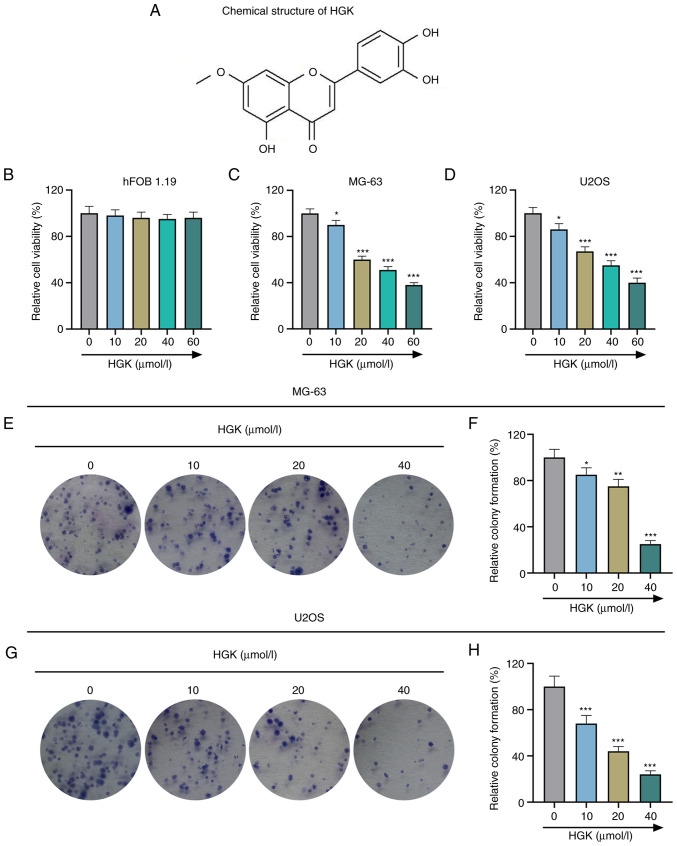
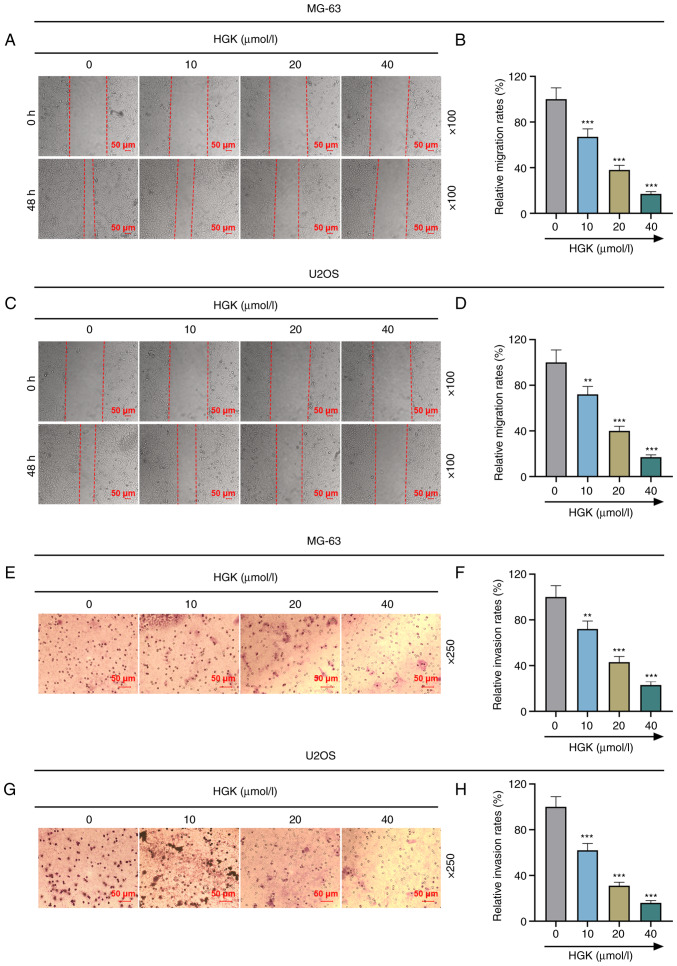
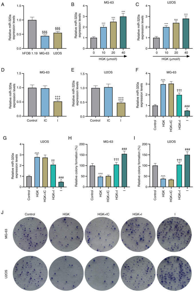
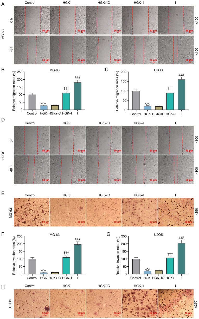
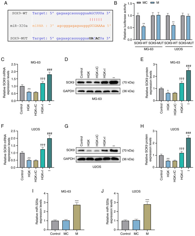
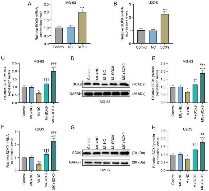
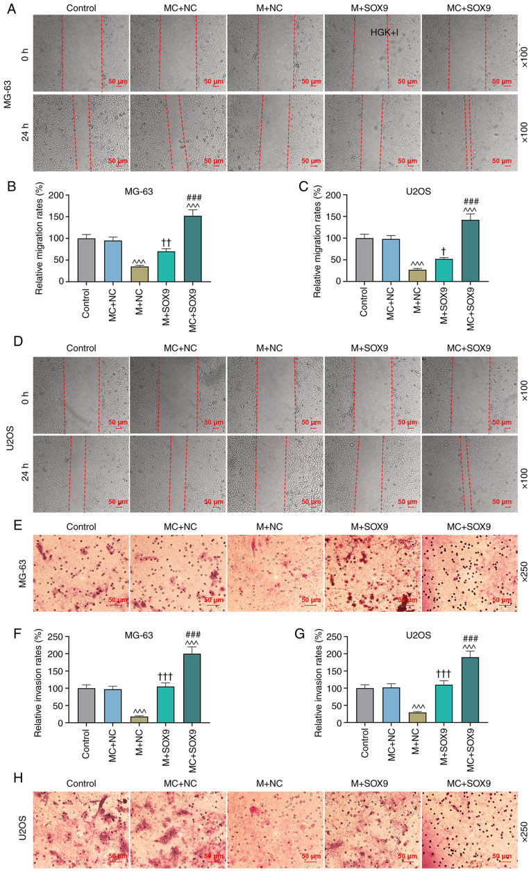
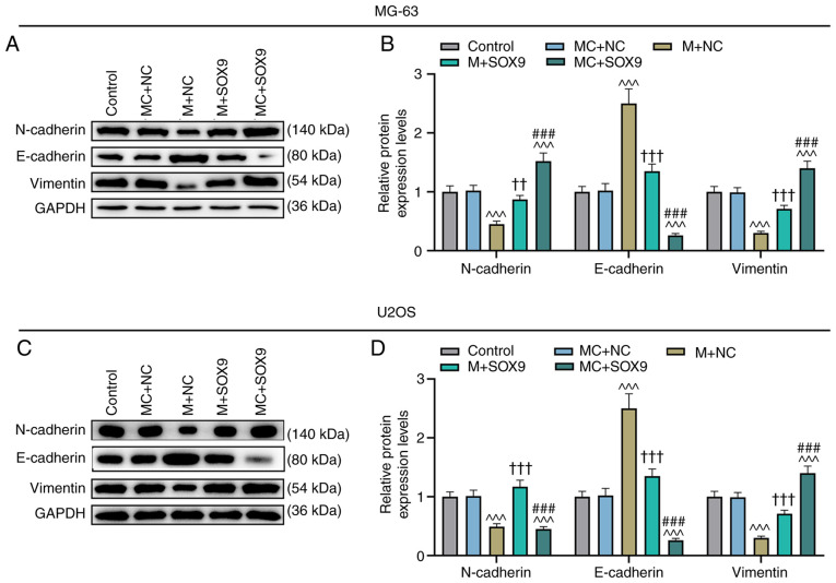
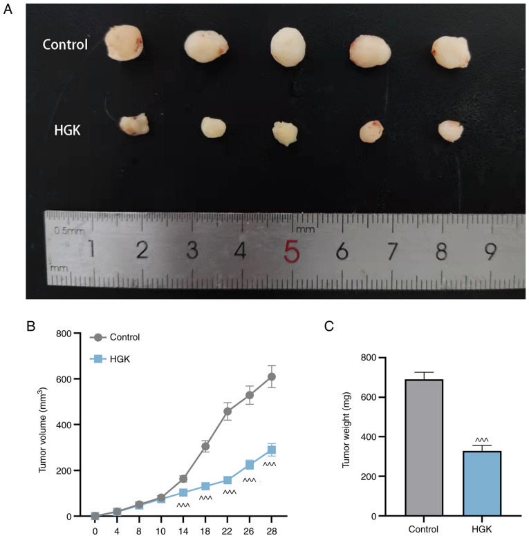
Hydroxygenkwanin (HGK) has an anticancer effect in a variety of tumors, but its role in osteosarcoma has not been explored. The purpose of the present study was to investigate the therapeutic effect of HGK on osteosarcoma and its specific molecular mechanism. Osteosarcoma cells (MG‑63 and U2OS) treated with various concentrations of HGK were assigned to the treatment group. MTT, clone formation, wound healing and Transwell assays were performed to assess the viability, proliferation, migration, and invasion of MG‑63 and U2OS cells. RT‑qPCR was conducted to quantify the expression levels of of microRNA (miR)‑320a and SRY‑box transcription factor 9 (SOX9) in MG‑63 and U2OS cells. The binding sites of miR‑320a and SOX9 were predicted by starBase database, and verified using the dual‑luciferase reporter assay. The expression levels of SOX9 and EMT‑related proteins (N‑cadherin, E‑cadherin and vimentin) were detected by western blot analysis. HGK inhibited cell proliferation, migration, invasion, but promoted the expression of miR‑320a in MG‑63 and U2OS cells. Downregulation of miR‑320a reversed the effects of HGK on proliferation, migration and invasion of MG‑63 and U2OS cells, while upregulation of miR‑320a had the opposite effect. HGK inhibited the expression of SOX9 by promoting the expression of miR‑320a. Upregulation of SOX9 could partially reverse miR‑320a‑induced migration and invasion of MG‑63 and U2OS cells. In addition, upregulation of miR‑320a promoted E‑cadherin expression and inhibited the expression of N‑cadherin and vimentin, and the effect of miR‑320a was also reversed by SOX9. In conclusion, HGK inhibited proliferation, migration and invasion of MG‑63 and U2OS cells through the miR‑320a/SOX9 axis.

Keywords: hydroxygenkwanin; invasion; miR‑320a/SOX9 axis; migration; osteosarcoma; proliferation.


Conflict of interest statement

The authors declare that they have no competing interests.

Figures

Figure 1.
Figure 1.
HGK decreases the viability and proliferation of MG-63 and U2OS cells. (A) The chemical structure of HGK. (B) The cell viability of hFOB 1.19 cells treated or untreated with HGK was detected by MTT assay. (C and D) The cell viability of MG-63 and U2OS cells treated or untreated with HGK was determined by MTT assay. (E-H) The cell proliferation of MG-63 and U2OS cells treated or untreated with HGK was assessed using colony formation assay. *P<0.05, **P<0.01 and ***P<0.001 vs. 0 µmol/l. HGK, hydroxygenkwanin; MTT, 3-(4,5-dimethylthiazol-2-yl)-2,5-diphenyltetrazolium bromide.
Figure 2.
Figure 2.
HGK decreases the migration and invasion of MG-63 and U2OS cells. (A-D) The cell migration of MG-63 and U2OS cells treated or untreated with HGK was assessed by wound healing assay. Magnification, ×100. (E-H) The cell invasion of MG-63 and U2OS cells treated or untreated with HGK was assessed by Transwell assay. Magnification, ×250. **P<0.01 and ***P<0.001 vs. 0 µmol/l. HGK, hydroxygenkwanin.
Figure 3.
Figure 3.
HGK decreases the proliferation of MG-63 and U2OS cells by promoting miR-320a expression. (A) The expression of miR-320a in hFOB 1.19, MG-63 and U2OS cells was quantified by RT-qPCR. (B and C) The expression of miR-320a in MG-63 and U2OS cells treated or untreated with HGK was detected by RT-qPCR. (D and E) Transfection efficiency of miR-320a inhibitor in MG-63 and U2OS cells was evaluated by RT-qPCR. (F and G) The expression of miR-320a in MG-63 and U2OS cells treated or untreated with HGK and/or miR-320a inhibitor was determined by RT-qPCR. (H-J) The cell proliferation of MG-63 and U2OS cells treated or untreated with HGK and/or miR-320a inhibitor was examined by colony formation assay. §§§P<0.001 vs. hFOB 1.19; ***P<0.001 vs. 0 µmol/l; ‡‡‡P<0.001 vs. IC; ^P<0.05 and ^^^P<0.001 vs. Control; ††P<0.01 and †††P<0.001 vs. HGK + IC; ###P<0.001 vs. HGK + I. HGK, hydroxygenkwanin; miR-320a, microRNA-320a; RT-qPCR, reverse transcription-quantitative polymerase chain reaction; I, miR-320a inhibitor; IC, miR-320a inhibitor control.
Figure 4.
Figure 4.
HGK decreases the migration and invasion of MG-63 and U2OS cells by promoting miR-320a expression. (A-D) The cell migration of MG-63 and U2OS cells treated or untreated with HGK and/or miR-320a inhibitor was detected by wound healing assay. Magnification, ×100. (E-H) The cell invasion of MG-63 and U2OS cells treated or untreated with HGK and/or miR-320a inhibitor was assessed by Transwell assay. Magnification, ×250. ^^^P<0.001 vs. Control; †††P<0.001 vs. HGK + IC; ###P<0.001 vs. HGK + I. HGK, hydroxygenkwanin; miR-320a, microRNA-320a; I, miR-320a inhibitor; IC, miR-320a inhibitor control.
Figure 5.
Figure 5.
HGK decreases SOX9 expression by promoting miR-320a expression in MG-63 and U2OS cells. (A) starBase predicted the binding site of miR-320a and SOX9. (B) Dual luciferase reporter assay was employed to verify the targeted bindings of miR-320a and SOX9. (C-H) The expression of SOX9 in MG-63 and U2OS cells treated or untreated with HGK and/or miR-320a inhibitor was quantified by RT-qPCR and western blotting. (I and J) Transfection efficiency of miR-320a mimic in MG-63 and U2OS cells was evaluated by RT-qPCR. **P<0.01 and ***P<0.001 vs. MC; ^P<0.05 and ^^P<0.01 vs. Control; †††P<0.001 vs. HGK + IC; ###P<0.001 vs. HGK + I. HGK, hydroxygenkwanin; SOX9, SRY-box transcription factor 9; miR-320a, microRNA-320a; RT-qPCR, quantitative reverse transcription polymerase chain reaction; M, miR-320a mimic; MC, miR-320a mimic control; IC, miR-320a inhibitor control; I, miR-320a inhibitor.
Figure 6.
Figure 6.
MiR-320a decreases the expression of SOX9 in MG-63 and U2OS cells. (A and B) Transfection efficiency of SOX9 overexpression plasmid in MG-63 and U2OS cells was assessed by RT-qPCR. (C-H) The expression of SOX9 in MG-63 and U2OS cells transfected or untransfected with miR-320a mimic and/or SOX9 overexpression plasmid was quantified by RT-qPCR and western blotting. ***P<0.001 vs. NC; ^P<0.05, ^^P<0.01 and ^^^P<0.001 vs. MC + NC; ††P<0.01 and †††P<0.001 vs. M + NC; ##P<0.01 and ###P<0.001 vs. M + SOX9. MiR-320a, microRNA-320a; SOX9, SRY-box transcription factor 9; RT-qPCR, reverse transcription-quantitative polymerase chain reaction; NC, negative control; MC, miR-320a mimic control; M, miR-320a mimic.
Figure 7.
Figure 7.
MiR-320a decreases the cell migration and invasion abilities of MG-63 and U2OS cells by inhibiting the expression of SOX9. (A-D) The migration of MG-63 and U2OS cells transfected or untransfected with miR-320a mimic and/or SOX9 overexpression plasmid was detected by wound healing assay. Magnification, ×100. (E-H) The cell invasion of MG-63 and U2OS cells transfected or untransfected with miR-320a mimic and/or SOX9 overexpression plasmid was determined by Transwell assay. Magnification, ×250. ^^^P<0.001 vs. MC + NC; †P<0.05, ††P<0.01 and †††P<0.001 vs. M + NC; ###P<0.001 vs. M + SOX9. MiR-320a, microRNA-320a; SOX9, SRY-box transcription factor 9; MC, miR-320a mimic control; NC, negative control; M, miR-320a mimic.
Figure 8.
Figure 8.
MiR-320a increases E-cadherin expression while decreasing N-cadherin, and vimentin expression levels by inhibiting the expression of SOX9. (A-D) The expression levels of E-cadherin, N-cadherin and vimentin in MG-63 and U2OS cells transfected or untransfected with miR-320a mimic and/or SOX9 overexpression plasmid were detected by western blotting. ^^^P<0.001 vs. MC + NC; ††P<0.01 and †††P<0.001 vs. M + NC; ###P<0.001 vs. M + SOX9. MiR-320a, microRNA-320a; SOX9, SRY-box transcription factor 9; MC, miR-320a mimic control; NC, negative control; M, miR-320a mimic.
Figure 9.
Figure 9.
HGK suppresses tumor growth in mice. (A) Tumor images of control and HGK groups (n=5). (B) The effects of HGK treatment on the tumor volume of mice. (C) The effects of HGK treatment on the tumor weight of mice. ^^^P<0.001 vs. Control. HGK, hydroxygenkwanin.

Similar articles

Cited by

References

    1. Mannerström B, Kornilov R, Abu-Shahba AG, Chowdhury IM, Sinha S, Seppänen-Kaijansinkko R, Kaur S. Epigenetic alterations in mesenchymal stem cells by osteosarcoma-derived extracellular vesicles. Epigenetics. 2019;14:352–364. doi: 10.1080/15592294.2019.1585177. - DOI - PMC - PubMed
    1. Brown HK, Tellez-Gabriel M, Heymann D. Cancer stem cells in osteosarcoma. Cancer Lett. 2017;386:189–195. doi: 10.1016/j.canlet.2016.11.019. - DOI - PubMed
    1. Gross AC, Cam H, Phelps DA, Saraf AJ, Bid HK, Cam M, London CA, Winget SA, Arnold MA, Brandolini L, et al. IL-6 and CXCL8 mediate osteosarcoma-lung interactions critical to metastasis. JCI Insight. 2018;3:e99791. doi: 10.1172/jci.insight.99791. - DOI - PMC - PubMed
    1. Meazza C, Scanagatta P. Metastatic osteosarcoma: A challenging multidisciplinary treatment. Expert Rev Anticancer Ther. 2016;16:543–556. doi: 10.1586/14737140.2016.1168697. - DOI - PubMed
    1. Luetke A, Meyers PA, Lewis I, Juergens H. Osteosarcoma treatment-where do we stand? A state of the art review. Cancer Treat Rev. 2014;40:523–532. doi: 10.1016/j.ctrv.2013.11.006. - DOI - PubMed