

Escaping the symmetry trap in helical reconstruction
- PMID: 35929538
- PMCID: PMC9642006
- DOI: 10.1039/d2fd00051b
Escaping the symmetry trap in helical reconstruction
Abstract
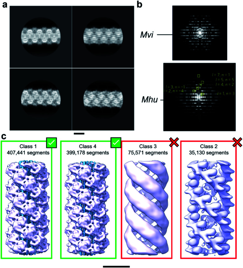
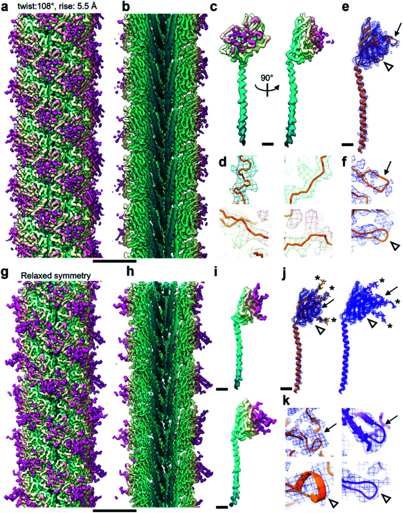
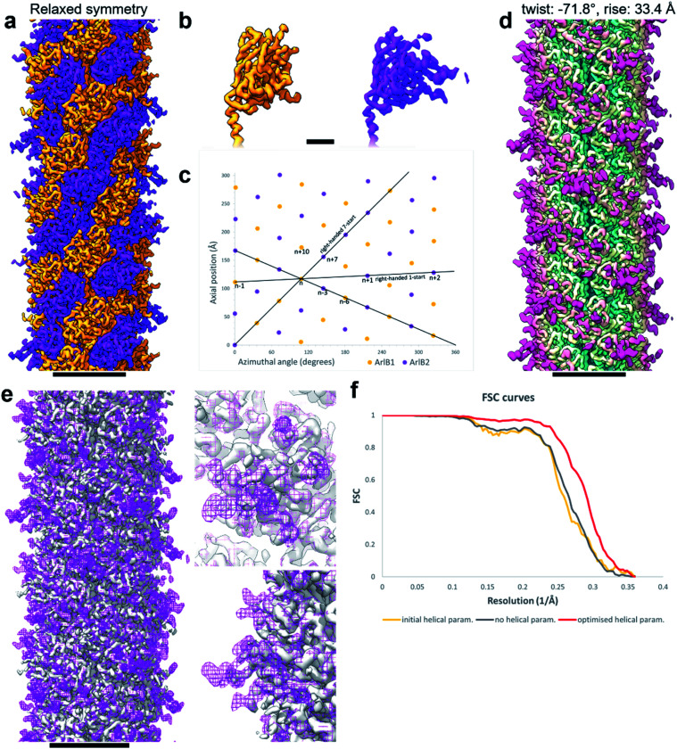
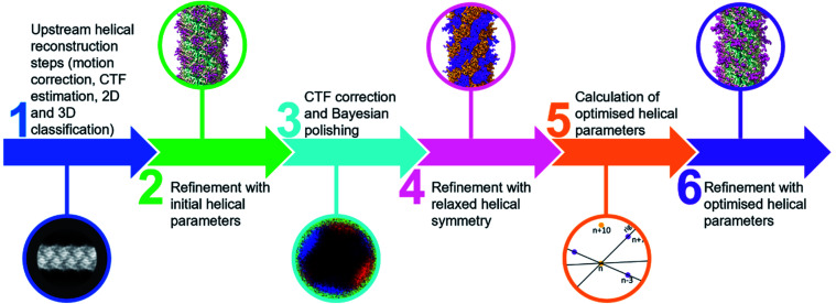
Helical reconstruction is the method of choice for obtaining 3D structures of filaments from electron cryo-microscopy (cryoEM) projections. This approach relies on applying helical symmetry parameters deduced from Fourier-Bessel or real space analysis, such as sub-tomogram averaging. While helical reconstruction continues to provide invaluable structural insights into filaments, its inherent dependence on imposing a pre-defined helical symmetry can also introduce bias. The applied helical symmetry produces structures that are infinitely straight along the filament's axis and can average out biologically important heterogeneities. Here, we describe a simple workflow aimed at overcoming these drawbacks in order to provide truer representations of filamentous structures.
Conflict of interest statement
There are no conflicts of interest to declare.
Figures
Similar articles
-
Cryo-EM Structure Determination Using Segmented Helical Image Reconstruction.Methods Enzymol. 2016;579:307-28. doi: 10.1016/bs.mie.2016.05.034. Epub 2016 Jun 28. Methods Enzymol. 2016. PMID: 27572732 Review.
-
Structure of HIV-1 capsid assemblies by cryo-electron microscopy and iterative helical real-space reconstruction.J Vis Exp. 2011 Aug 9;(54):3041. doi: 10.3791/3041. J Vis Exp. 2011. PMID: 21860371 Free PMC article.
-
SPRING - an image processing package for single-particle based helical reconstruction from electron cryomicrographs.J Struct Biol. 2014 Jan;185(1):15-26. doi: 10.1016/j.jsb.2013.11.003. Epub 2013 Nov 21. J Struct Biol. 2014. PMID: 24269218
-
Helical Indexing in Real Space.Sci Rep. 2022 May 17;12(1):8162. doi: 10.1038/s41598-022-11382-7. Sci Rep. 2022. PMID: 35581231 Free PMC article.
-
Cryo-Electron Tomography and Subtomogram Averaging.Methods Enzymol. 2016;579:329-67. doi: 10.1016/bs.mie.2016.04.014. Epub 2016 Jun 22. Methods Enzymol. 2016. PMID: 27572733 Review.
Cited by
-
Perturbed N-glycosylation of Halobacterium salinarum archaellum filaments leads to filament bundling and compromised cell motility.Nat Commun. 2024 Jul 11;15(1):5841. doi: 10.1038/s41467-024-50277-1. Nat Commun. 2024. PMID: 38992036 Free PMC article.
References
-
- Kreutzberger M. A. B. Ewing C. Poly F. Wang F. Egelman E. H. Atomic structure of the Campylobacter flagellar filament reveals how ε Proteobacteria escaped Toll-like receptor 5 surveillance. Proc. Natl. Acad. Sci. U. S. A. 2020;117(29):16985–16991. doi: 10.1073/pnas.2010996117. doi: 10.1073/pnas.2010996117. - DOI - DOI - PMC - PubMed
-
- Wang F. Baquero D. P. Beltran L. C. Su Z. Osinski T. Zheng W. Prangishvili D. Krupovic M. Egelman E. H. Structures of filamentous viruses infecting hyperthermophilic archaea explain DNA stabilization in extreme environments. Proc. Natl. Acad. Sci. U. S. A. 2020;117(33):19643–19652. doi: 10.1073/pnas.2011125117. doi: 10.1073/pnas.2011125117. - DOI - DOI - PMC - PubMed
Publication types
MeSH terms
Grants and funding
LinkOut - more resources
Full Text Sources