. 2022 Sep 30;12(10):jkac200.
doi: 10.1093/g3journal/jkac200.

Discovery of nonautonomous modulators of activated Ras

Affiliations

Discovery of nonautonomous modulators of activated Ras

Marcos Corchado-Sonera et al. G3 (Bethesda). .

Abstract

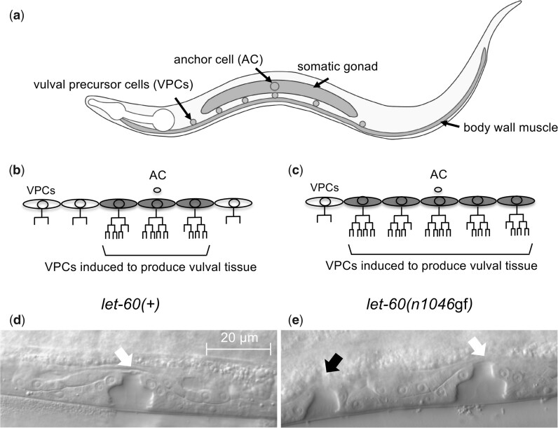
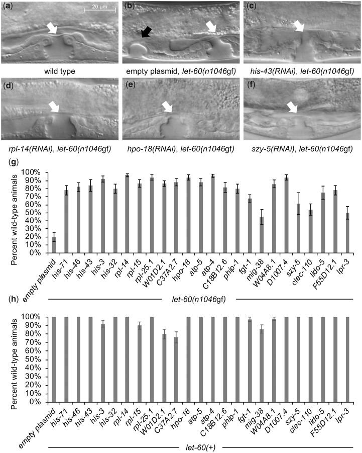
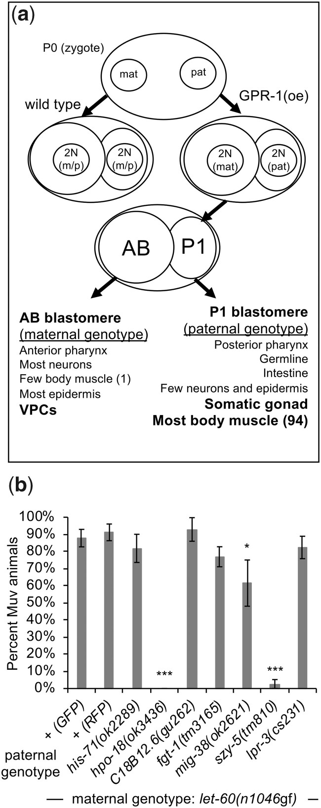
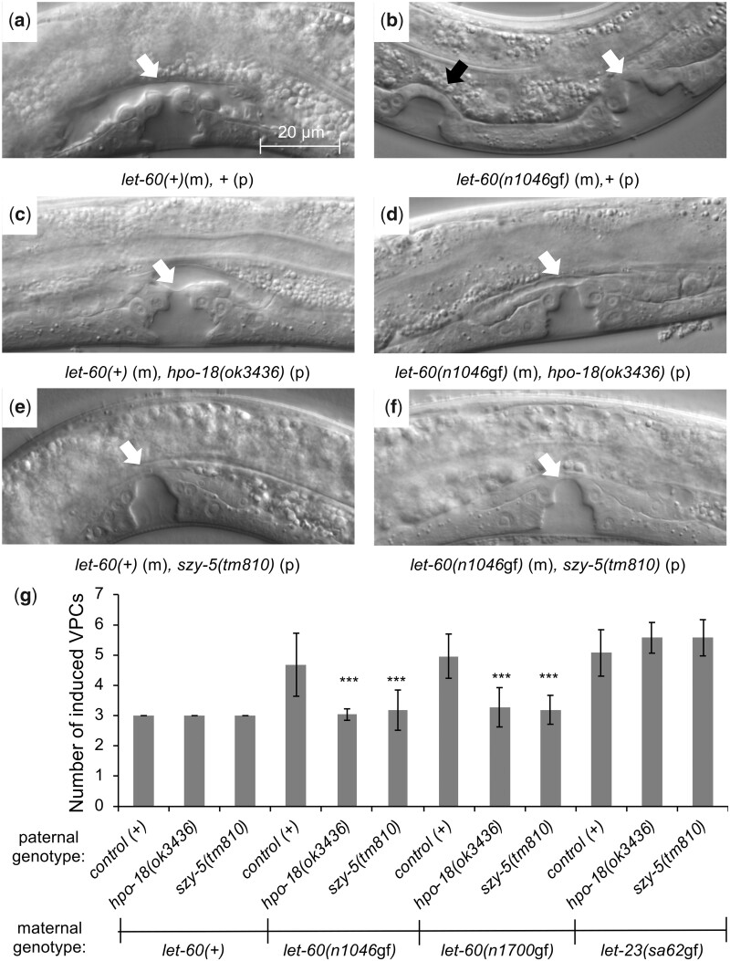
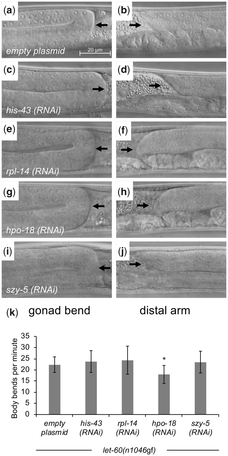
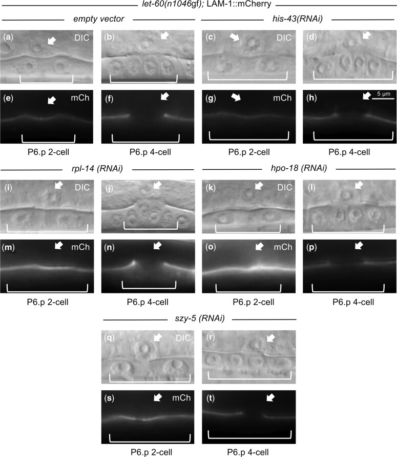
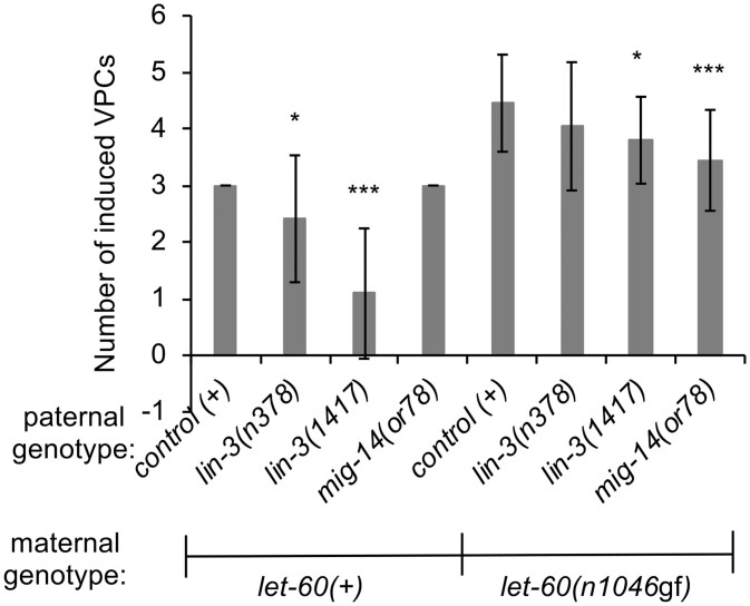
Communication between mesodermal cells and epithelial cells is fundamental to normal animal development and is frequently disrupted in cancer. However, the genes and processes that mediate this communication are incompletely understood. To identify genes that mediate this communication and alter the proliferation of cells with an oncogenic Ras genotype, we carried out a tissue-specific genome-wide RNAi screen in Caenorhabditis elegans animals bearing a let-60(n1046gf) (RasG13E) allele. The screen identifies 24 genes that, when knocked down in adjacent mesodermal tissue, suppress the increased vulval epithelial cell proliferation defect associated with let-60(n1046gf). Importantly, gene knockdown reverts the mutant animals to a wild-type phenotype. Using chimeric animals, we genetically confirm that 2 of the genes function nonautonomously to revert the let-60(n1046gf) phenotype. The effect is genotype restricted, as knockdown does not alter development in a wild type (let-60(+)) or activated EGF receptor (let-23(sa62gf)) background. Although many of the genes identified encode proteins involved in essential cellular processes, including chromatin formation, ribosome function, and mitochondrial ATP metabolism, knockdown does not alter the normal development or function of targeted mesodermal tissues, indicating that the phenotype derives from specific functions performed by these cells. We show that the genes act in a manner distinct from 2 signal ligand classes (EGF and Wnt) known to influence the development of vulval epithelial cells. Altogether, the results identify genes with a novel function in mesodermal cells required for communicating with and promoting the proliferation of adjacent epithelial cells with an activated Ras genotype.

Keywords: Caenorhabditis elegans; ATP synthase; Ras; cell communication; vulval development.


Figures

Fig. 1.
Fig. 1.
Vulval development in C. elegans. a) A cartoon of a C. elegans hermaphrodite in the third larval (L3) stage, illustrating the relative position of VPCs and some adjacent mesodermal cells: somatic gonad (including the AC) and (ventral) body wall muscle. VPC lineages in wild type (let-60(+)) (b) and a representative animal with an activated Ras genotype (let-60(n1046gf)) (c). Normally, through coordination of several cell signals, 3 of 6 VPCs are “induced” to divide and produce cells of the vulva. In most let-60(n1046gf) animals, more than 3 (and up to 6) VPCs divide to produce vulval cells. Mid-body region of fourth larval stage (L4) hermaphrodites, identifying the vulval structures produced from VPC descendants in let-60(+) (d) and let-60(n1046gf) (e) animals. A single vulval structure forms from the offspring of 3 induced VPCs (white arrow), and additional structures result if ectopic VPCs divide to produce vulval cells (black arrow).
Fig. 2.
Fig. 2.
Gene knockdown reverts let-60(n1046gf) to a wild-type phenotype but does not disrupt vulva development in a let-60(+) background. a–f) DIC images of the ventral epidermis in L4 stage. a) In wild-type animals, the offspring of 3 ventral epithelial cells (VPCs) organize to form a single vulval opening, identified with a white arrow, as in Fig. 1. b) let-60(n1046gf) mutants bearing the mesodermal-RNAi system, treated with control RNAi, produce a Muv phenotype with a primary vulval opening (white arrow) as well as ectopic vulval tissue (black arrow) resulting from the division of additional VPCs. c–f) Images of mesodermal-RNAi knockdown of representative genes. RNAi knockdown reverts the let-60(n1046gf) Muv to a wild-type phenotype. Uncropped images are provided in Supplementary Fig. 1. g) Mesodermal-RNAi knockdown of all genes identified in the screen revert let-60(n1046gf) to a wild-type phenotype. Data are indicated as % wild type (rather than % Muv, as in Table 1) to highlight this reversion to wild type effect. In both control and experimental knockdown, the nonwild-type animals were predominantly Muv (as is also seen in chimeras, Fig. 4). The empty plasmid is L4440. n ≥ 40 for each RNAi knockdown except for his-43, C18B12.6, mig-38, szy-5, lido-5 (full data and sample numbers provided in Supplementary tables). All experimental values statistically different from negative control, except for mig-38, szy-5, and lpr-3 (P < 0.05, 2-tailed proportional Z test, with Bonferroni correction). h) Mesodermal-RNAi knockdown of all genes identified in the screen have minimal effect on vulval development in a let-60(+) background. Knockdown of some genes results in a low percentage (<25%) of animals with nonwild-type vulva development. These abnormal animals include a mixture of abnormal vulva morphology, and possible reduced vulva induction, but no animals were vulvaless (Vul). Error bars correspond to standard error. n ≥ 40 for each RNAi knockdown except for his-32, fgt-1, szy-5, lido-5, and lpr-3 (full data and sample numbers provided in Supplementary tables).
Fig. 3.
Fig. 3.
Genetic chimeras demonstrate that hpo-18 and szy-5 act nonautonomously to revert the let-60(n1046gf) phenotype. a) Schematic representation of the genetic chimera method, after Artiles et al. (2019). GPR-1 is overexpressed (GPR-1(oe)) in the maternal germline and oocytes, causing the pronuclei to segregate to daughter cells without fusing in the zygote. The chimera class evaluated in this experiment is the case where replicated maternal chromosomes segregate to the AB blastomere (precursor to the VPCs), while replicated paternal chromosomes segregate to the P1 blastomere (precursor to most mesodermal cells). b) Chimeric animals with paternal (P1) mutant alleles for each of 7 genes (with let-60(+)) in the P1 cell lineage and maternal let-60(n1046gf) (with wild type for each paternal gene) in the AB cell lineage. Alleles of szy-5 and hpo-18 confer a strong suppression of the Muv phenotype. ril-1(ok2492) and the strain VC2839 (with the ok2678 allele that deletes a portion of atp-4 plus the adjacent gene T05H4.11) were also tested, but resulted in chimeras that do not survive to adulthood. Animals with mig-38(ok2621) exhibit a modest modulatory effect in adults, but subsequent analysis of L4 chimeric animals suggest the phenotype derives from delayed development or altered morphogenesis rather than reduced vulval development (data not shown). + (GFP) and + (RFP) represent 2 control experiments, using a paternally supplied myo-2::GFP or myo-2::mCherry/RFP transgene (umnIs7 or hjSi20, respectively), to contrast with the fluorescent marker in the maternal strains, as in Artiles et al. (2019). These paternal genotypes are wild type except for the transgene. Error bars correspond to standard error. Asterisks indicate statistically different from control (Z-test, *P < 0.05; ***P < 0.001). Full data and sample numbers are provided in Supplementary tables.
Fig. 4.
Fig. 4.
Loss of hpo-18 or szy-5 in the P1 lineage does not disrupt normal vulva development and revert let-60(gf) to a wild-type phenotype. a–f) DIC images of the ventral epidermis in L4 stage, as in Fig. 2. Chimeric animals generated as in Fig. 3. Genotype of animal in each image indicated, with (m) identifying the relevant maternally derived genotype, and (p) the relevant paternally derived genotype. a, b) Control chimeric animals, showing wild-type vulval morphology in let-60(+) and ectopic vulval tissue in let-60(n1046gf). c–f) Animals homozygous for hpo-18(ok3436) or szy-5(tm810) in P1 exhibit normal vulval morphology when AB genotype is let-60(+) and revert let-60(n1046gf) to a wild-type phenotype. g) Quantification of the effect. In wild-type animals, 3 of 6 VPCs divide to produce vulval tissue and are identified as induced to produce vulval tissue, and this is unaltered in hpo-18(ok3436) or szy-5(tm810) chimeras. In animals with maternally provided let-60(n1046gf), let-60(n1700gf), or let-23(sa62gf) in AB, more than 3 VPCs divide and are induced (detailed in Fig. 1). This phenotype is suppressed to wild type if mutations in hpo-18 or szy-5 are present in P1 for both let-60 alleles, but not for let-23(sa62gf). Error bars correspond to standard deviation. Asterisks indicate statistically different from control (t-test, ***P < 0.001). All conditions include 21 or more animals. Full data and sample numbers provided in Supplementary tables.
Fig. 5.
Fig. 5.
Gross gonad and muscle functions are maintained upon mesodermal-RNAi knockdown of identified genes. a–j) Hermaphrodite animals from CM2453 (mesodermal-RNAi strain) treated with RNAi under conditions that produce a phenotype were selected as L4s and evaluated for gonad morphology, including appropriate bending at the 2 ends of the animal, and presence of the distal ends in the middle of the animal, suggestive of normal somatic gonad anatomy and distal tip cell migration. RNAi knockdown of representative genes identified in the screen do not disrupt normal somatic gonad morphology. Black arrows in the figures from the left column indicate dorsal-to-ventral bend of gonad arm, while black arrows from the right column show presence of a distal gonad arm dorsal to the uterus. Representative animals shown, with n = 10 evaluated for each RNAi knockdown and no defects observed. k) Muscle function was evaluated using a locomotion (body bends per minute) assay. RNAi knockdown of representative genes generally does not alter muscle function, although knockdown of hpo-18 had a modest effect. n = 10 for each RNAi knockdown. Error bars correspond to standard deviation. Asterisks indicate statistically different from control (2-tailed t-test, *P < 0.05). Raw data provided in Supplementary tables.
Fig. 6.
Fig. 6.
AC anatomy and functions are maintained upon mesodermal-RNAi knockdown of identified genes. Hermaphrodite animals with the mesodermal-RNAi system and a LAM-1::mCherry reporter transgene were treated with RNAi under conditions that produce a phenotype and were selected as L3s and evaluated for AC morphology and capacity to mediate breakdown of the basement membrane between the AC and the dividing VPCs. The presence and timing of basement membrane breakdown (as indicated by clearance of LAM-1::mCherry) between the tissues was maintained under RNAi knockdown of all representative genes from each functional group. n = 10 for each RNAi knockdown. Images illustrate representative animals, and no defects were observed.
Fig. 7.
Fig. 7.
The nonautonomous modulators of let-60(n1046gf) act in a unique manner, distinct from known mesodermally derived signals that influence vulval development. Chimeric animals generated as in Fig. 3. VPC induction data for L4 chimeric animals, as in Fig. 4. Chimeric animals bearing reduction-of-function alleles of lin-3/EGF in P1 and let-60(+) in AB exhibit reduced production of vulval cell types. In animals with let-60(n1046gf) in AB, disruption of either lin-3 or mig-14 moderately reduces the number of induced VPCs. However, neither genotype exhibits an extensive reversion to wild type as observed with hpo-18 or szy-5 (Fig. 4). Error bars correspond to standard deviation. Asterisks indicate statistically different from control (t-test, *P < 0.05; ***P < 0.001). Full data and sample numbers provided in Supplementary tables.

Similar articles

Cited by

References

    1. Alexander J, Cukierman E.. Cancer associated fibroblast: mediators of tumorigenesis. Matrix Biol. 2020;91–92:19–34. 10.1016/j.matbio.2020.05.004 - DOI - PMC - PubMed
    1. Antonioli L, Fornai M, Pellegrini C, D'Antongiovanni V, Turiello R, Morello S, Haskó G, Blandizzi C.. Adenosine signaling in the tumor microenvironment. Adv Exp Med Biol. 2021;1270:145–167. 10.1007/978-3-030&ndash;47189-7_9 - DOI - PubMed
    1. Ardizzi JP, Epstein HF.. Immunochemical localization of myosin heavy chain isoforms and paramyosin in developmentally and structurally diverse muscle cell types of the nematode Caenorhabditis elegans. J Cell Biol. 1987;105(6 Pt 1):2763–2770. 10.1083/jcb.105.6.2763 - DOI - PMC - PubMed
    1. Artiles KL, Fire AZ, Frøkjær-Jensen C.. Assessment and maintenance of unigametic germline inheritance for C. elegans. Dev Cell. 2019;48(6):827–839.e9. 10.1016/j.devcel.2019.01.020 - DOI - PMC - PubMed
    1. Bänziger C, Soldini D, Schütt C, Zipperlen P, Hausmann G, Basler K.. Wntless, a conserved membrane protein dedicated to the secretion of Wnt proteins from signaling cells. Cell. 2006;125(3):509–522. 10.1016/j.cell.2006.02.049 - DOI - PubMed

Publication types

LinkOut - more resources