

Musculoskeletal ultrasound in children: Current state and future directions
- PMID: 35929859
- PMCID: PMC7004269
- DOI: 10.5152/eurjrheum.2019.19170
Musculoskeletal ultrasound in children: Current state and future directions
Abstract
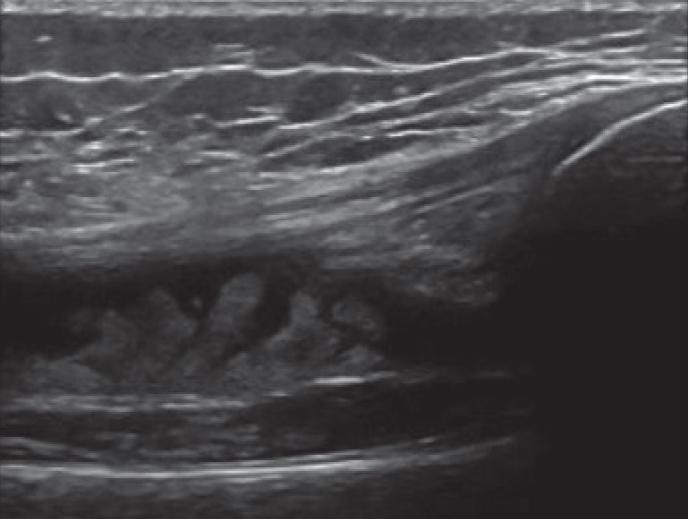
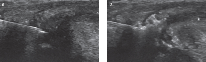
Juvenile idiopathic arthritis (JIA) is a heterogeneous group of chronic inflammatory arthritides that if inadequately treated, may be associated with chronic disability and deformity. Early diagnosis and treatment initiation is essential in the management of patients with JIA. Conventional means of evaluation of disease presence, disease activity and response to therapy including physical exam, labs and x-rays are at times limited and may be insufficient in making an accurate assessment. Musculoskeletal ultrasound (MSUS) is a well-established modality that is patient and family-friendly, non-invasive, does not require sedation and can be performed at the bedside in real-time. MSUS offers information that cannot be attained by standard outcome measures, and may help to advance both diagnosis and treatment of patients with JIA ultimately improving patient outcomes. This review explores the background of MSUS and the current evidence to support its potential role as a diagnostic, disease activity monitoring and interventional tool.
Conflict of interest statement
Figures

Similar articles
-
Ultrasound in juvenile idiopathic arthritis.Pediatr Rheumatol Online J. 2016 May 27;14(1):33. doi: 10.1186/s12969-016-0096-2. Pediatr Rheumatol Online J. 2016. PMID: 27234966 Free PMC article. Review.
-
Contribution of Ultrasound in Current Practice for Managing Juvenile Idiopathic Arthritis.J Clin Med. 2022 Dec 22;12(1):91. doi: 10.3390/jcm12010091. J Clin Med. 2022. PMID: 36614888 Free PMC article. Review.
-
Ultrasound in pediatric rheumatology: Highlighting the differences with adults.Eur J Rheumatol. 2022 Mar 11;11(3):S348-S357. doi: 10.5152/eujrheum.2022.21119. Online ahead of print. Eur J Rheumatol. 2022. PMID: 35943455 Free PMC article.
-
Utilizing ultrasound findings of a single indicator joint to assess non-systemic juvenile idiopathic arthritis.Pediatr Rheumatol Online J. 2021 Apr 29;19(1):60. doi: 10.1186/s12969-021-00550-0. Pediatr Rheumatol Online J. 2021. PMID: 33926518 Free PMC article.
-
The new role of musculoskeletal ultrasound in the treat-to-target management of juvenile idiopathic arthritis.Rheumatology (Oxford). 2021 May 14;60(5):2046-2053. doi: 10.1093/rheumatology/keab004. Rheumatology (Oxford). 2021. PMID: 33493330 Review.
Cited by
-
Low prevalence of subclinical synovitis in patients with juvenile idiopathic arthritis (JIA) in long-term clinical remission on medication.Clin Rheumatol. 2024 Jan;43(1):393-398. doi: 10.1007/s10067-023-06729-y. Epub 2023 Aug 5. Clin Rheumatol. 2024. PMID: 37542584
-
Fat, flames and ultrasounds: the effects of obesity on pediatric joint inflammation.Ital J Pediatr. 2025 Mar 24;51(1):96. doi: 10.1186/s13052-025-01937-5. Ital J Pediatr. 2025. PMID: 40128829 Free PMC article. Review.
-
Pediatric rheumatology: A special issue from the European Journal of Rheumatology.Eur J Rheumatol. 2020 Feb;7(Suppl1):S1-S2. doi: 10.5152/eurjrheum.2020.090120. Eur J Rheumatol. 2020. PMID: 35929858 Free PMC article. No abstract available.
-
A clinical perspective on imaging in juvenile idiopathic arthritis.Pediatr Radiol. 2024 Apr;54(4):490-504. doi: 10.1007/s00247-023-05815-2. Epub 2023 Nov 28. Pediatr Radiol. 2024. PMID: 38015293 Free PMC article. Review.
-
Basic Differences and Most Common Findings in Ultrasound Examinations of Musculoskeletal System in Children: A Narrative Literature Review.Healthcare (Basel). 2022 Oct 12;10(10):2010. doi: 10.3390/healthcare10102010. Healthcare (Basel). 2022. PMID: 36292459 Free PMC article. Review.
References
-
- Filippou G, Cantarini L, Bertoldi I, Picerno V, Frediani B, Galeazzi M. Ultrasonography vs. clinical examination in children with suspected arthritis. Does it make sense to use poliarticular ultrasonographic screening? Clin Exp Rheumatol. 2011;29:345–50. - PubMed
LinkOut - more resources
Full Text Sources
