

doi: 10.5152/tud.2021.21004.
Simplified PI-RADS (S-PI-RADS) for biparametric MRI to detect and manage prostate cancer: What urologists need to know
Affiliations
- PMID: 35929870
- PMCID: PMC8260088
- DOI: 10.5152/tud.2021.21004
Item in Clipboard
Simplified PI-RADS (S-PI-RADS) for biparametric MRI to detect and manage prostate cancer: What urologists need to know
Turk J Urol.
2021 May.
Abstract
Biparametric magnetic resonance imaging (bpMRI) of the prostate has emerged as an alternative to multiparametric MRI (mpMRI) for the detection of clinically significant prostate cancer (csPCa). However, while the Prostate Imaging Reporting and Data System (PI-RADS) is widely known for mpMRI, a proper PI-RADS for bpMRI has not yet been adopted. In this review, we report the current status and the future directions of bpMRI, and propose a simplified PI-RADS (S-PI-RADS) that could help radiologists and urologists in the detection and management of PCa.
Conflict of interest statement
Figures
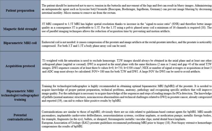
Recommendations for the acquisition of prostate biparametric magnetic resonance imaging.
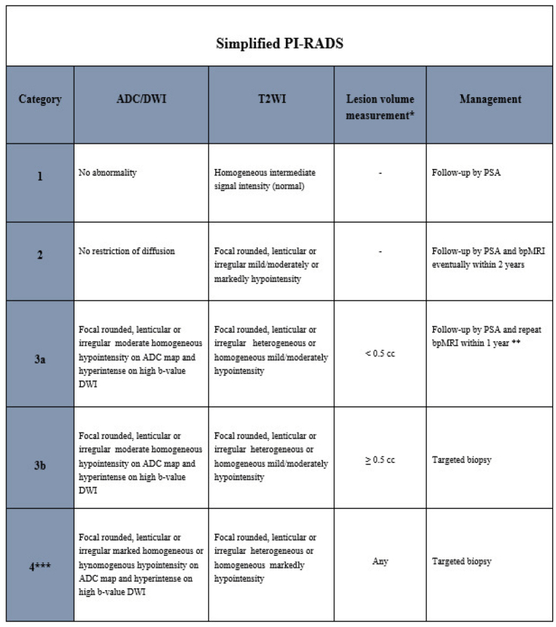
Simplified prostate imaging reporting and data system version according to biparametric magnetic resonance imaging. (Abbreviations: ADC: Apparent diffusion coefficient; DWI: Diffusion weighted imaging; T2WI: T2-weighted imaging. PSA: Prostate Specific Antigen; bpMRI: biparametric magnetic resonance imaging. Note: * Lesion volume is calculate by ellissoidal formula. ** Accurate evaluation of age and clinical informations are need. *** Category 4 includes lesions with volume < and > 0.5 cc and intra- and extraglandular lesions.
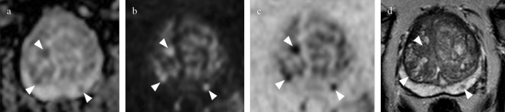
Biparametric MRI of the prostate at 3T in 63-year-old, PSA=6.1 ng/mL. Round lesions located in the left posteromedial peripheral zone and in the left anterior and posterior transition zone of the midgland (arrowheads), is assigned to category 3a (volume<0.5 cc) by S-PI-RADS, no biopsy is indicated. (arrowheads in a) Moderately hypointense lesion on axial ADC map, (arrowheads in b) hypointense on axial DWI at high b-value, (arrowheads in c), axial DWI at high b-value inverted, and (arrowheads in d) hypointense on axial T2WI.
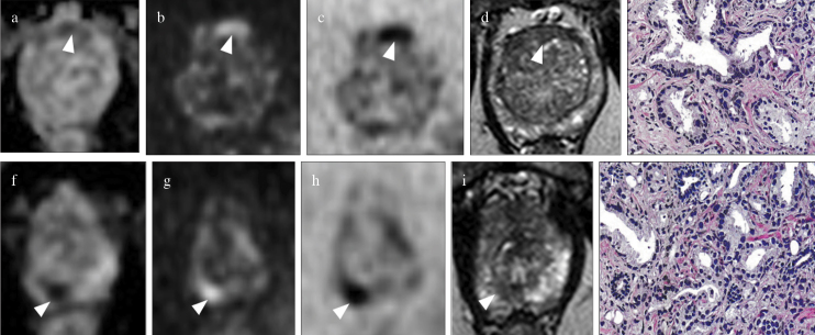
Biparametric MRI of the prostate at 3T in a 71-year-old patient with PSA=11 ng/mL with multifocal prostate cancer. Ovalar lesion located in the anterior transition zone at midgland is assigned to S-PI-RADS category 3b lesion (volume>0.5 cc, targeted biopsy is indicated); (arrowhead in a) the lesion is moderately hypointense on axial ADC map, (arrowhead in b) hyperintense on axial DWI at high b-value, (arrowhead in c) hypointense on axial DWI at high b-value inverted, and (arrowhead in d) hypointense on axial T2WI: (e) Gleason score 6 on histology after targeted TRUS/MRI transperineal biopsy. The smallest round (index lesion) in the peripheral posteromedial zone at the apex is assigned to S-PI-RADS category 4 lesion (volume<0.5 cc, targeted biopsy is indicated); (arrowhead in f) the lesion is markedly hypointense on axial ADC map, (arrowhead in g) hyperintense on axial DWI at high b value, (arrowhead in h) hypointense on axial DWI at high b-value inverted, and (arrowhead in i) hypointense on axial T2WI: (l) Gleason score 7 on histology after targeted TRUS/MRI transperineal biopsy.
References
-
- Drost FH, Osses D, Nieboer D, Bangma CH, Steyerberg EW, Roobol MJ, et al. Prostate magnetic resonance imaging, with or without magnetic resonance imaging-targeted biopsy, and systematic biopsy for detecting prostate cancer: a Cochrane systematic review and meta-analysis. Eur Urol. 2020;77:78–94. doi: 10.1016/j.eururo.2019.06.023. - DOI - PubMed
-
- Bjurlin MA, Carroll PR, Eggener S, Fulgham PF, Margolis DJ, Pinto PA, et al. Update of the standard operating procedure on the use of multiparametric magnetic resonance imaging for the diagnosis, staging and management of prostate cancer. J Urol. 2020;203:706–12. doi: 10.1097/JU.0000000000000617. - DOI - PMC - PubMed
LinkOut - more resources
Full Text Sources
Research Materials
Miscellaneous