. 2022 Nov;49(13):4736-4747.
doi: 10.1007/s00259-022-05925-3. Epub 2022 Aug 5.

[89Zr]Zr-PSMA-617 PET/CT in biochemical recurrence of prostate cancer: first clinical experience from a pilot study including biodistribution and dose estimates

Affiliations

[89Zr]Zr-PSMA-617 PET/CT in biochemical recurrence of prostate cancer: first clinical experience from a pilot study including biodistribution and dose estimates

Florian Rosar et al. Eur J Nucl Med Mol Imaging. 2022 Nov.

Abstract

Purpose: Prostate-specific membrane antigen (PSMA)-targeted PET/CT has become increasingly important in the management of prostate cancer, especially in localization of biochemical recurrence (BCR). PSMA-targeted PET/CT imaging with long-lived radionuclides as 89Zr (T1/2 = 78.4 h) may improve diagnostics by allowing data acquisition on later time points. In this study, we present our first clinical experience including preliminary biodistribution and dosimetry data of [89Zr]Zr-PSMA-617 PET/CT in patients with BCR of prostate cancer.

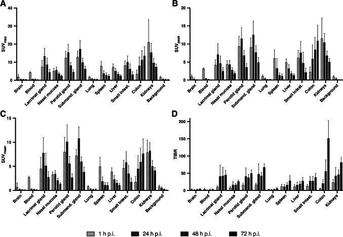
Methods: Seven patients with BCR of prostate cancer who revealed no (n = 4) or undetermined (n = 3) findings on [68Ga]Ga-PSMA-11 PET/CT imaging were referred to [89Zr]Zr-PSMA-617 PET/CT. PET/CT imaging was performed 1 h, 24 h, 48 h, and 72 h post injection (p.i.) of 111 ± 11 MBq [89Zr]Zr-PSMA-617 (mean ± standard deviation). Normal organ distribution and dosimetry were determined. Lesions visually considered as suggestive of prostate cancer were quantitatively analyzed.

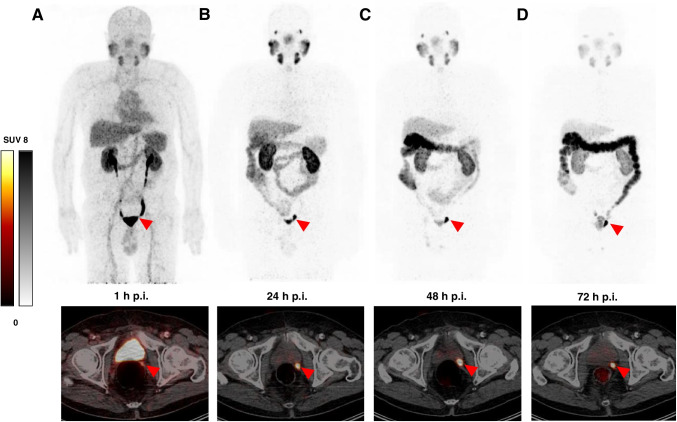
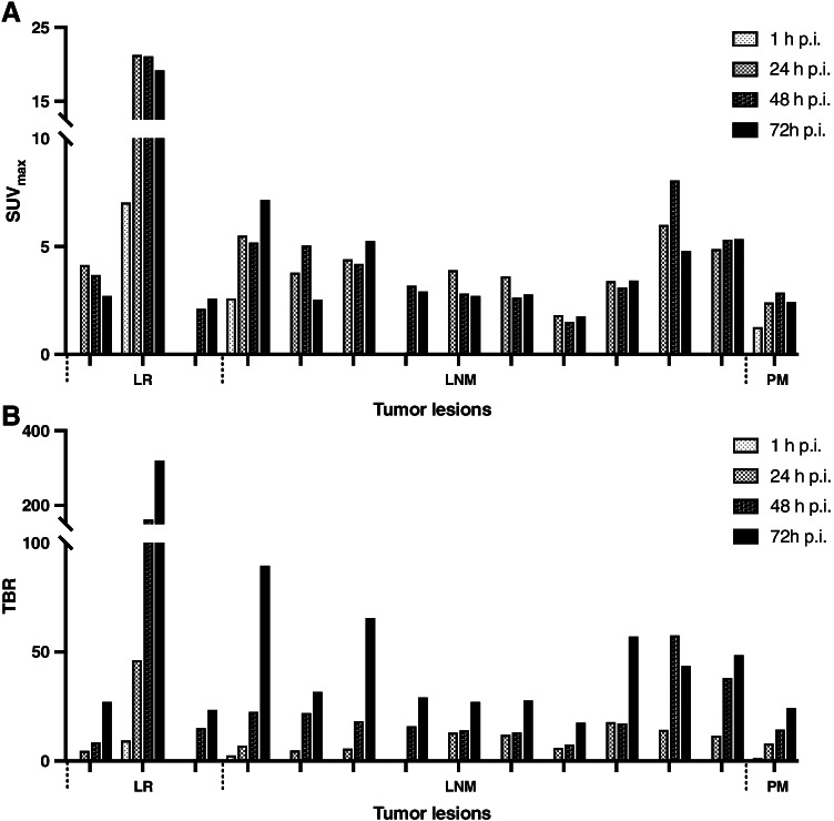
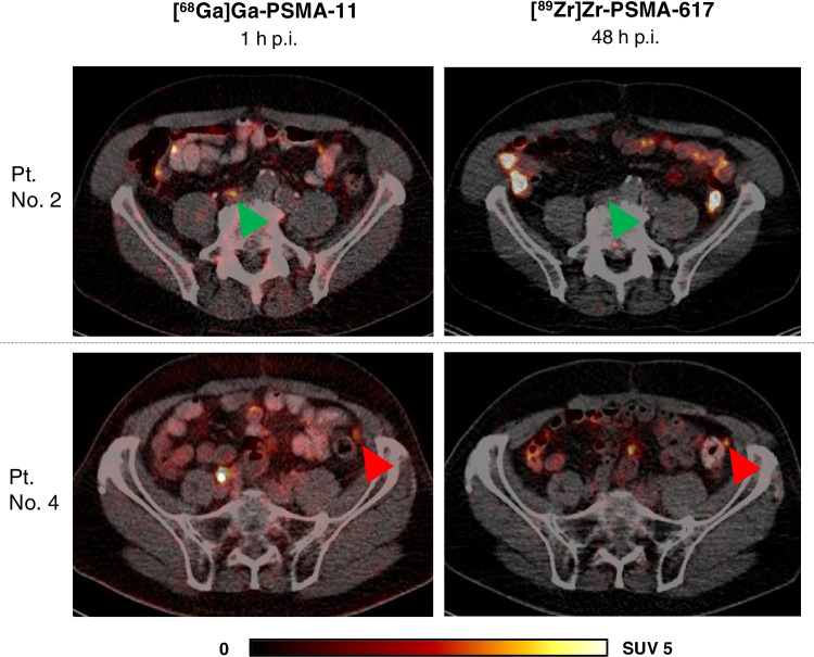
Results: Intense physiological uptake was observed in the salivary and lacrimal glands, liver, spleen, kidneys, intestine and urinary tract. The parotid gland received the highest absorbed dose (0.601 ± 0.185 mGy/MBq), followed by the kidneys (0.517 ± 0.125 mGy/MBq). The estimated overall effective dose for the administration of 111 MBq was 10.1 mSv (0.0913 ± 0.0118 mSv/MBq). In 6 patients, and in particular in 3 of 4 patients with negative [68Ga]Ga-PSMA-11 PET/CT, at least one prostate cancer lesion was detected in [89Zr]Zr-PSMA-617 PET/CT imaging at later time points. The majority of tumor lesions were first visible at 24 h p.i. with continuously increasing tumor-to-background ratio over time. All tumor lesions were detectable at 48 h and 72 h p.i.

Conclusion: [89Zr]Zr-PSMA-617 PET/CT imaging is a promising new diagnostic tool with acceptable radiation exposure for patients with prostate cancer especially when [68Ga]Ga-PSMA-11 PET/CT imaging fails detecting recurrent disease. The long half-life of 89Zr enables late time point imaging (up to 72 h in our study) with increased tracer uptake in tumor lesions and higher tumor-to-background ratios allowing identification of lesions non-visible on [68Ga]Ga-PSMA-11 PET/CT imaging.

Keywords: Biochemical recurrence; PET/CT; PSMA; Prostate cancer; Zirconium-89.


Conflict of interest statement

The authors declare no competing interests.

Figures

Fig. 1
Fig. 1
Representative example (patient no. 2) A Maximum intensity projections at 4 time points post injection (p.i.) of [89Zr]Zr-PSMA-617, B Respective SUVmax, SUVpeak and SUVmean kinetics in normal organs (kidneys, liver, spleen, parotid gland, submandibular gland) and background (gluteal muscle)
Fig. 2
Fig. 2
Biodistribution of [89Zr]Zr-PSMA-617 with descriptive statistics (mean ± standard deviation) of A SUVmax, B SUVpeak, and C SUVmean and D tissue-to-background ratio (TiBR) in normal organs at 1 h, 24 h, 48 h, and 72 h p.i.
Fig. 3
Fig. 3
Maximum intensity projections (MIP) and transversal PET/CT slices of patient no. 6 (PSA 0.43 ng/ml) at 1 h (A), 24 h (B), 48 h (C), and 72 h (D) post injection (p.i.) of [89Zr]Zr-PSMA-617. Red arrows point to a suspicious focal uptake in the left seminal vesicle bed showing increased tracer uptake at 24 h p.i., 48 h p.i., and 72 h p.i. compared to 1 h p.i., therefore considered as local recurrence. SUVmax at 1 h / 24 h / 48 h and 72 h p.i.: 7.04 / 21.27 / 21.09 / 19.19
Fig. 4
Fig. 4
SUVmax (A) and tumor-to-background ratio (TBR) (B) of all tumor lesions at 1 h, 24 h, 48 h, and 72 h post injection (p.i.) of [89Zr]Zr-PSMA-617. LR, local recurrence; LNM, lymph node metastasis; PM, peritoneal metastasis
Fig. 5
Fig. 5
Transversal slices of [89Zr]Zr-PSMA-617 (right) and [68Ga]Ga-PSMA-11 (left) PET/CT of 2 patients (patient no. 1 and 7) with PSMA-positive lesions detected by [89Zr]Zr-PSMA-617 but unidentified by [68Ga]Ga-PSMA-11. Red arrows point to suspected focal uptake. Patient no. 1 (PSA 1.92 ng/ml) with focal uptake (SUVmax 3.68) in the prostate bed considered as local recurrence. Patient no. 7 (PSA 0.68 ng/ml) with focal uptake (SUVmax 8.06) in a pelvic lymph node considered as lymph node metastasis
Fig. 6
Fig. 6
Transversal slices of [89Zr]Zr-PSMA-617 (right) and [68Ga]Ga-PSMA-11 (left) PET/CT of 2 patients (patient no. 2 and 4) with undetermined PSMA-positive findings on [68Ga]Ga-PSMA-11 PET/CT clarified by [89Zr]Zr-PSMA-617 PET/CT. Green arrows point to pelvic tracer uptake (SUVmax 4.47) on [68Ga]Ga-PSMA-11 PET/CT 1 h p.i. with no corresponding uptake on [89Zr]Zr-PSMA-617 PET/CT, considered as unspecific uptake. Red arrows point to faint tracer uptake (SUVmax 2.06) in a peritoneal lesion on [68Ga]Ga-PSMA-11 PET/CT 1 h p.i. with corresponding uptake (SUVmax 2.87) on [89Zr]Zr-PSMA-617 PET/CT 48 h p.i. considered as peritoneal metastasis

References

    1. Hofman MS, Iravani A, Nzenza T, Murphy DG. Advances in urologic imaging: prostate-specific membrane antigen ligand PET imaging. Urol Clin North Am. 2018;45:503–524. doi: 10.1016/j.ucl.2018.03.016. - DOI - PubMed
    1. Ghosh A, Heston WDW. Tumor target prostate specific membrane antigen (PSMA) and its regulation in prostate cancer. J Cell Biochem. 2004;91:528–539. doi: 10.1002/jcb.10661. - DOI - PubMed
    1. Schwarzenboeck SM, Rauscher I, Bluemel C, Fendler WP, Rowe SP, Pomper MG, et al. PSMA ligands for PET imaging of prostate cancer. J Nucl Med. 2017;58:1545–1552. doi: 10.2967/jnumed.117.191031. - DOI - PubMed
    1. Fendler WP, Rahbar K, Herrmann K, Kratochwil C, Eiber M. 177Lu-PSMA Radioligand therapy for prostate cancer. J Nucl Med. 2017;58:1196–1200. doi: 10.2967/jnumed.117.191023. - DOI - PubMed
    1. Cytawa W, Seitz AK, Kircher S, Fukushima K, Tran-Gia J, Schirbel A, et al. 68Ga-PSMA I&T PET/CT for primary staging of prostate cancer. Eur J Nucl Med Mol Imaging. 2020;47:168–177. doi: 10.1007/s00259-019-04524-z. - DOI - PubMed