. 2022 Aug 5;79(8):467.
doi: 10.1007/s00018-022-04507-8.

Global phosphoproteomic analysis identified key kinases regulating male meiosis in mouse

Affiliations

Global phosphoproteomic analysis identified key kinases regulating male meiosis in mouse

Haojie Li et al. Cell Mol Life Sci. .

Abstract

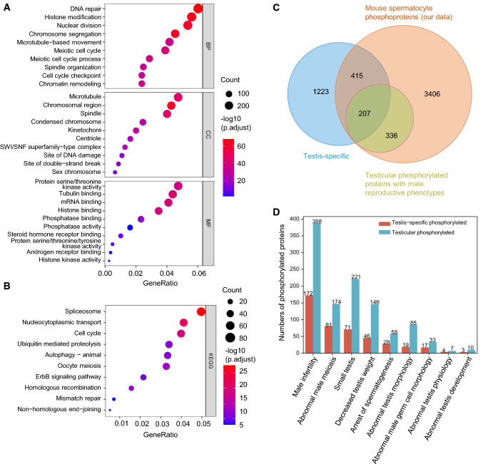
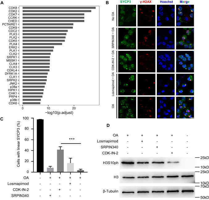
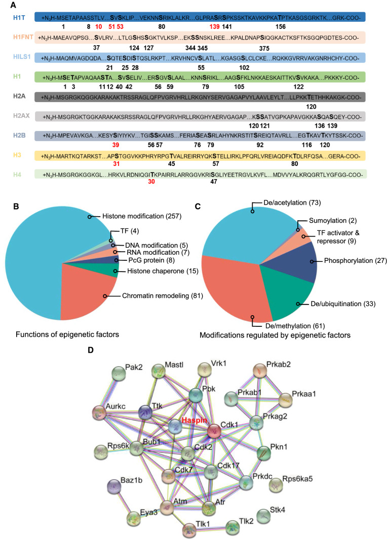
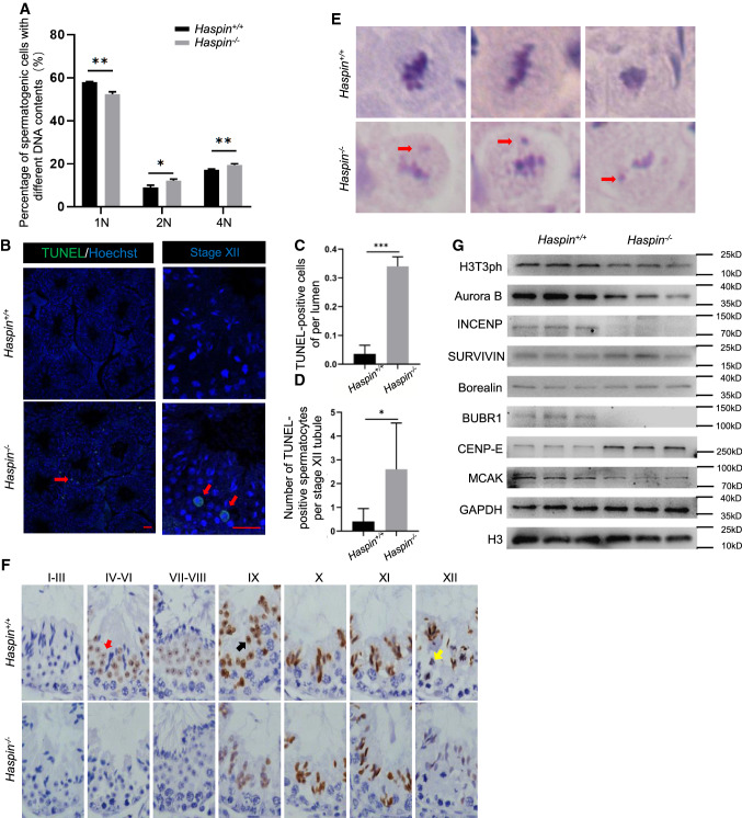
Meiosis, a highly conserved process in organisms from fungi to mammals, is subjected to protein phosphorylation regulation. Due to the low abundance of phosphorylation, there is a lack of systemic characterization of phosphorylation regulation of meiosis in mammals. Using the phosphoproteomic approach, we profiled large-scale phosphoproteome of purified primary spermatocytes undergoing meiosis I, and identified 14,660 phosphorylation sites in 4419 phosphoproteins. Kinase-substrate phosphorylation network analysis followed by in vitro meiosis study showed that CDK9 was essential for meiosis progression to metaphase I and had enriched substrate phosphorylation sites in proteins involved in meiotic cell cycle. In addition, histones and epigenetic factors were found to be widely phosphorylated. Among those, HASPIN was found to be essential for male fertility. Haspin knockout led to misalignment of chromosomes, apoptosis of metaphase spermatocytes and a decreased number of sperm by deregulation of H3T3ph, chromosomal passenger complex (CPC) and spindle assembly checkpoint (SAC). The complicated protein phosphorylation and its important regulatory functions in meiosis indicated that in-depth studies of phosphorylation-mediated signaling could help us elucidate the mechanisms of meiosis.

Keywords: CDK9; Chromosomal passenger complex; Haspin; Histone phosphorylation; Phosphoproteome; Spermatocyte; Spindle assembly checkpoint.


Conflict of interest statement

The authors declare that they have no competing interests.

Figures

Fig. 1
Fig. 1
Purification and phosphoproteomic analysis of the mouse spermatocytes. A Mouse spermatocytes purified by STA-PUT were stained by SYCP3 (green), γ-H2AX (red) and Hoechst (blue) with Rabbit-IgG (green) and Mouse-IgG (red) as negative controls. Bar = 20 μm, each tested in three replicates. B and C The overlap of identified phosphorylation sites (B) and phosphoproteins (C) among three biological replicates. D The distribution of amino acids (S/T/Y) identified to be phosphorylated in mouse spermatocytes. E and F The overlap of phosphorylation sites among our study (spermatocytes), mouse testis, spermatogonia and spermatid (E), and among our study (spermatocytes), Gygi’s data and PhosphoSitePlus database (F). G Sequence logo of 15 amino acids flanking identified phosphorylation sites of mouse spermatocytes
Fig. 2
Fig. 2
Gene ontology, KEGG pathway and phenotype annotation of the mouse spermatocyte phosphoproteome. A and B The top 10 enriched terms of biological processes, cellular components and molecular functions (A) and KEGG pathways (B) in the mouse spermatocyte phosphoproteome. C The Overlap among testis-specific proteins, the mouse spermatocyte phosphoproteins (our data) and those with male reproductive phenotypes in MGI database. D The distribution of the number of mouse spermatocyte phosphoproteins with MGI phenotype terms related to reproductive abnormalities with and without testis-specific expression
Fig. 3
Fig. 3
The enrichment and functional analysis of kinases in the meiosis of spermatocytes. A The top 30 kinases with enriched phosphorylation substrates in mouse spermatocyte phosphoproteome. BD Mouse spermatocytes were cultured and treated with SRPIN340, CDK-IN-2 or losmapimod for 12 h, and with or without okadaic acid for 5 h, and subjected to immunofluorescence staining by SYCP3 (green), γ-H2AX (red) and Hoechst (blue) (B) with the percentages of metaphase I spermatocytes according to linear distribution of SYCP3 shown in Bar graph (n = 3) (C), represent variation (SEM) among biological triplicates and Western blot analysis of H3 and H3S10ph with β-Tubulin as a loading control (D) (n = 3). For cell number analysis, at least 400 nuclei were counted in each group. Bar = 40 μm. All data are presented as mean ± SEM and analyzed by unpaired 2-tailed Student’s t test. ***P < 0.001
Fig. 4
Fig. 4
Epigenetic analysis of the spermatocyte phosphoproteins. A Histone phosphorylation sites identified in this study on 9 histone proteins, and 7 newly identified histone phosphorylation sites were marked with red. B and C Functional classifications (B) and modification annotations (C) of 377 epigenetic factors in mouse spermatocyte phosphoproteins according to the Epifactors database. D Network analysis of phosphorylated epigenetic factors in the mouse spermatocyte phosphoproteome
Fig. 5
Fig. 5
Phenotype analysis of Haspin knockout mice. A Expression of Haspin in different stages of spermatogenic cells according to scRNA-seq data. (A1: type A1 spermatogonia, In: intermediate spermatogonia, BS: S phase type B spermatogonia, BG2: G2/M phase type B spermatogonia, G1: G1 phase preleptotene, ePL: early S phase preleptotene, mPL: middle S phase preleptotene, lPL: late S phase preleptotene, L: leptotene, Z: zygotene, eP: early pachytene, mP: middle pachytene, lP: late pachytene, D: diplotene, MI: metaphase I, MII: metaphase II, RS2: steps 1–2 spermatids, RS4: steps 3–4 spermatids, RS6: steps 5–6 spermatids, RS8: steps 7–8 spermatids) (GEO, GSE107644). B Schematic of Haspin knockout using CRISPR-Cas9 system with sgRNAs colored in green and yellow and PAM sequences in red. C Genotype analysis of Haspin+/+ and Haspin−/− mice (n = 3). D Western blot analysis of expression of Haspin in Haspin+/+ and Haspin−/− testes (n = 3). EK The litter sizes (E), testis weights (F), sperm count (G), the motility (H) and progressive motility (I) of sperm, the morphology of cauda epididymis and sperm by H&E staining (J), and percentages of sperm with abnormal morphologies (K) in Haspin+/+ and Haspin−/− mice. Bar = 20 μm. L, M In vitro fertilization and early embryonic development to blastocysts. The representative pictures of zygotes, 2-cell embryos and blastocysts (L), and rate statistics of zygotes, 2-cell and 8-cell embryos, morulae, and blastocysts for Haspin+/+ and Haspin−/− sperm (M) (n = 3). Bar = 100 μm. All data are presented as mean ± SEM and analyzed by unpaired 2-tailed Student’s t-test. ns, not significant; *P < 0.05, **P < 0.01, ***P < 0.001
Fig. 6
Fig. 6
Meiotic abnormalities in testes of Haspin knockout mice. A The distribution of the ratio of spermatogenic cells with different DNA contents by FACS. BD TUNEL staining (B) and statistics of TUNEL-positive cells of per lumen (C) (n = 3) and TUNEL-positive spermatocytes per stage XII tubule (D) (n = 3) in Haspin+/+ and Haspin−/− mice. Bar = 100 μm. E The metaphase spermatocytes with misaligned chromosomes marked by red arrow after H&E staining in Haspin+/+ and Haspin−/− mice. Bar = 10 μm. F Immunohistochemistry of Haspin+/+ and Haspin−/− testes stained by anti-H3T3ph antibody. Bar = 40 μm. G Western blot analysis of the levels of H3T3ph, Aurora B, INCENP, SURVIVIN, Borealin, BUBR1, CENP-E and MCAK in Haspin+/+ and Haspin−/− testes with H3 and GAPDH as controls (n = 3). Data are presented as mean ± SEM and analyzed by unpaired 2-tailed Student’s t-test. ns, not significant; *P < 0.05, **P < 0.01, ***P < 0.001

References

    1. Marston AL, Amon A. Meiosis: cell-cycle controls shuffle and deal. Nat Rev Mol Cell Biol. 2004;5:983–997. doi: 10.1038/nrm1526. - DOI - PubMed
    1. Zickler D, Kleckner N. Recombination, pairing, and synapsis of homologs during meiosis. Cold Spring Harb Perspect Biol. 2015;7:a016626. doi: 10.1101/cshperspect.a016626. - DOI - PMC - PubMed
    1. Liu D, Liao C, Wolgemuth DJ. A role for cyclin A1 in the activation of MPF and G2-M transition during meiosis of male germ cells in mice. Dev Biol. 2000;224:388–400. doi: 10.1006/dbio.2000.9776. - DOI - PubMed
    1. Guo FZ, Zhang LS, Wei JL, Ren LH, Zhang J, Jing L, et al. Endosulfan inhibiting the meiosis process via depressing expressions of regulatory factors and causing cell cycle arrest in spermatogenic cells. Environ Sci Pollut Res Int. 2016;23:20506–20516. doi: 10.1007/s11356-016-7195-y. - DOI - PubMed
    1. Du M, Yuan L, Zhang Z, Zhang C, Zhu M, Zhang Z, et al. PPP2R1B is modulated by ubiquitination and is essential for spermatogenesis. FASEB J. 2021;35:e21564. doi: 10.1096/fj.202002810R. - DOI - PubMed

LinkOut - more resources