. 2023 Feb;45(1):249-264.
doi: 10.1007/s11357-022-00621-4. Epub 2022 Aug 5.

Aging compromises oligodendrocyte precursor cell maturation and efficient remyelination in the monkey brain

Affiliations

Aging compromises oligodendrocyte precursor cell maturation and efficient remyelination in the monkey brain

Christina Dimovasili et al. Geroscience. 2023 Feb.

Abstract

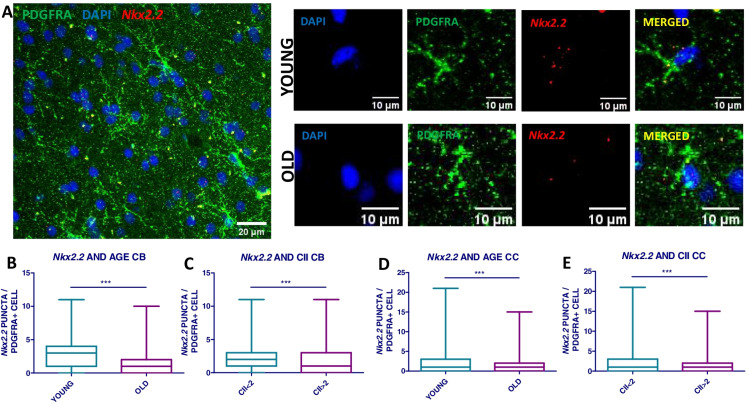
Age-associated cognitive decline is common among otherwise healthy elderly people, even in the absence of Alzheimer's disease and neuron loss. Instead, white matter loss and myelin damage are strongly associated with cognitive decline. Myelin is subject to lifelong oxidative stress that damages the myelin sheath, which is repaired by cells of the oligodendrocyte lineage. This process is mediated by oligodendrocyte precursor cells (OPCs) that sense the damage and respond by proliferating locally and migrating to the region, where they differentiate into mature myelinating oligodendrocytes. In aging, extensive myelin damage, in combination with inefficient remyelination, leads to chronically damaged myelin and loss of efficient neuronal conduction. This study used the rhesus monkey model of normal aging to examine how myelin regeneration capacity is affected by age. Results show that older subjects have reduced numbers of new BCAS1 + myelinating oligodendrocytes, which are newly formed cells, and that this reduction is associated with poorer cognitive performance. Interestingly, this does not result from limited proliferation of progenitor OPCs. Instead, the transcription factor NKX2.2, which regulates OPCs differentiation, is significantly decreased in aged OPCs. This suggests that these OPCs have a diminished potential for differentiation into mature oligodendrocytes. In addition, mature oligodendrocytes have reduced RNA expression of two essential myelin protein markers, MBP and PLP. These data collectively suggest that in the normal aging brain, there is a reduction in regenerative OPCs as well as myelin production that impairs the capacity for remyelination.

Keywords: Aging; Myelination; Oligodendrocyte; RNAscope.


Conflict of interest statement

The authors declare no competing interests.

Figures

Fig. 1
Fig. 1
Subjects’ characteristics and study design. A Table of subjects’ IDs, demographics, and cognitive impairment index (CII). All subjects were used for the BCAS1/BrdU staining except for the two marked with *. The 12 subjects used for the Nkx2.2 RNAscope analysis are marked with #, and the 17 subjects used for the Mbp/Plp RNAscope are marked with &. B Study flowchart of behavioral tests followed by two thionin-stained whole hemisphere sections where the anterior cingulum bundle ROI is outlined in red (CB), and the corpus callosum ROI is outlined in green (CC). C This shows the significant effect of age on increasing cognitive impairment (CII) in the total cohort where the dashed line marks 2 standard deviations of impairment such that subjects below are in the normal range while those above are significantly impaired
Fig. 2
Fig. 2
BCAS1 + cell comparison across monkey lifespan. A Images of representative types of BCAS1 + cells including type 1 pre-myelinating (left) and type 2 myelinating cells (right) from an old monkey brain. B Images of clusters of BCAS1 type 1 cells in a young (left) and an old (right) brain. Arrows highlight BCAS1 + cell clusters in the young versus sparse single cells in the old monkey. C Representative image of a BCAS1 + /BRDU + double-stained cell (BCAS1 in brown, BrdU in black). DG Quantification of BCAS1 cell types by age (N = 23). HO Quantification of BCAS1 + cell types by cognitive test performances (N = 23). PQ Quantification of BCAS1 + /BRDU + cell density by age, normalized against BRDU days of survival (N = 23) (young < 10 years, middle aged 10–20 years, old > 20 years; CB, cingulum bundle; CC, corpus callosum; *p < 0.05)
Fig. 3
Fig. 3
Nkx2.2 expression in OPCs in young and old monkeys. A Representative tile scan and close ups of Nkx2.2 in situ hybridization (RNAscope) combined with anti-PDGFRA immunofluorescence. B and C Quantification of Nkx2.2 expression between young and old monkeys and cognitive status in the anterior cingulum bundle. D and E Quantification of Nkx2.2 expression across age groups and cognitive status in the anterior corpus callosum (CII < 2 normal cognition) (N = 12, approx. 200 cells counted per age group per ROI). ***p < 0.0001
Fig. 4
Fig. 4
Mbp and Plp expression in oligodendrocytes from young and old monkeys. A and B Representative images of Mbp/Plp RNAscope in young (top panels) and old (bottom panels) subjects. C and D Quantification of Mbp expression between age groups. E and F Quantification of Plp expression across age groups and HJ by cognitive test performance (young group < 10 years, old group > 20 years; CII < 2 normal cognition; N = 12, approx. 100 cells counted per age group per ROI; CB, cingulum bundle, CC, corpus callosum). ***p < 0.0001
Fig. 5
Fig. 5
Mbp and Plp expression in newly formed oligodendrocytes. A Representative close-up image of Mbp/Plp RNAscope in a newly formed oligodendrocyte marked by BCAS1 staining in the cingulum bundle of a young animal. B and C Quantification of Mbp and Plp expression in BCAS1 + cells across age groups and (C and D; F and G) by cognitive test performance in the anterior cingulum bundle (young group < 10 years, old group > 20 years; CII < 2 normal cognition; N = 12, approx. 100 cells counted per age group per ROI). For D and G, AM294 data point has been removed as an outlier (> 2 standard deviations from mean value). ***p < 0.0001

References

    1. Park DC, Reuter-Lorenz P. The adaptive brain: aging and neurocognitive scaffolding. Annu Rev Psychol. 2009;60:173–196. - PMC - PubMed
    1. Harada CN, Natelson MC, Love KL. Triebel, Normal cognitive aging. Clin Geriatr Med. 2013;29(4):737–52. - PMC - PubMed
    1. Freeman SH, et al. Preservation of neuronal number despite age-related cortical brain atrophy in elderly subjects without Alzheimer disease. J Neuropathol Exp Neurol. 2008;67(12):1205–1212. - PMC - PubMed
    1. Guttmann CR, et al. White matter changes with normal aging. Neurology. 1998;50(4):972–978. - PubMed
    1. Fjell AM, Walhovd KB. Structural brain changes in aging: courses, causes and cognitive consequences. Rev Neurosci. 2010;21(3):187–221. - PubMed

Publication types