. 2022 Aug 19;17(8):2296-2303.
doi: 10.1021/acschembio.2c00407. Epub 2022 Aug 5.

Characterization of a Nanobody-Epitope Tag Interaction and Its Application for Receptor Engineering

Affiliations

Characterization of a Nanobody-Epitope Tag Interaction and Its Application for Receptor Engineering

Chino C Cabalteja et al. ACS Chem Biol. .

Abstract

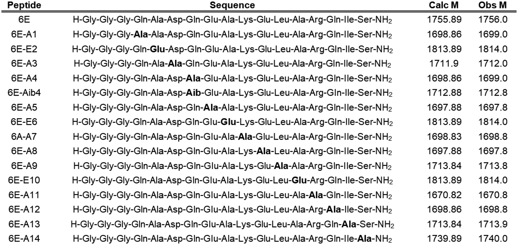
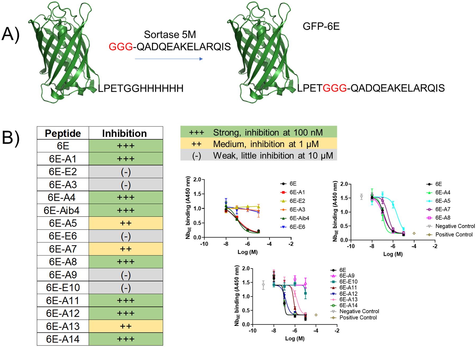
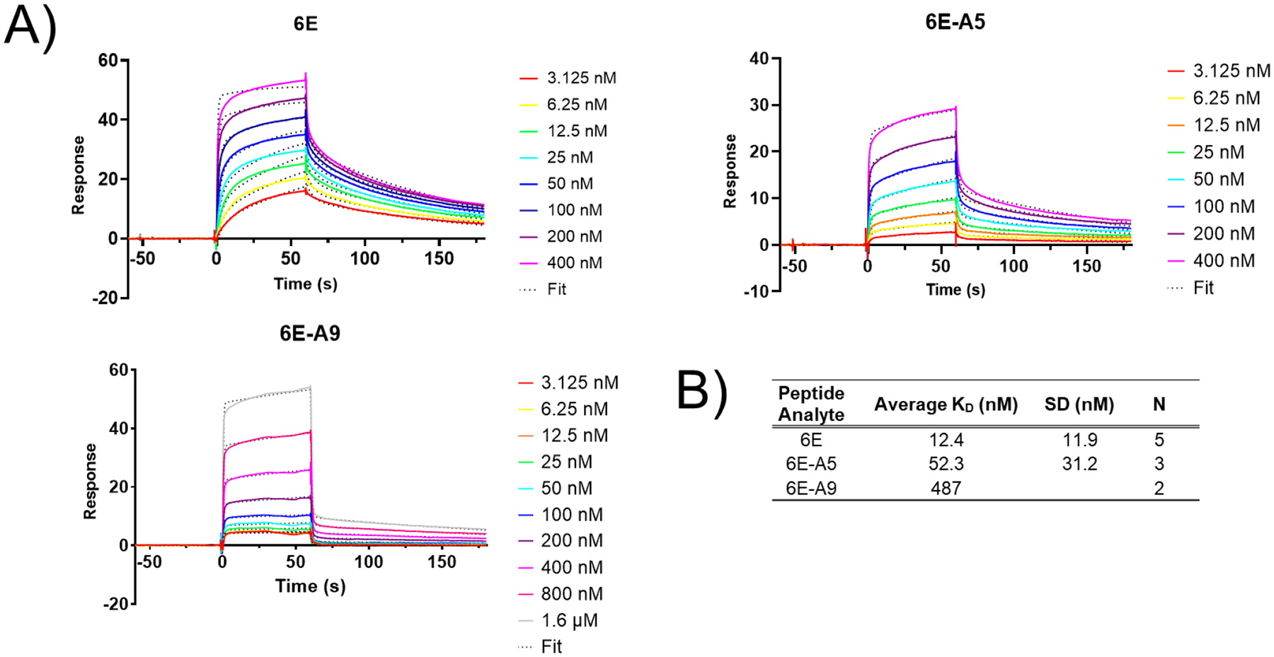
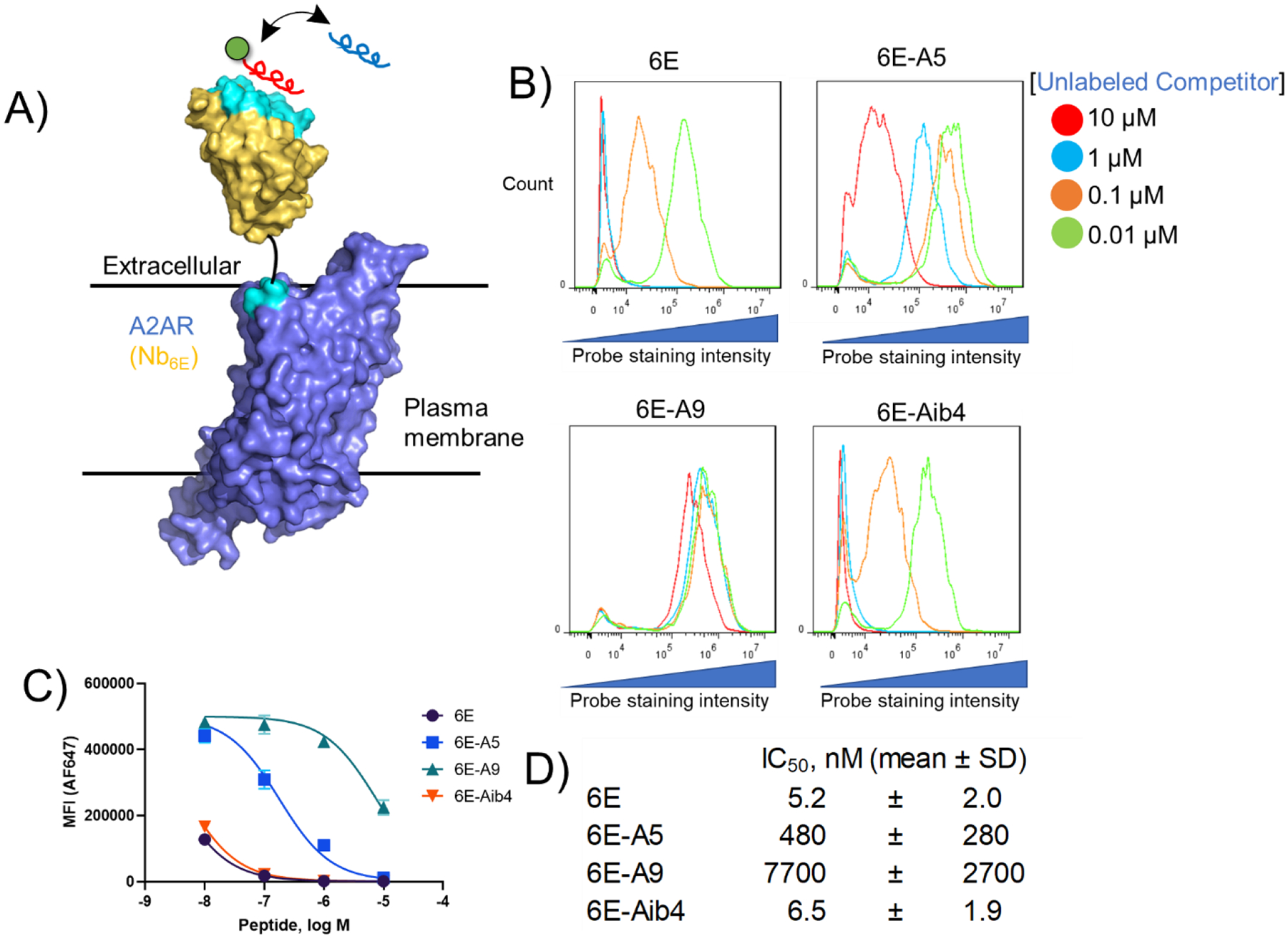
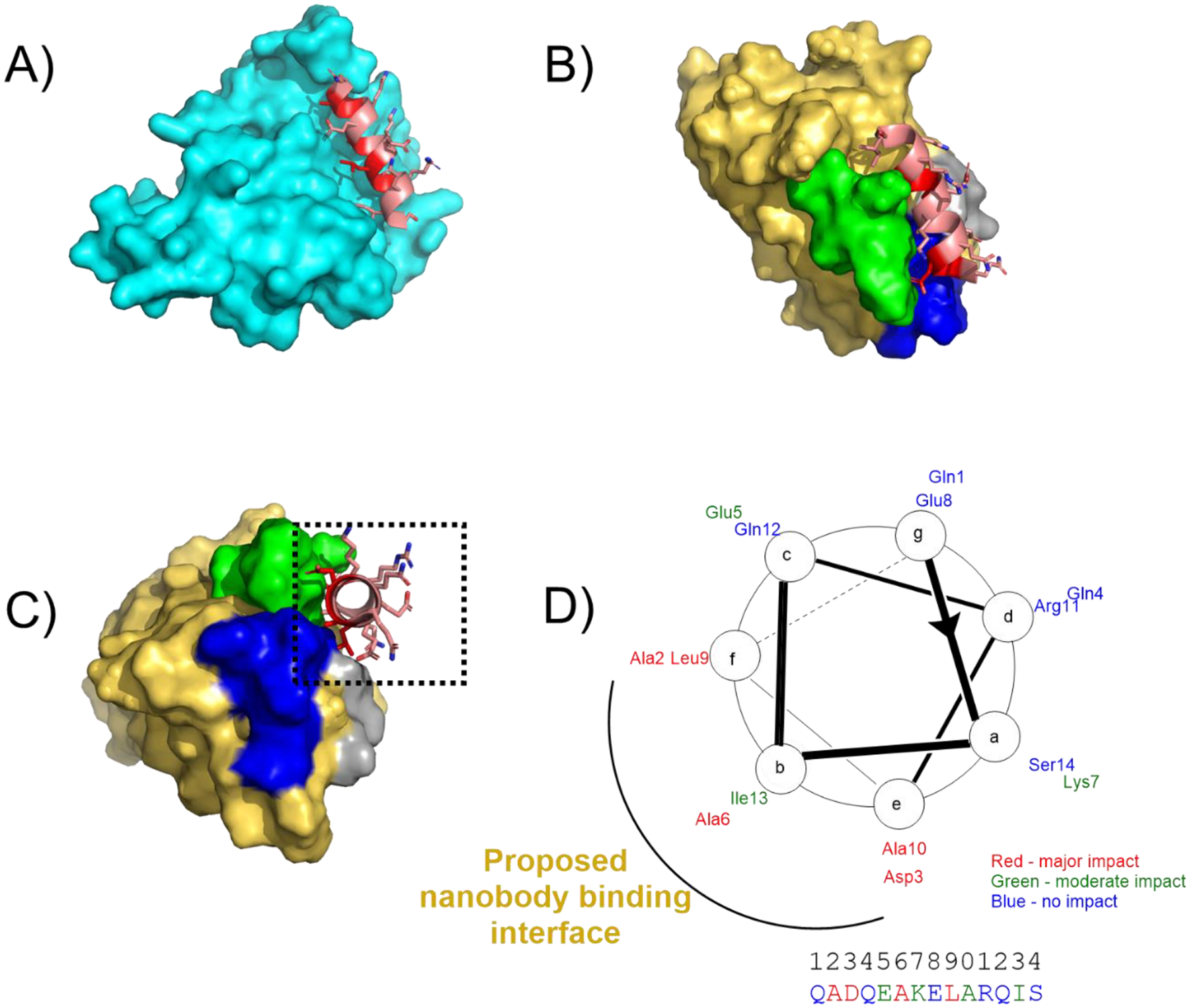
Peptide epitope tags offer a valuable means for detection and manipulation of protein targets for which high quality detection reagents are not available. Most commonly used epitope tags are bound by conventional, full-size antibodies (Abs). The complex architecture of Abs complicates their application in protein engineering and intracellular applications. To address these shortcomings, single domain antibodies (nanobodies, Nbs) that recognize short peptide epitopes have become increasingly prized. Here, we characterize the interaction between a Nb (Nb6E) and a 14-mer peptide epitope. We identify residues in the peptide epitope essential for high affinity binding. Using this information in combination with computational modeling we propose a mode of interaction between Nb6E and this epitope. We apply this nanobody-epitope pair to augment the potency of a ligand at an engineered adenosine A2A receptor. This characterization of the nanobody-epitope pair opens the door to diverse applications including mechanistic studies of the G protein-coupled receptor function.


Figures

Figure 1.
Figure 1.. Sequences of peptides used in this study.
“Obs M” represents the neutral mass corresponding to the most abundant ion peak recorded in characterization using liquid chromatography/mass spectrometry (Supporting Figure 1). Neutral mass M was determined from M = (p*z) – z where z is the charge state of the most abundant ion peak p.
Figure 2.
Figure 2.. Characterization of 6E peptide-Nb6E binding by competition ELISA.
A) Schematic for the preparation of GFP-6E conjugates using sortagging for immobilization in ELISA. See methods for reaction details. B) Summary data for analysis of peptide analogue binding using ELISA. Peptides were categorized into three groups based on their performance in these assays. Representative data from individual assays are shown at right. Data points show mean ± SD from technical replicates in a single experiment. Positive control data points correspond to signal measured in the absence of Nb6E-biotin. Negative control data points correspond to signal observed in the absence of competitor peptide (but with Nb6E-biotin). Curves result from fitting a four-parameter sigmoidal dose-response model to the data (except for weak (−) compounds, which show connected points). Independent replicate data are shown in Supporting Figure 3. Each analogue was tested in ≥ 3 independent experiments. If peptide inhibition categories varied between independent experiments, we categorized compounds in this table based on their modal performance in assays.
Figure 3.
Figure 3.. Characterization of Nb6E-6E peptide association and dissociation by surface plasmon resonance assays.
A) Representative sensorgrams showing binding and dissociation of 6E peptides to immobilized Nb6E. Nb6E was conjugated to biotin at its C-terminus using sortagging and immobilized onto sensor chips functionalized with streptavidin. Peptides (at indicated concentrations) were flowed over functionalized sensor chips for 60 s (association) followed by 120 s of buffer flow with no peptide (dissociation). Data were fit a two-state binding model with local R-max with curve fits shown as dotted lines. B) Tabulation of binding parameters from independent replicate experiments. KD averages (mean) and SD were calculated using KD values derived for the indicated number of independent experiments (“N”).
Figure 4.
Figure 4.. Assessment of Nb6E-6E peptide interaction on the surface of mammalian cells.
A) Schematic of a competition binding assay involving cells expressing A2AR(Nb6E), a fluorescein-labeled tracer peptide (red), and unlabeled competitor peptides (blue). B) Representative data for flow cytometry analysis of the inhibition of fluorescein-labelled tracer peptide binding by indicated concentrations of unlabeled competitor peptides. Tracer peptide (10 nM) and unlabeled competitor peptides were incubated with cells expressing A2AR(Nb6E), followed by washing, and detection with anti-fluorescein secondary antibody labeled with AF647. Data is presented as a histogram of the intensity of AF647 staining of live cells. Data from independent replicate experiments are shown in Supporting Figure 5. C) Median fluorescence intensity (MFI) quantification of the histograms presented in panel 4B. Curves correspond to application of a sigmoidal dose-response response model to the data. Data points and error bars correspond to mean ± SD from triplicate measurements. Data from independent replicate experiments are shown in Supporting Figure 7. D) Mean IC50 values for inhibition of FAM-6E-C14 binding for each unlabeled peptide tested. IC50 values are reported as mean ± SD from three independent experiments.
Figure 5.
Figure 5.. Models of 6E peptide structure and interactions.
A) Model of the 6E peptide portion of UBC6e (cartoon, shown in salmon with residues important for binding to Nb6E in red) in the context of UBC6e (remainder of the modeled protein shown in cyan surface). The structural model was generated using Alphafold 2. The structure of the C-terminal portion (residues 183 onward) of UBC6e was predicted with low confidence and is omitted from this structure. The color-coded sequence is shown in Supporting Figure 10. B) Model of 6E peptide (salmon/red cartoon) bound to Nb6E (gold surface) with Nb complementary determining regions (CDRs) highlighted in contrasting colors (CDR1: gray, CDR2: green, CDR3: blue). The structure was generated by inputting a sequence consisting of Nb6E fused to 6E peptide by a flexible linker peptide. The color-coded input sequence is shown in Supporting Figure 10. Note that the 6E peptide used for modeling does not contain the GGG extension at the N-terminus. C) Reoriented perspective of hypothetical complex between 6E (red/salmon) and Nb6E (gold). D) Helical wheel diagram of 6E peptide with ELISA data summarized using color coding. The putative nanobody binding interface is proposed based on the structural model shown in panel C and structure-activity relationship data.
Figure 6.
Figure 6.. Synthesis and application of a GPCR ligand-6E peptide conjugate for enhancing ligand potency.
A) Synthetic scheme for the preparation of conjugates consisting of an A2AR agonist (CGS21680) and 6E peptide using “click” chemistry. Detailed synthetic methodology and characterization is provided in the methods section and Supporting Figures 1–2. B) Evaluation of pharmacological properties of CGS-6E. (left) A representative dose-response curve for the action of indicated compounds for inducing cAMP responses on cells expressing A2AR(Nb6E). Data points represent readings from individual wells in a single representative experiment. Additional dose-response data sets are shown in Supporting Figure 11. (right) Tabulation of compound agonist potency parameters. EC50 values are reported as mean ± SD from three independent experiments.

References

    1. Carter PJ; Lazar GA Next Generation Antibody Drugs: Pursuit of the “High-Hanging Fruit.” Nat. Rev. Drug Discov 2018, 17 (3), 197–223. 10.1038/nrd.2017.227. - DOI - PubMed
    1. Hutchings CJ A Review of Antibody-Based Therapeutics Targeting G Protein-Coupled Receptors: An Update. Expert Opin. Biol. Ther 2020, 20 (8), 925–935. 10.1080/14712598.2020.1745770. - DOI - PubMed
    1. Vandemoortele G; Eyckerman S; Gevaert K Pick a Tag and Explore the Functions of Your Pet Protein. Trends Biotechnol. 2019, 37 (10), 1078–1090. 10.1016/j.tibtech.2019.03.016. - DOI - PubMed
    1. Chodorge M; Celeste AJ; Grimsby J; Konkar A; Davidsson P; Fairman D; Jenkinson L; Naylor J; White N; Seaman JC; et al. Engineering of a GLP-1 Analogue Peptide/Anti-PCSK9 Antibody Fusion for Type 2 Diabetes Treatment. Sci. Rep 2018, 8 (1), 17545. 10.1038/s41598-018-35869-4. - DOI - PMC - PubMed
    1. Nelson AL Antibody Fragments: Hope and Hype. mAbs 2010, 2 (1), 77–83. 10.4161/mabs.2.1.10786. - DOI - PMC - PubMed

Publication types