. 2022 Aug 5;16(8):e0010651.
doi: 10.1371/journal.pntd.0010651. eCollection 2022 Aug.

FXR deficiency in hepatocytes disrupts the bile acid homeostasis and inhibits autophagy to promote liver injury in Schistosoma japonicum-infected mice

Affiliations

FXR deficiency in hepatocytes disrupts the bile acid homeostasis and inhibits autophagy to promote liver injury in Schistosoma japonicum-infected mice

Beibei Zhang et al. PLoS Negl Trop Dis. .

Abstract

Background: Schistosomiasis, with 250 million people affected, is characterized by its serious hepatic inflammatory response and fibrosis formation, which could lead to dangerous complications, such as portal hypertension, splenomegaly and even ascites. But until now, the pathogenesis of schistosomiasis remains largely unknown. Farnesoid X Receptor (FXR), a bile acid-activated nuclear transcription factor mainly expresses in hepatocytes in the liver, can regulate liver diseases by controlling bile acid metabolism.

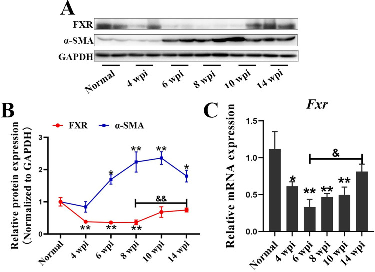
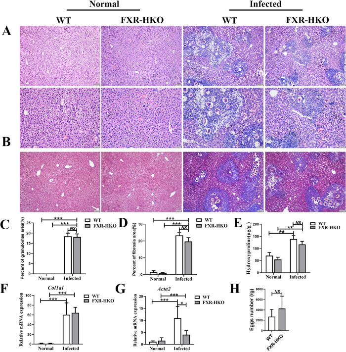
Methodology/principal findings: In this study, we found that the expression of FXR was decreased in the liver of infected mice as shown by western blot and RT-qPCR assays. Furthermore, hepatocyte-specific FXR-deficient mice (FXRflox/floxAlbCre, FXR-HKO) were generated and infected with ~16 cercariae of S. japonicum for five weeks. We found that FXR deficiency in hepatocytes promoted the progression of liver injury, aggravated weight loss and death caused by infection, and promoted inflammatory cytokines production, such as IL-6, IL-1β, TNF-α, IL-4, IL-10, and IL-13. Surprisingly, hepatic granulomas and fibrosis were not affected. In addition, using UPLC-MS/MS spectrometry, it was found that S. japonicum infection resulted in elevated bile acids in the liver of mice, which was more obvious in FXR-deficient mice. Meanwhile, autophagy was induced in littermate control mice due to the infection, but it was significantly decreased in FXR-HKO mice.

Conclusions/significance: All these findings suggest that FXR deficiency in hepatocytes disrupts bile acid homeostasis and inhibits autophagy, which may aggravate the damages of hepatocytes caused by S. japonicum infection. It highlights that FXR in hepatocytes plays a regulatory role in the progression of schistosomiasis.


Conflict of interest statement

The authors have declared that no competing interests exist.

Figures

Fig 1
Fig 1. FXR decreases in mice livers with pathological injuries induced by S. japonicum infection.
(A) Expression levels of hepatic FXR and α-SMA in mice infected with S. japonicum at different time-points were measured by western blot. (B) The relative expression levels of FXR and α-SMA were normalized to GAPDH, and the fold changes were calculated by comparing with the normal group. (C) The hepatic mRNA level of Fxr in mice infected with S. japonicum was detected by RT-qPCR. Data are presented as mean ± SEM. * P <0.05, infected groups vs normal group; ** P <0.01, infected groups vs normal group; &P <0.05, &&P <0.01.
Fig 2
Fig 2. FXR deficiency promotes the loss of body weight, death of mice, and liver injury caused by S. japonicum infection.
(A) The change ratio of body weight for mice in each group. (B) Survival rate of each group. (C) Serum analysis of ALT for mice in each group. (D) Serum analysis of AST for mice in each group. Data are presented as mean ± SEM. * P < 0.05, ** P < 0.01, *** P < 0.001.
Fig 3
Fig 3. FXR deficiency does not affect hepatic granulomas and fibrosis caused by S. japonicum infection for 5 weeks post-infection.
(A) Representative images of HE staining for each group. (B) Representative images of Masson staining for each group. (C) Percent of granulomas area was analyzed according to HE staining by Image Pro Plus 6.0 software. (D) Percent of fibrosis area was analyzed according to Masson staining by Image Pro Plus 6.0 software. (E) Liver hydroxyproline was detected by the alkaline lysis assay. The expression of Col1a1 (F) and Acta2 (for encoding protein α-SMA) (G) in liver were detected by RT-qPCR. (H) Eggs per gram in mice liver infected with S. japonicum. Data are presented as mean ± SEM. NS means no significance, * P <0.05, ** P <0.01, *** P <0.001.
Fig 4
Fig 4. FXR deficiency promotes inflammatory cytokines production caused by S. japonicum infection.
The levels of interleukin-6 (IL-6) (A), Interleukin-1β (IL-1β) (B), Tumor necrosis factor-α (TNF-α) (C), Interleukin-4 (IL-4) (D), Interleukin-10 (IL-10) (E), and Interleukin-13 (IL-13) (F) in liver from each group were detected by ELISA. Data are presented as mean ± SEM. *P <0.05, ** P <0.01, *** P <0.001.
Fig 5
Fig 5. Bile acid homeostasis is disrupted in FXR-deficient mice with S. japonicum infection.
The content of TBA (A), TTBA (B), TCDCA (C), and T-β-MCA (D) in mice liver from each group. The hepatic mRNA levels of Cyp7a1 (E), Bsep (F), Ntcp (G), and Ostβ (H) in each group were detected by RT-qPCR. Data are expressed as mean ± SEM. * P <0.05, ** P <0.01.
Fig 6
Fig 6. Autophagy is inhibited in FXR-deficient mice with S. japonicum infection.
(A) Hepatic LC3, Beclin-1, and P62 in the mice of each group were measured by western blot. The densitometry analysis of LC3 (B), Beclin-1 (C), and P62 (D) was screened and measured by Image Pro Plus 6.0 software. (E) Immunofluorescene analysis of LC3 in liver tissue from each group, and the representative images were shown. (F) Counts of cells with LC3 dots were analyzed in mice for each group. Data are expressed as mean ± SEM. * P <0.05, *** P <0.001.
Fig 7
Fig 7. A schematic model shows that FXR deficient in hepatocytes increases bile acids toxicity and inhibits hepatocellular autophagy, which may therefore accelerate the progression of schistosomiasis by promoting hepatocytes injuries.

Similar articles

Cited by

References

    1. Nono JK, Kamdem SD, Musaigwa F, Nnaji CA, Brombacher F. Influence of schistosomiasis on host vaccine responses. Trends in Parasitology. 2022; 38(1): 67–79. doi: 10.1016/j.pt.2021.07.009 - DOI - PubMed
    1. Tamarozzi F, Fittipaldo VA, Orth HM, Richter J, Buonfrate D, Riccardi N, et al.. Diagnosis and clinical management of hepatosplenic schistosomiasis: A scoping review of the literature. PLoS Neglected Tropical Diseases. 2021; 15(3): e0009191. doi: 10.1371/journal.pntd.0009191 - DOI - PMC - PubMed
    1. Chuah C, Jones MK, Burke ML, McManus DP, Gobert GN. Cellular and chemokine-mediated regulation in schistosome-induced hepatic pathology. Trends in Parasitology. 2014; 30(3): 141–150. doi: 10.1016/j.pt.2013.12.009 - DOI - PubMed
    1. Carson JP, Ramm GA, Robinson MW, McManus DP, Gobert GN. Schistosome-Induced Fibrotic Disease: The Role of Hepatic Stellate Cells. Trends in Parasitolology. 2018; 34(6): 524–540. doi: 10.1016/j.pt.2018.02.005 - DOI - PubMed
    1. Anthony BJ, Ramm GA, McManus DP. Role of resident liver cells in the pathogenesis of schistosomiasis. Trends in Parasitology. 2012; 28(12): 572–579. doi: 10.1016/j.pt.2012.09.005 - DOI - PubMed

Publication types

Substances