

Magnetic bioassembly platforms for establishing craniofacial exocrine gland organoids as aging in vitro models
- PMID: 35930565
- PMCID: PMC9355193
- DOI: 10.1371/journal.pone.0272644
Magnetic bioassembly platforms for establishing craniofacial exocrine gland organoids as aging in vitro models
Abstract
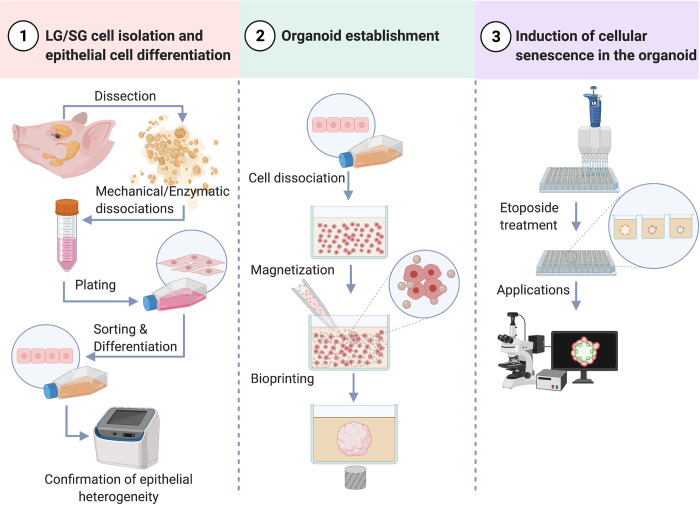
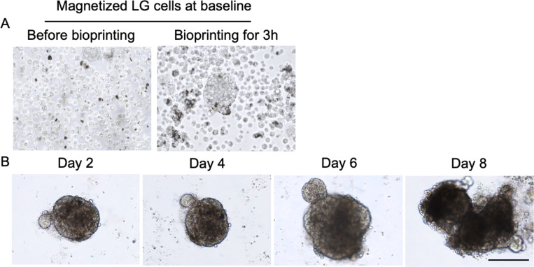
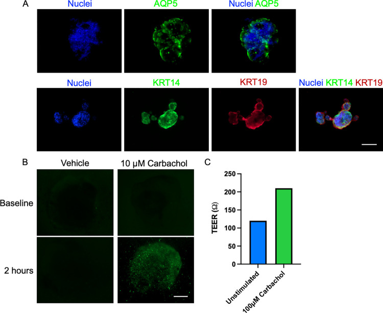
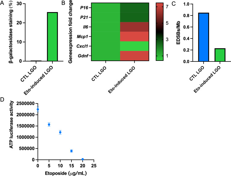
A multitude of aging-related factors and systemic conditions can cause lacrimal gland (LG) or salivary gland (SG) hypofunction leading to degenerative dry eye disease (DED) or dry mouth syndrome, respectively. Currently, there are no effective regenerative therapies that can fully reverse such gland hypofunction due to the lack of reproducible in vitro aging models or organoids required to develop novel treatments for multi-omic profiling. Previously, our research group successful developed three-dimensional (3D) bioassembly nanotechnologies towards the generation of functional exocrine gland organoids via magnetic 3D bioprinting platforms (M3DB). To meet the needs of our aging Asian societies, a next step was taken to design consistent M3DB protocols to engineer LG and SG organoid models with aging molecular and pathological features. Herein, a feasible step-by-step protocol was provided for producing both LG and SG organoids using M3DB platforms. Such protocol provided reproducible outcomes with final organoid products resembling LG or SG native parenchymal epithelial tissues. Both acinar and ductal epithelial compartments were prominent (21 ± 4.32% versus 42 ± 6.72%, respectively), and could be clearly identified in these organoids. Meanwhile, these can be further developed into aging signature models by inducing cellular senescence via chemical mutagenesis. The generation of senescence-like organoids will be our ultimate milestone aiming towards high throughput applications for drug screening and discovery, and for gene therapy investigations to reverse aging.
Conflict of interest statement
The authors declare the following financial interests/personal relationships which may be considered as potential competing interests: Glauco R. Souza is employed by Greiner Bio-One International GmbH which produces NanoShuttle™ magnetic nanoparticles.
Figures
Similar articles
-
Unveiling senescence-associated ocular pathogenesis via lacrimal gland organoid magnetic bioassembly platform and HMGB1-Box A gene therapy.Sci Rep. 2024 Sep 18;14(1):21784. doi: 10.1038/s41598-024-73101-8. Sci Rep. 2024. PMID: 39294273 Free PMC article.
-
Bioprinting salivary gland models and their regenerative applications.BDJ Open. 2024 May 30;10(1):39. doi: 10.1038/s41405-024-00219-2. BDJ Open. 2024. PMID: 38816372 Free PMC article. Review.
-
Development of high-throughput lacrimal gland organoid platforms for drug discovery in dry eye disease.SLAS Discov. 2022 Apr;27(3):151-158. doi: 10.1016/j.slasd.2021.11.002. Epub 2021 Dec 4. SLAS Discov. 2022. PMID: 35058190 Review.
-
Salivary gland regeneration: from salivary gland stem cells to three-dimensional bioprinting.SLAS Technol. 2023 Jun;28(3):199-209. doi: 10.1016/j.slast.2023.03.004. Epub 2023 Apr 3. SLAS Technol. 2023. PMID: 37019217
-
Engineering innervated secretory epithelial organoids by magnetic three-dimensional bioprinting for stimulating epithelial growth in salivary glands.Biomaterials. 2018 Oct;180:52-66. doi: 10.1016/j.biomaterials.2018.06.011. Epub 2018 Jun 12. Biomaterials. 2018. PMID: 30025245
Cited by
-
Unveiling senescence-associated ocular pathogenesis via lacrimal gland organoid magnetic bioassembly platform and HMGB1-Box A gene therapy.Sci Rep. 2024 Sep 18;14(1):21784. doi: 10.1038/s41598-024-73101-8. Sci Rep. 2024. PMID: 39294273 Free PMC article.
-
Biomimetic Gland Models with Engineered Stratagems.Research (Wash D C). 2023 Sep 15;6:0232. doi: 10.34133/research.0232. eCollection 2023. Research (Wash D C). 2023. PMID: 37719047 Free PMC article. Review.
-
Bioprinting salivary gland models and their regenerative applications.BDJ Open. 2024 May 30;10(1):39. doi: 10.1038/s41405-024-00219-2. BDJ Open. 2024. PMID: 38816372 Free PMC article. Review.
References
Publication types
MeSH terms
LinkOut - more resources
Full Text Sources
