. 2022 Aug 5;18(8):e1010750.
doi: 10.1371/journal.ppat.1010750. eCollection 2022 Aug.

The TPR domain of PgaA is a multifunctional scaffold that binds PNAG and modulates PgaB-dependent polymer processing

Affiliations

The TPR domain of PgaA is a multifunctional scaffold that binds PNAG and modulates PgaB-dependent polymer processing

Roland Pfoh et al. PLoS Pathog. .

Abstract

The synthesis of exopolysaccharides as biofilm matrix components by pathogens is a crucial factor for chronic infections and antibiotic resistance. Many periplasmic proteins involved in polymer processing and secretion in Gram-negative synthase dependent exopolysaccharide biosynthetic systems have been individually characterized. The operons responsible for the production of PNAG, alginate, cellulose and the Pel polysaccharide each contain a gene that encodes an outer membrane associated tetratricopeptide repeat (TPR) domain containing protein. While the TPR domain has been shown to bind other periplasmic proteins, the functional consequences of these interactions for the polymer remain poorly understood. Herein, we show that the C-terminal TPR region of PgaA interacts with the de-N-acetylase domain of PgaB, and increases its deacetylase activity. Additionally, we found that when the two proteins form a complex, the glycoside hydrolase activity of PgaB is also increased. To better understand structure-function relationships we determined the crystal structure of a stable TPR module, which has a conserved groove formed by three repeat motifs. Tryptophan quenching, mass spectrometry analysis and molecular dynamics simulation studies suggest that the crystallized TPR module can bind PNAG/dPNAG via its electronegative groove on the concave surface, and potentially guide the polymer through the periplasm towards the porin for export. Our results suggest a scaffolding role for the TPR domain that combines PNAG/dPNAG translocation with the modulation of its chemical structure by PgaB.


Conflict of interest statement

The authors have declared that no competing interests exist.

Figures

Fig 1
Fig 1. Residues 32–220 of PgaA and the glycoside hydrolase domain of PgaB are dispensable for PgaA/B interaction.
A) Generalized synthase-dependent schematic for the production and secretion of exopolysaccharides in Gram-negative bacteria. B) PgaA and PgaB domain topology. The regions determined to interact are highlighted in red. C) Western blot analysis of Ni-pulldown assays with various N-terminally His-tagged PgaA constructs. PgaA and PgaB were co-expressed with each protein construct containing a signal sequence for periplasmic translocation. PgaA and PgaB constructs are depicted in the anticipated orientation with the membrane anchor and the porin positioned in the outer membrane, indicated in cyan. Lysate fractions (red L) and elution fractions (green E) are indicated for each lane. Western blots from the pulldown experiments were probed with a monoclonal primary mouse anti-his (Abgent) or a rabbit polyclonal primary antibody raised against PgaB. The molecular weight markers (kDa) are indicated on the left of each panel.
Fig 2
Fig 2. PgaA increases the deacetylase and glycoside hydrolase activities of PgaB.
A) Coomassie gels of protein samples used in this experiment, with molecular weight markers (kDa). B) Fluorescamine-base deacetylation assay using 30 μM enzyme and 30 mM PNAG hexamer. The concentration of GlcN produced was as following: PgaA, 73.8 ± 6.3 μM; PgaB, 988.6 ± 104.7 μM; PgaA/B, 2709.0 ± 271.5 μM; PgaA+B, 1288 ± 131.8 μM; PgaAΔ220/B), 2291.0 ± 217.3 μM; PgaA/B_D115A, 132.1 ± 16.4 μM; BSA, 31.0 ± 18.1 μM; PgaB+BSA, 932.4 ± 53.4 μM. C) Crystal violet-based biofilm disruption assay using S. epidermidis SE801. The calculated EC50’s for each of the enzymes are as follows: PgaB, 302 ± 82.3 nM; PgaA/B, 22.8 ± 5.1 nM; PgaA+B, 241.1 ± 16.3 nM; PgaAΔ220/B, 279.8 ± 19.6 nM; PgaA/B_D115A, 14.2 ± 1.1 nM; ND: not determined. PgaA: PgaA-32-807; PgaB: PgaB-22-672; PgaA/B: complex obtained by co-expression of PgaA-32-807 and PgaB-1-672; PgaA + PgaB: PgaA-32-807 and PgaB-22-672 individually purified and then mixed. Statistical significance is given for comparison against PgaB unless indicated by brackets. ****P ≤ 0.0001, *P ≤ 0.1, NS: no significant difference. Statistical significance was evaluated using two-way analysis of variance and Tukey’s multiple comparison test. Error bars represent the standard error from n = 3 technical replicates.
Fig 3
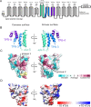
Fig 3. Crystal structure of PgaA 220–367 reveals a curved TPR-like module formed by two TPR motifs and one α/α-repeat.
A. Topology model of PgaA including the crystallized module shown in shades of blue. TPR motifs and α/α-repeats were predicted by TPRpred and HHpred [22,23], respectively. B. Crystal structure of PgaA-220-367 shown in cartoon representation and coloured as in panel A. The TPR5 helix is part of a domain swap and has been omitted for clarity. C. Surface representation of the structure mapped with conservation levels as calculated in ConSurf [57] (yellow: insufficient data for conservation analysis of this residue). D. Surface representation with color-coded electrostatics calculated with PyMOL (DeLano Scientific, http://www.pymol.org/).
Fig 4
Fig 4. Tryptophan quenching suggests dPNAG interacts with the concave surface of the TPR.
A) TPR 220–367 contains three tryptophan residues, shown in orange stick representation. Left is a view at the concave surface and right is a view along the superhelical axis. B) Tryptophan quenching of PgaA-220-367 with dPNAG, PNAG, and chitin oligomers. C) Tryptophan quenching with TPR variants suggests that dPNAG-interacts with residues W314 and W318. ****P ≤ 0.0001, ***P ≤ 0.001, *P ≤ 0.1, NS: no significant difference. Statistical significance was evaluated using two-way analysis of variance and Tukey’s multiple comparison test. Error bars represent the standard error (n = 3). D) Tryptophan quenching PgaA-220-367 wild-type as a function of dPNAG concentration. The dissociation constant and S.E. were obtained by fitting to single-site binding equation with nonlinear regression analysis, with R2 = 0.93 and a standard deviation of the residuals Sy.x = 8.3%. The analysis was performed with GraphPad Prism version 6.0 for Mac OS X.
Fig 5
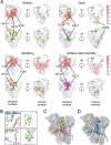
Fig 5. MD simulations reveal GlcNAc and GlcN binding sites along the concave TPR surface.
A) Statistical analysis of MD simulations of PgaA 220–340 with 50 mM GlcNAc monomers, 50 mM GlcN monomers, 7 mM (GlcNAc)3 trimers, and 7 mM GlcNAc-GlcN-GlcNAc trimers. Two types of illustrations are used to show interaction regions: The upper illustration using a white-to-red color scheme indicates the fraction of time that a protein residue is bound to a sugar over the course of the simulation, while the lower illustration depicts the spatial density distribution for each bound sugar at 0.25 occupancy. B) Snapshots from MD simulations with monomers. C) Snapshot of MD simulations with trimers. D) Potential dPNAG pathway on PgaA 220–340 based on trimer binding orientations. The star indicates the reducing end of the polymer. The superhelical axis of the TPR domain is oriented vertically with the C-terminal end leading to the porin pointing upwards.
Fig 6
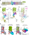
Fig 6. Homology and AlphaFold2 models of PgaA reveal a potential pathway for dPNAG into the outer-membrane porin.
A. Topology model of PgaA based on structure prediction servers HHpred and TPRpred [22,23]. B. Left side: Composite model of PgaA. The TPR region is a Phyre2 model [21] based on human O-linked GlcNAc transferase (OGT, PDB ID 1W3B)[18] combined with the crystal structure of PgaA-220-367 (this work). The overall sequence identity of the TPR domain and OGT is only 14%. We expect the accuracy of the model to be higher in regions with a continuous TPR repeat (eg TPRs 5–8) since OGT shows continuous superhelical TPR fold. We cannot exclude the possibility of deviations from a continuous TPR superhelix, as for example seen in BcsC (PDB 5XW7) [4], which for PgaA seems more likely near the unclassified α/α-repeats. Right side: Alphafold2 (AF2) model of PgaA. C. Proposed dPNAG pathway shown together with a semi-transparent view of the AF2 model of PgaA. The proposed pathway runs along the labeled residues. Residues R237, W314, Y317 and F240 were selected based on the MD simulation in this work; residues E353, Y360, Y413 and W477 were selected based on their position on the concave TPR surface in the AF2 model; residues E741, D777, E800 and E802 were selected based on a complementation study with single-point mutants showing reduced biofilm formation [6]. D) Surface views of the AF2 model of PgaA-223-807.
Fig 7
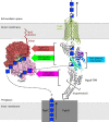
Fig 7. The TPR domain is a central scaffold in the PNAG processing and secretion mechanism.
Summary of the functions of the TPR domain in PNAG processing and secretion. Colored signs/arrows indicate different TPR functions within the complex. The PNAG/dPNAG polymer likely runs continuously from synthesis to secretion as indicated by dotted lines. Where data supports, we have displayed the location of the polymer using a box, coloured using the standard nomenclature for GlcNAc and GlcN. The box sizes are not to scale. A star indicates the expected reducing end of the polymer. The direction of polymerization by PgaC/D is based on polymerization truncation experiments [32], which suggests polymer extension occurs at the non-reducing end. The direction of polymer being processed by PgaB-GH is based on a co-crystal structure of PagB and a PNAG hexamer (4P7R), and at the TPR domain it is based on MD simulation (this work). PgaB (using coordinates from PDB ID 4FD9) is shown in surface representation. The composite PgaA model (using coordinates for porin from PDB ID 4Y25) is shown in cartoon representation, with the crystallized TPR model (this work) is shown in green, blue, and purple.

Similar articles

Cited by

References

    1. Whitney JC, Howell PL. Synthase-dependent exopolysaccharide secretion in Gram-negative bacteria. Trends Microbiol. 2013;21: 63–72. doi: 10.1016/j.tim.2012.10.001 - DOI - PMC - PubMed
    1. Mittl PRE, Schneider-Brachert W. Sel1-like repeat proteins in signal transduction. Cell Signal. 2007;19: 20–31. doi: 10.1016/j.cellsig.2006.05.034 - DOI - PubMed
    1. Keiski C-L, Harwich M, Jain S, Neculai AM, Yip P, Robinson H, et al.. AlgK is a TPR-containing protein and the periplasmic component of a novel exopolysaccharide secretin. Struct Lond Engl 1993. 2010;18: 265–73. doi: 10.1016/j.str.2009.11.015 - DOI - PMC - PubMed
    1. Nojima S, Fujishima A, Kato K, Ohuchi K, Shimizu N, Yonezawa K, et al.. Crystal structure of the flexible tandem repeat domain of bacterial cellulose synthesis subunit C. Sci Rep-uk. 2017;7: 13018. doi: 10.1038/s41598-017-12530-0 - DOI - PMC - PubMed
    1. Marmont LS, Whitfield GB, Rich JD, Yip P, Giesbrecht LB, Stremick CA, et al.. PelA and PelB form a modification and secretion complex essential for Pel polysaccharide-dependent biofilm formation in Pseudomonas aeruginosa. J Biological Chem. 2017;292: 19411–19422. doi: 10.1074/jbc.M117.812842 - DOI - PMC - PubMed

Publication types