. 2022 Aug 5;8(31):eabn9181.
doi: 10.1126/sciadv.abn9181. Epub 2022 Aug 5.

lncRNA-GM targets Foxo1 to promote T cell-mediated autoimmunity

Affiliations

lncRNA-GM targets Foxo1 to promote T cell-mediated autoimmunity

Yali Chen et al. Sci Adv. .

Abstract

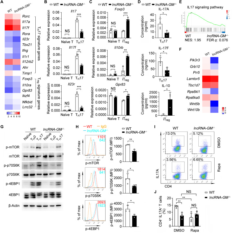
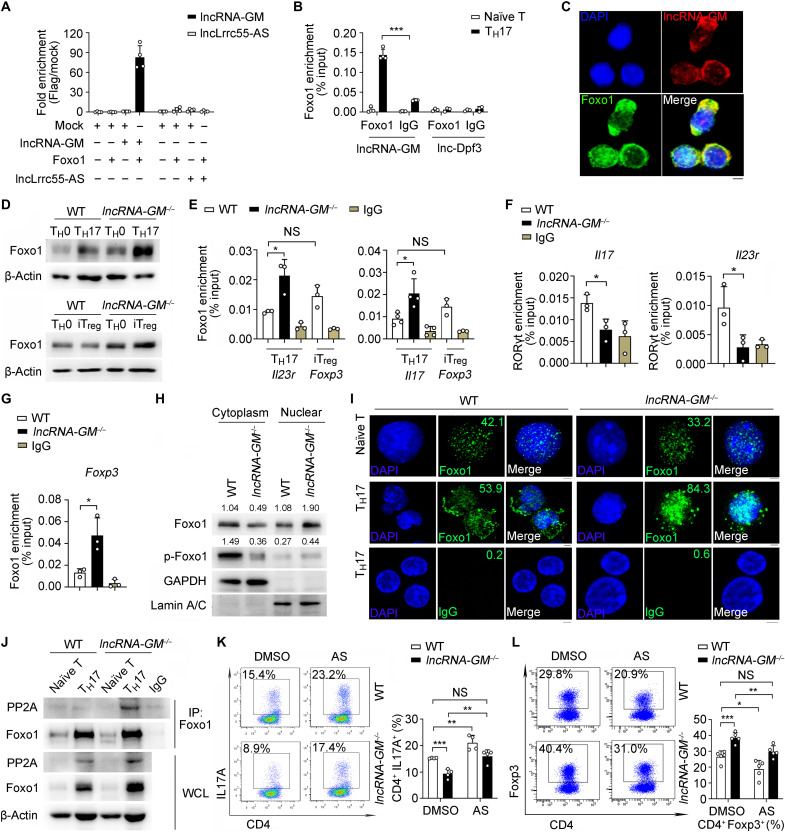
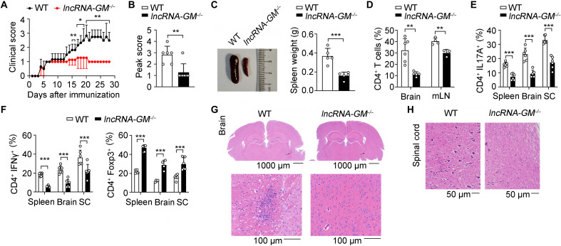
RNA-RBP interaction is important in immune regulation and implicated in various immune disorders. The differentiation of proinflammatory T cell subset TH17 and its balance with regulatory T cell (Treg) generation is closely related to autoimmune pathogenesis. The roles of RNA-RBP interaction in regulation of TH17/Treg differentiation and autoinflammation remain in need of further investigation. Here we report that lncRNA-GM polarizes TH17 differentiation but inhibits iTreg differentiation by reducing activity of Foxo1, a transcriptional factor that is important in inhibiting TH17 differentiation but promoting Treg generation. lncRNA-GM-deficient mice were protected from experimental autoimmune encephalomyelitis. Mechanistically, lncRNA-GM directly binds to cytoplasmic Foxo1, thus inhibiting its activity through blocking dephosphorylation of Foxo1 by phosphatase PP2A to promote Il23r transcription. The human homolog of lncRNA-GM (AK026392.1) also polarizes human TH17 differentiation. Our study provides mechanistic insight into the interaction of lncRNA and transcriptional factor in determining T cell subset differentiation during T cell-mediated autoimmune diseases.


Figures

Fig. 1.
Fig. 1.. lncRNA-GM is selectively up-regulated in TH17 cells.
(A) Quantitative polymerase chain reaction (qPCR) analysis of lncRNA-GM mRNA expression in TH1, TH2, nonpathogenic TH17 (IL-6 + TGF-β), pathogenic TH17 (IL-6 + IL-1β + IL-23), optimal pathogenic TH17 (IL-6 + TGF-β + IL-1β + IL-23) cells, and iTreg cells from mice spleen (n = 3 to 4). (B) qPCR of nTreg cells (CD4+ CD25+) in thymocytes and iTreg cells in splenocytes for lncRNA-GM mRNA expression (n = 3 to 4). (C) qPCR analysis of human lncRNA-GM mRNA expression in TH1, TH2, TH17, and iTreg cells from human peripheral blood mononuclear cells (n = 3). (D) Copy number analysis of lncRNA-GM determined by qPCR in TH1 and TH17 (optimal pathogenic induction) cells (n = 3). (E) Chromatin immunoprecipitation (ChIP)–qPCR analysis of the enrichment of H3K9ac, H3K27ac, and H4K4me3 in lncRNA-GM promoter region in naïve T, TH1, TH17, and iTreg cells (n = 3 to 6). (F) qPCR analysis of lncRNA-GM mRNA expression in TH17 cells treated with 20 μM CPI-637 (CPI; n = 4). Results are presented as means ± SD (A to F). *P < 0.05; **P < 0.01; ***P < 0.001; ****P < 0.0001. NS, not significant; DMSO, dimethyl sulfoxide.
Fig. 2.
Fig. 2.. lncRNA-GM promotes TH17 cell differentiation but inhibits Treg cell differentiation in vivo and in vitro.
(A) Flow cytometric analysis of CD44+ and CD62L+ in splenic CD4+ T cells from WT and lncRNA-GM−/− mice (n = 4). (B and C) Flow cytometric analysis IFN-γ+, IL-4+, IL-17A+, and Foxp3+ cells in splenic CD4+ T cells (B) and differentiated TH1, TH2, nonpathogenic TH17 (IL-6 + TGF-β), pathogenic TH17 (IL-6 + IL-1β + IL-23), optimal pathogenic TH17 (IL-6 + TGF-β + IL-1β + IL-23), and iTreg cells for 3 days (C) (n = 4 to 5). (D) Quantification of IFN-γ+, IL-4+, IL-17A+, and Foxp3+ T cells in differentiated TH1, TH2, nonpathogenic TH17, pathogenic TH17, optimal pathogenic TH17, and iTreg cells (n = 3 to 5). (E) Flow cytometric analysis and quantification of IL-17A+ cells transfected with control lentivirus and lncRNA-GM overexpression lentivirus in TH17 cells (n = 4). Results are presented as means ± SD (A, B, D, and E). One of three representative experiments is shown. *P < 0.05; **P < 0.01; ***P < 0.001.
Fig. 3.
Fig. 3.. lncRNA-GM promotes TH17 cell differentiation by activating mTORC1 signaling.
(A) Heatmap showing the representative up-regulated or down-regulated genes from WT and lncRNA-GM−/− TH17 cells (fold change > 1.5, P < 0.05). (B) qPCR analysis of Il17, Il17f, and Il23r mRNA expression in naïve T and TH17 cells (n = 3). (C) qPCR analysis of Foxp3, Il10rb, and Gpr83 mRNA expression in naïve T and iTreg cells (n = 3). (D) Cytometric bead array analysis of IL-17A and IL-17F in TH17 cell supernatants and IL-10 in iTreg cell supernatants from WT and lncRNA-GM−/− mice (n = 3 to 4). (E) Gene set enrichment analysis of TH17 cells from WT and lncRNA-GM−/− mice. (F) Heatmap showing down-regulated mTOR pathway–related genes in lncRNA-GM−/− TH17 cells (P < 0.05). (G) Immunoblot analysis of mTORC1 and downstream signaling pathway in WT and lncRNA-GM−/− naïve T, TH0 (α-CD3/CD28), and TH17 (α-CD3/CD28 plus IL-6 + TGF-β + IL-1β + IL-23) cells for 3 days. (H) Phosphorylation of mTOR, p70S6K, and 4EBP1 was measured by flow cytometry from WT and lncRNA-GM−/− TH17 cells (n = 3 to 4). (I and J) Flow cytometric analysis (I) and quantification (J) of CD4+ IL-17A+ cells in WT and lncRNA-GM−/− TH17 cells after treatment with DMSO and 50 nM rapamycin (Rapa; n = 4). Naïve CD4+ T cells were cultured in vitro under optimal pathogenic TH17 cell polarizing conditions for 3 days. Results are presented as means ± SD (B, C, D, H, and J). One of three representative experiments is shown. *P < 0.05; **P < 0.01; ***P < 0.001. FDR, false discovery rate. NES, normalized enrichment scores; MFI, mean fluorescence intensity.
Fig. 4.
Fig. 4.. lncRNA-GM directly interacts with Foxo1 and attenuates Foxo1 dephosphorylation by PP2A.
(A and B) RIP-qPCR analysis of lncRNA-GM and lncLrrc55-AS immunoprecipitated by Flag antibody in HEK293T (A) and lncRNA-GM and lnc-Dpf3 immunoprecipitated by Foxo1 or IgG in T cells (B) (n = 3 to 4). (C) RNA FISH analysis of lncRNA-GM (red) and Foxo1 (green) in TH17 cells. DNA (blue) was stained with 4′,6-diamidino-2-phenylindole (DAPI). Scale bar, 2.5 μm. (D) Immunoblot analysis of Foxo1 levels in TH17 and iTreg cells. (E and F) ChIP-qPCR analysis of the recruitment of Foxo1 to Il23r and Il17 promoter regions (E) and RORγt to Il23r and Il17 promoter regions (F) in TH17 cells and the recruitment of Foxo1 to Foxp3 promoter regions in WT iTreg cells (n = 3 to 4). (G) ChIP-qPCR analysis of the recruitment of Foxo1 to Foxp3 promoter regions in iTreg cells (n = 3). (H) Immunoblot analysis of Foxo1 and p-Foxo1 (Ser256) levels in cytoplasm and nuclear extracts from TH17 cells. (I) Immunofluorescence analysis of Foxo1 (green) or IgG (green) in naïve T or TH17 cells. DNA (blue) was stained with DAPI. Scale bar, 2.5 μm. (J) Immunoblot analysis of PP2A in T cells after IP with anti-Foxo1 antibody. (K and L) Flow cytometric analysis of IL-17A+ cells in TH17 (K) after treatment with DMSO or 50 nM AS1842856 (AS) and Foxp3+ cells in iTreg (L) after treatment with 200 nM AS1842856 (n = 4 to 5). Results are presented as means ± SD (A, B, E, F, G, K, and L). One of three representative experiments is shown. *P < 0.05; **P < 0.01; ***P < 0.001.
Fig. 5.
Fig. 5.. Deficiency of lncRNA-GM attenuates pathogenesis of active EAE.
(A and B) Mean clinical score (A) and peak clinical score (B) of WT and lncRNA-GM−/− mice after induction of EAE with MOG35–55 and pertussis toxin (n = 6). (C to H) At day 28 after EAE induction, WT and lncRNA-GM−/− mice were analyzed for spleen size and weight (n = 5) (C), quantification of CD4+ T cells in the brain and mesenteric lymph node (mLN; n = 3 to 6) (D), quantification of IL-17A+, IFN-γ+, and Foxp3+ T cells in the brain, spleen, and spinal cord (SC; n = 4 to 6) (E and F), and histology of hematoxylin and eosin–stained brain (G) and spinal cord (H). Results are presented as means ± SD (A, B, C, D, E, and F). One of three representative experiments is shown. *P < 0.05; **P < 0.01; ***P < 0.001.
Fig. 6.
Fig. 6.. Deficiency of lncRNA-GM reduces severity of EAE by T cell adoptive transfer.
(A and B) Mean clinical score (A) and peak clinical score (B) of Rag2−/− recipients of WT and lncRNA-GM−/− CD4+ T cells after induction of EAE (n = 7). (C and D) At day 28 after EAE induction, Rag2−/− recipient mice were analyzed for quantification of CD4+ and CD8+ T cells in the spleen (n = 4) (C) and quantification of CD4+ IL-17A+, IFN-γ+, and Foxp3+ T cells in the spleen, brain, and spinal cord (n = 3 to 6) (D). Results are presented as means ± SD (A to D). One of three representative experiments is shown. *P < 0.05; **P < 0.01; ***P < 0.001; ****P < 0.0001.

Similar articles

Cited by

References

    1. Fugger L., Jensen L. T., Rossjohn J., Challenges, progress, and prospects of developing therapies to treat autoimmune diseases. Cell 181, 63–80 (2020). - PubMed
    1. Chataway J., Tackling progression in multiple sclerosis. Lancet Neurol. 17, 489–491 (2018). - PubMed
    1. Theofilopoulos A. N., Kono D. H., Baccala R., The multiple pathways to autoimmunity. Nat. Immunol. 18, 716–724 (2017). - PMC - PubMed
    1. Gaublomme J. T., Yosef N., Lee Y., Gertner R. S., Yang L. V., Wu C., Pandolfi P. P., Mak T., Satija R., Shalek A. K., Kuchroo V. K., Park H., Regev A., Single-cell genomics unveils critical regulators of Th17 cell pathogenicity. Cell 163, 1400–1412 (2015). - PMC - PubMed
    1. Ghoreschi K., Laurence A., Yang X.-P., Tato C. M., McGeachy M. J., Konkel J. E., Ramos H. L., Wei L., Davidson T. S., Bouladoux N., Grainger J. R., Chen Q., Kanno Y., Watford W. T., Sun H.-W., Eberl G., Shevach E. M., Belkaid Y., Cua D. J., Chen W., O’Shea J. J., Generation of pathogenic TH17 cells in the absence of TGF-β signalling. Nature 467, 967–971 (2010). - PMC - PubMed