. 2022 Aug 5;7(74):eabl3995.
doi: 10.1126/sciimmunol.abl3995. Epub 2022 Aug 5.

Size-dependent activation of CAR-T cells

Affiliations

Size-dependent activation of CAR-T cells

Qian Xiao et al. Sci Immunol. .

Abstract

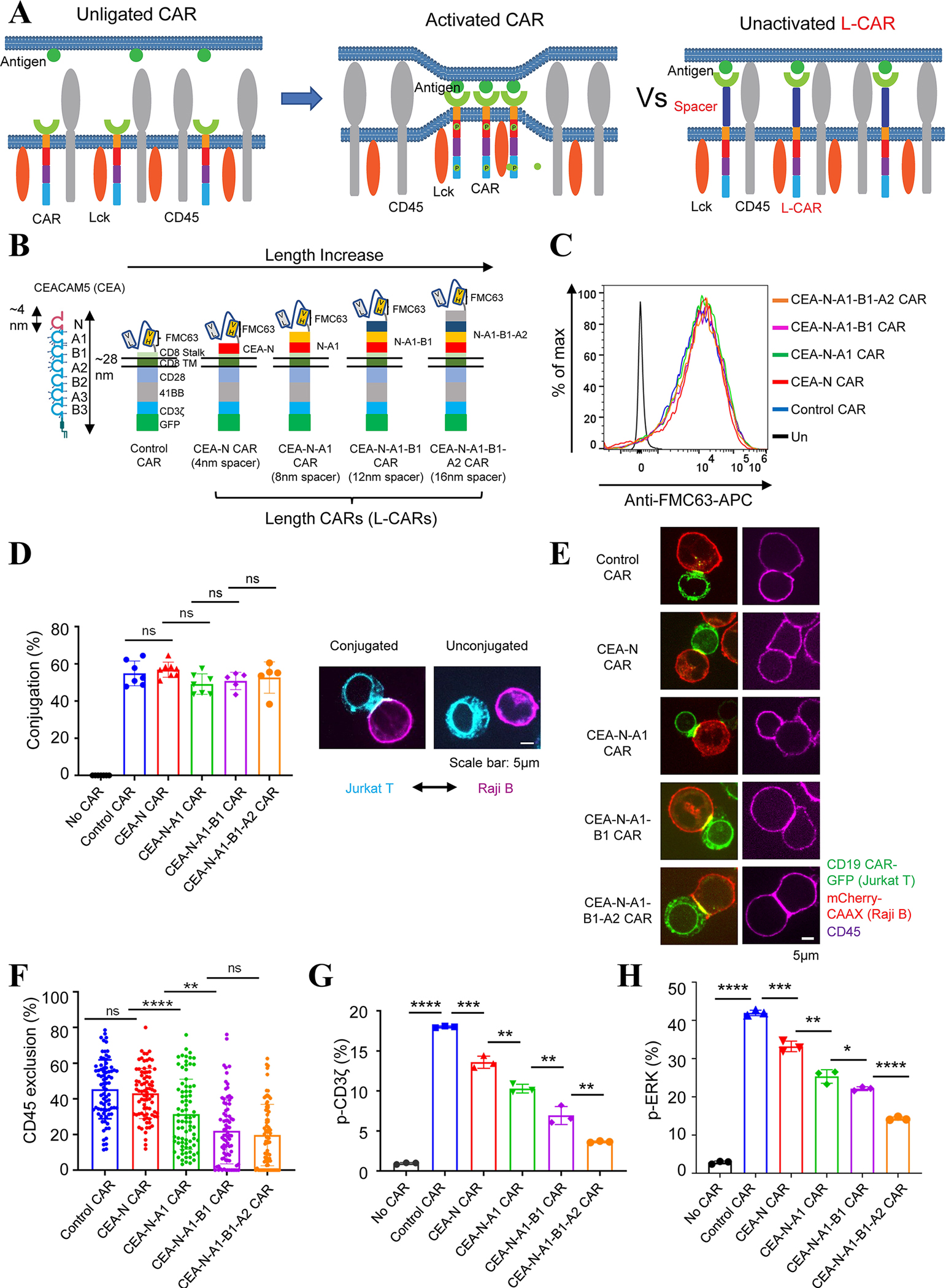
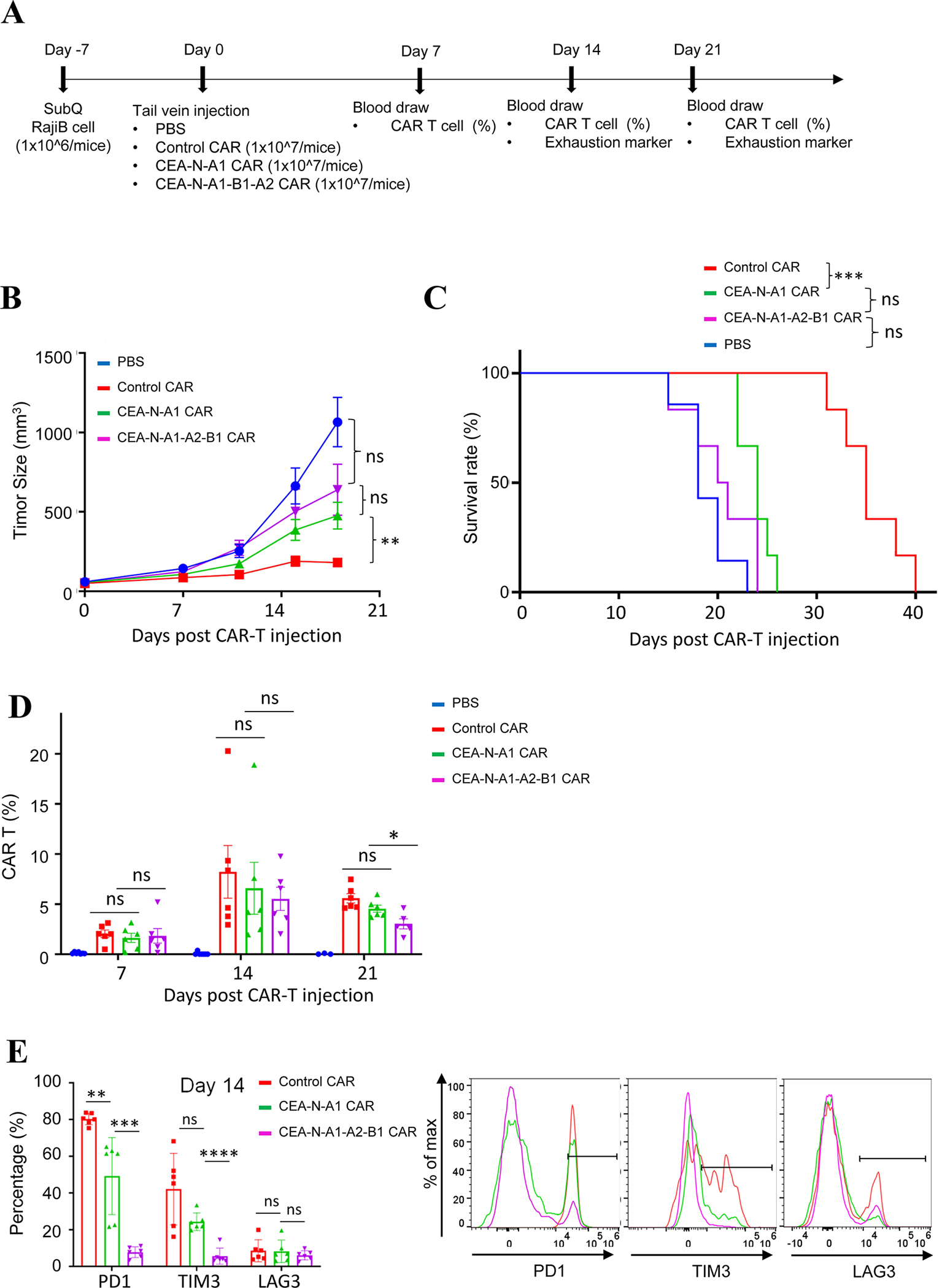
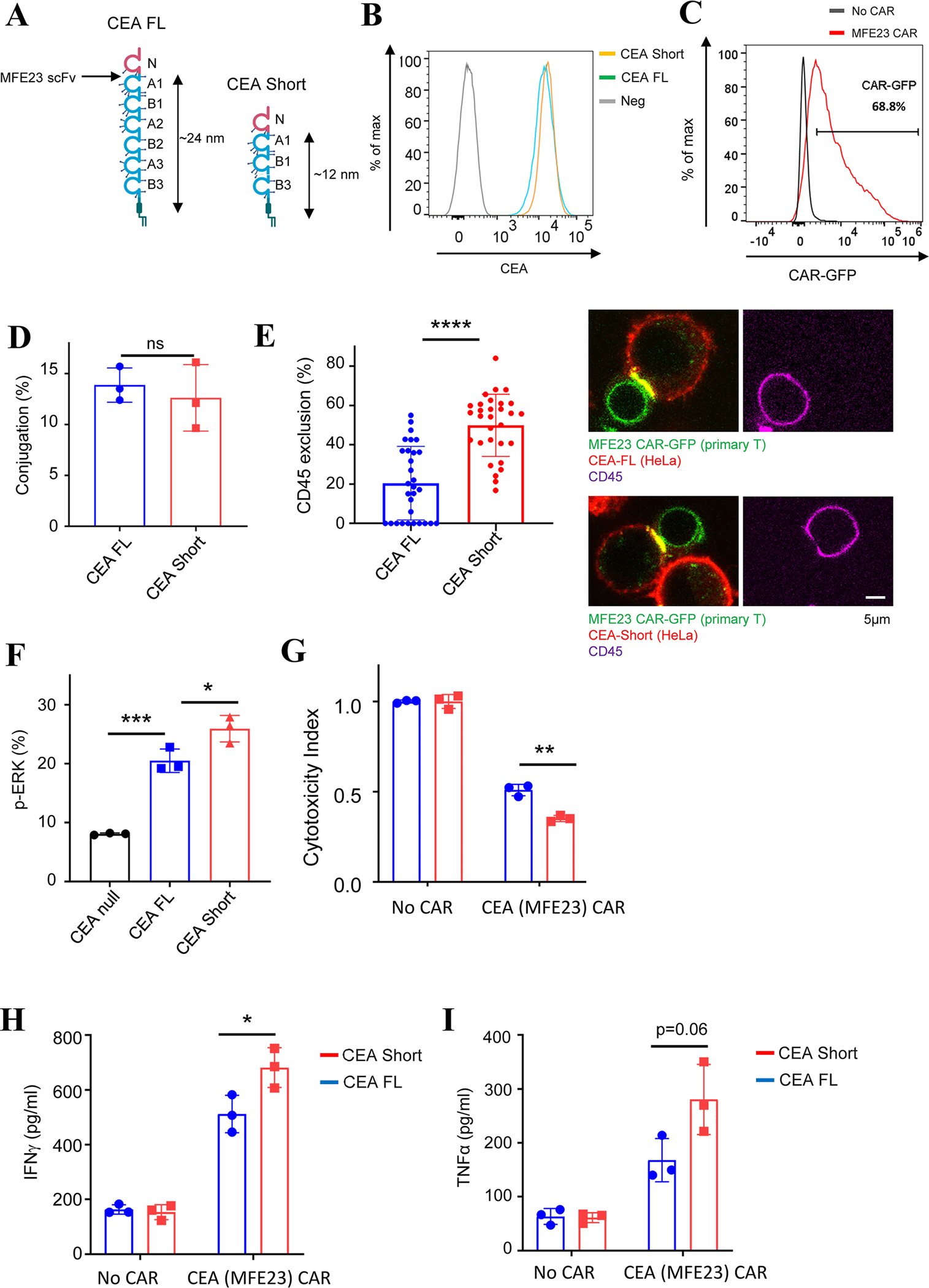
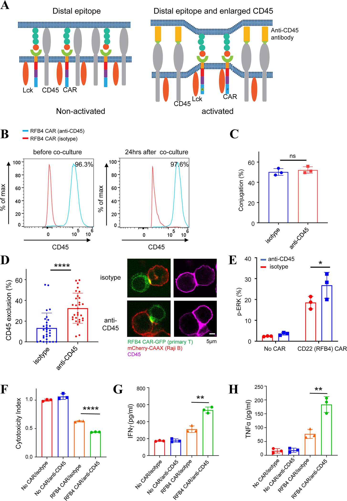
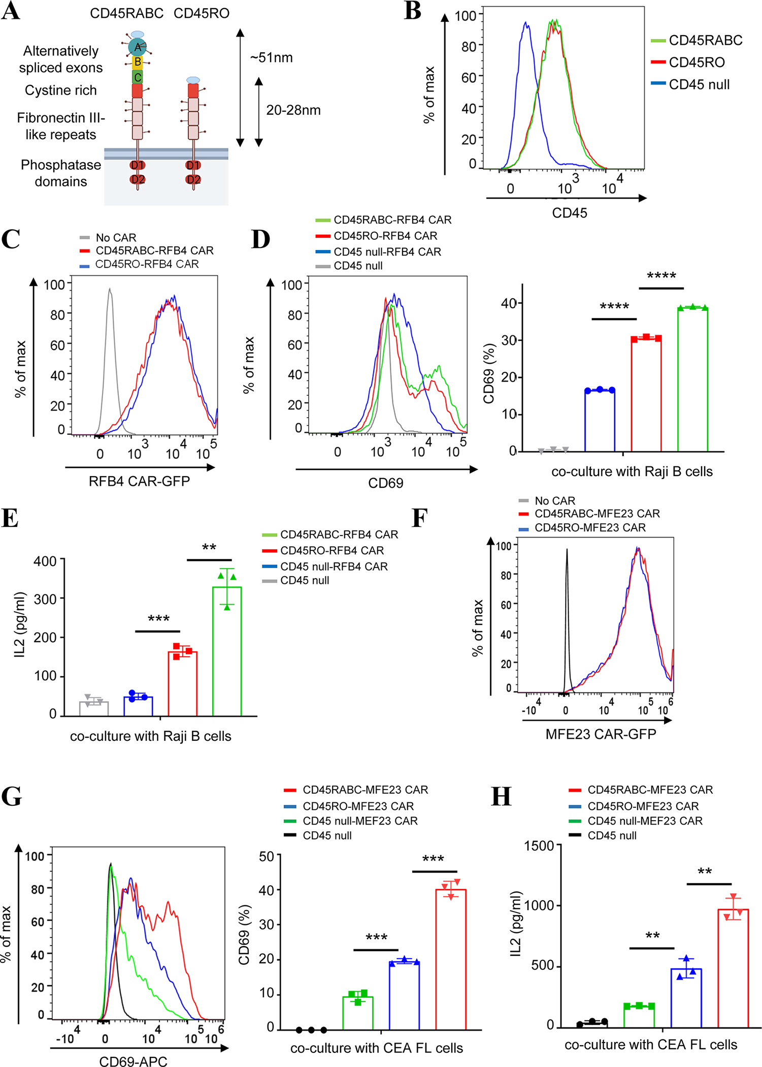
As the targets of chimeric antigen receptor (CAR)-T cells expand to a variety of cancers, autoimmune diseases, viral infections, and fibrosis, there is an increasing demand for identifying new antigens and designing new CARs that can be effectively activated. However, the rational selection of antigens and the design of CARs are limited by a lack of knowledge regarding the molecular mechanism by which CARs are activated by antigens. Here, we present data supporting a "size exclusion" model explaining how antigen signals are transmitted across the plasma membrane to activate the intracellular domains of CARs. In this model, antigen engagement with CAR results in a narrow intermembrane space that physically excludes CD45, a bulky phosphatase, out of the CAR zone, thus favoring CAR phosphorylation by kinases, which further triggers downstream pathways leading to T cell activation. Aligned with this model, increasing the size of CAR extracellular domains diminished CAR-T activation both in vitro and in a mouse lymphoma model; membrane-proximal epitopes activated CAR-Ts better than membrane-distal epitopes. Moreover, increasing the size of CD45 by antibody conjugation enhanced the activation of CARs that recognize membrane-distal epitopes. Consistently, CAR-Ts expressing CD45RABC, the larger isoform, were activated to a higher level than those expressing a smaller isoform CD45RO. Together, our work revealed that CAR-T activation depends on the size difference between the CAR-antigen pair and CD45; the size of CAR, antigen, and CD45 can thus be targets for tuning CAR-T activation.


Conflict of interest statement

Competing interests:

CH provides consulting for GlaxoSmithKine and PACT Pharma, holds NIH patents and royalties in the field of immunotherapy and cell therapy, and receives research funding from Neogene Therapeutics and T-Cure Bioscience. XS is a co-applicant for a provisional patent on CAR, which is not based on the specific results in this manuscript.

Figures

Fig. 1.
Fig. 1.. The length of CAR extracellular domain regulated CD45 exclusion and CAR signaling in Jurkat T cells.
(A) Schematic representation of the ‘size exclusion’ hypothesis explaining antigen-dependent CAR activation. LEFT: In resting cells, the phosphorylation of CAR by the kinase Lck is antagonized by the phosphatase CD45. MIDDLE: The binding of antigen to CAR creates a narrowed intermembrane space (synapse), from which CD45 is excluded, leading to the phosphorylation of CAR. RIGHT: The binding of antigen to a CAR with a longer extracellular domain (L-CAR) does not exclude CD45. Consequently, CAR is not activated (phosphorylated). (B) Generation of CD19 CARs with increasing lengths (L-CARs) by inserting tandem immunoglobulin domains of CEACAM5 (CEA) into the extracellular part of the CAR. Each immunoglobulin domain of CEA is around 4 nm long. (C) CAR variants expression on the surface of Jurkat T cells, as determined by staining with an anti-FMC63 (CD19 scFv) antibody. (D) Effects of inserted Ig domains on CAR’s binding to antigen. Jurkat T cells expressing CAR-GFP and Raji B cells expressing mCherry-CAAX (E:T=1:1) were co-cultured for 30 mins and the cell-cell conjugation was imaged by confocal microscopy. LEFT: Quantification of conjugation percentage (%), which was calculated by dividing the number of CAR T cells that bound to Raji B cells by the total number of CAR T cells. Each dot indicates one independent experiment. Data were presented as mean with SD (Unpaired T test, p>0.05 (ns)). N=5–7 co-culture experiments. RIGHT: representative images of conjugated cells versus unconjugated cells. (E) Effects of CAR length on the exclusion of CD45 from the CAR synapse. Jurkat T cells expressing CAR-GFP and Raji B cells expressing mCherry-CAAX (E:T =1:1) were co-cultured for 30 mins, fixed, and stained with an anti-CD45 antibody conjugated with APC. Images were acquired by confocal microscopy. The exclusion percentage = (1 − ICD45 in car zone/ICD45out car zone) *100%. Data were presented as mean with SD (Mann-Whitney U test, p>0.05 (ns), p<0.01 (**), p<0.0001 (****)). N=80 synapses. Representative images were shown. (F) Phosphorylation of the CD3ζ domain in Jurkat T cells expressing CAR variants. pCD3ζ in GFP+ CAR Jurkat T cells was detected by flow cytometry 5 min after co-culture with Raji B cells (E:T =1:1). Data were presented as mean with SD (Unpaired T test, p<0.0001 (****), p<0.001 (***), p<0.01 (**)). N=3 co-culture experiments. (G) ERK activation in Jurkat T cells expressing CAR variants. pERK in GFP+ CAR Jurkat T cells was detected by flow cytometry 5 min after co-culture with Raji B cells (E:T =1:1). Data were presented as mean with SD (Unpaired T test, p<0.0001 (****), p<0.001 (***), p<0.01 (**), p<0.05 (*)). N=3 co-culture experiments.
Fig. 2.
Fig. 2.. The length of CAR extracellular domain regulated CAR-triggered human primary T cell activation.
(A) Human T cells were purified from PMBC from anonymous healthy donors, expanded, and engineered with lentivirus to express CD19 CARs. CAR variants were expressed in a similar level on the surface of primary T cells, as determined by an anti-FMC63 (CD19 scFv) antibody. (B) Antigen binding capacity after insertion of Ig domains into the CAR. Human T cells expressing CAR-GFP and Raji B cells expressing mCherry-CAAX (E:T =1:1) were co-cultured for 30 mins and the cell conjugation was imaged by confocal microscopy. Conjugation percentage (%) was calculated by dividing the number of CAR T cells that bound to Raji B cells by the total number of CAR T cells. Data were presented as mean with SD (Unpaired T test, p>0.05 (ns)). N=3 co-culture experiments. (C) Effects of increasing CAR length on the exclusion of CD45 from the CAR synapse. Human T cells expressing CAR-GFP and Raji B cells expressing mCherry-CAAX (E:T =1:1) were co-cultured for 30 mins, fixed, and stained with an anti-CD45 antibody conjugated with APC. Images were acquired by confocal microscopy. Data were presented as mean with SD (Mann-Whitney U test, p>0.05 (ns), p<0.05 (*), p<0.01 (**), p<0.0001 (****)). N=33~34 synapses. Representative images were shown. (D) Proliferative capacity of CAR-T cells after exposure to Raji B cells (E:T =1:1). The human CD8+ CAR-T cell number upon Raji B cell stimulation was calculated with counting beads by flow cytometry at indicated time points. Data were presented as mean with SD (Unpaired T test, p<0.0001 (****)). N=3 co-culture experiments. (E) The tumor cell-killing capacity of CAR-T. Human CD8+ CAR T cells were co-cultured with Raji B-LUC-mCherry cells (E:T =5:1) for 24 hours. The Raji B cells remained were quantified by luciferase-generated fluorescence as detected by microplate photoreaders. Data were presented as mean with SD (Unpaired T test, p>0.05 (ns), p<0.05 (*), p<0.01 (**), p<0.0001 (****)). N=3 co-culture experiments. (F-G) The co-cultured medium from (E) was used for the measurement of IFNγ and TNFα by ELISA. Data were presented as mean with SD (Unpaired T test, p>0.05 (ns), p<0.05 (*), p<0.01 (**), p<0.001 (***)). N=3 co-culture experiments.
Fig. 3.
Fig. 3.. Length-dependent anti-tumor activity of CAR-T in vivo.
(A) Experimental settings of CAR-T mediated killing of xenografted tumors. (B) Tumor growth monitored in NSG mice inoculated with Raji B cells and treated with PBS, control CAR T, CEA-N-A1 CAR T, or CEA-N-A1-B1-A2 CAR T cells. Data were presented as mean with SD (Unpaired T test was performed on data of day 18; P >0.05 (ns), P < 0.01 (**)). N = 6–7 mice. (C) Mice survival with CAR-T treatment. Log-rank (Mantel-Cox) test was performed on data at day 28; P>0.05 (ns); P < 0.001 (***). N = 6–7 mice. (D) CAR T percentage in circulation at day 7, 14, 21 post injection was determined via flow cytometry. Data were presented as mean with SD (Unpaired T test; P>0.05 (ns)). N= 6–7 mice. (E) Flow cytometry analysis of exhaustion marker PD1, TIM3, LAG3 on CAR T cells at day 14. Data were presented as mean with SD (Unpaired T test; P>0.05 (ns), P < 0.01 (**), P < 0.001 (***), P < 0.0001 (****)). N=6–7 mice.
Fig. 4
Fig. 4. Effects of epitope position on human primary CAR-T targeting CD22.
(A) Schematics of CAR activation by membrane-proximal versus membrane-distal epitopes. LEFT: CARs bind membrane proximal epitopes to create a narrow intermembrane space that excludes phosphatase CD45 and induces CAR activation. Right: CARs recognize membrane-distal epitopes, which cannot form a close-contact zone to exclude CD45 effectively. (B) Schematics of the binding sites of two scFvs, m971 and RFB4, that recognize CD22. CD22 contains seven extracellular immunoglobulin domains. RFB4 recognizes distal epitopes on Domain 3 whereas m971 recognizes proximal epitopes on Domain 7. A short version of CD22 was constructed by removing Domain 5–7 so that the epitopes on Domain 3 become membrane proximal. An mCherry tag replaced the intracellular part of both the full-length and short CD22. (C) Expression of the full-length or short extracellular domain of CD22 in KU812 cells, as detected by western blot. (D) Human primary T cells were purified from the peripheral blood of anonymous healthy donors, expanded, and engineered with lentivirus to express RFB4 CARs. CAR expression was evaluated by flow cytometry. (E) Conjugation of RFB4 CAR-T cells with KU812 cells expressing the full-length (FL) or short CD22. Human primary T cells expressing RFB4 CAR were co-cultured with KU812 cells expressing FL or short CD22 extracellular domain (E:T =1:1) for 30 min before being imaged by confocal microscopy. Data were presented as mean with SD (Unpaired T test, p>0.05(ns)). N=3 co-culture experiments. (F) Effects of epitope position on CD45 exclusion from the RFB4 CAR synapses. Primary T cells expressing RFB4 CAR were co-cultured with KU812 cells expressing the FL or short CD22 (E:T =1:1) for 30 min, followed by staining with an anti-CD45 antibody. Imaging was acquired by confocal microscopy. Data were presented as mean with SD (Mann-Whitney U test, p<0.01 (**)). N=50 synapses. (G) ERK phosphorylation in RFB4 CAR-T cells engaged with the full-length or short CD22. Primary RFB4 CAR-T cells were cocultured with KU812 cells (E:T =1:1) for 10 minutes. ERK phosphorylation was assessed by flow cytometry. Data were presented as mean with SD (Unpaired T test, p<0.01 (**), p<0.05 (*)). N=3 co-culture experiments. (H) Cytotoxicity of RFB4 CAR-T cells to cells expressing the full-length or short CD22. Primary RFB4 CAR T cells were co-cultured with FL or short CD22 cells (E:T =5:1) for 24 hours, followed by flow cytometry analysis. Data were presented as mean with SD (Unpaired T test, p<0.05 (*), p<0.01 (**)). N=3 co-culture experiments. (I-J) TNFα and IFNγ production in RFB4 CAR-T cells. The co-cultured medium from (H) was used for the measurement of IFNγ and TNFα by ELISA. Data were presented as mean with SD (Unpaired T test, p>0.05 (ns), p<0.05 (*), p<0.01 (**), p<0.0001 (****)). N=3 co-culture experiments.
Fig. 5
Fig. 5. Effects of epitope position on human primary CAR-T targeting CEA.
(A) The full-length (FL) CEA contains multiple immunoglobulin domains. scFv MFE23 recognize epitopes between the N and A1 domain. A short CEA was constructed by deleting domain A2, B2, and A3 so that the epitope for MFE23 was positioned closer to the membrane. (B) Comparing the expression level of the FL and short CEA in HeLa cells, as determined by flow cytometry. (C) Primary T cells expressing MFE23 CAR that recognizes CEA. CAR expression was evaluated by flow cytometry. (D) Binding of primary MEF23 CAR-T cells to HeLa cells expressing the FL or short CEA. Primary T cells expressing MFE23 CAR were co-cultured with HeLa cells expressing the FL or short CEA (E:T =1:1) for 30 min before being imaged by confocal microscopy. Data were presented as mean with SD (Unpaired T test, p>0.05 (ns)). N=3 co-culture experiments. (E) Effects of epitope position on CD45 exclusion from the MFE23 CAR synapses. Primary T cells expressing MFE23 CAR were co-cultured with HeLa cells expressing the FL or short CEA (E:T =1:1) for 30 min, followed by staining with an anti-CD45 antibody. Imaging was acquired by confocal microscopy. Data were presented as mean with SD (Mann-Whitney U test, p<0.0001 (****)). N=30 synapses. (F) ERK phosphorylation in MFE23 CAR-T cells engaged with the full-length or short CEA. Primary MFE23 CAR-T cells were cocultured with HeLa cells (E:T =1:1) for 10 minutes. pERK was assessed by flow cytometry. Data were presented as mean with SD (Unpaired T test, p<0.001 (***), p<0.05 (*)). N=3 co-culture experiments. (G) Cytotoxicity of MFE23 CAR-T cells to HeLa cells expressing the full-length or short CEA. Primary MFE23 CAR T cells were co-cultured with HeLa cells expressing the FL CEA or short CEA cells (E:T =5:1) for 24 hours, followed by flow cytometry analysis. Data were presented as mean with SD (Unpaired T test, p<0.01 (**)). N=3 co-culture experiments. (H-I) TNFα and IFNγ production in MFE23 CAR-T cells. The co-cultured medium from (G) was used for the measurement of IFNγ and TNFα by ELISA. Data were presented as mean with SD (Unpaired T test, p>0.05 (ns), p<0.05 (*)). N=3 co-culture experiments.
Fig. 6
Fig. 6. Effects of a CD45 antibody on the activation of human primary CAR-T cells recognizing membrane-distal antigens.
(A) Illustration of a size increase in CD45 by conjugation with an anti-CD45 antibody. LEFT: CARs recognize membrane-distal epitopes, which does not result in sufficient CD45 exclusion and CAR activation. RIGHT: An ani-CD45 antibody increases the size of CD45 and promotes CD45 exclusion from the CAR synapse, which results in CAR activation. (B) Human primary RFB4 (CD22) CAR-T cells were labeled with a biotinylated anti-CD45 antibody, crosslinked with streptavidin, and co-cultured with Raji B cells. Flow cytometry analysis showed that the CD45 antibody remained bound with CAR-T cells 24 hrs after co-culture with Raji B cells. The isotype antibody served as a negative control. (C) Effects of the CD45 antibody on the RFB4 CAR-T conjugation with Raji B cells. Primary T cells expressing RFB4 CAR were co-cultured with Raji B cells expressing mCherry-CAAX (E:T =1:1) for 30 min before being imaged by confocal microscopy. Data were presented as mean with SD (Unpaired T test, p>0.05(ns)). N=3 co-culture experiments. (D) Effects of the CD45 antibody on CD45 exclusion from the CAR synapse. Primary RFB4 CAR-T cells were labeled with CD45 or isotype antibodies, and cocultured with Raji B cells expressing mCherry-CAAX (E:T =1:1) for 30 min, followed by fixation and staining. Images were acquired by confocal microscopy. Data were presented as mean with SD (Mann Whitney U test, p<0.0001 (****). N=60 synapses. (E) ERK phosphorylation affected by the CD45 antibody. RFB4 CAR-T cells were labeled with CD45 or isotype antibodies and cocultured with Raji B cells (E:T=1:1) for 10 minutes. ERK phosphorylation was assessed by flow cytometry. Data were presented as mean with SD (Unpaired T test, p<0.05 (*)). N=3 co-culture experiments. (F) Cytotoxicity of RFB4 CAR T cells against Raji B cells in the presence of the CD45 antibody. Primary RFB4 CAR T cells were co-cultured with Raji B-LUC-mCherry cells (E:T =5:1) for 24 hours. The Raji B cells remained were quantified by luciferase-generated fluorescence as detected by a microplate photoreader. Data were presented as mean with SD (Unpaired T test, p<0.0001 (****)). N=3 co-culture experiments. (G-H) Cytokine production in primary RFB4 CAR-T cells with the CD45 antibody. The co-cultured medium from (F) was used for the measurement of IFNγ and TNFα by ELISA. Data were presented as mean with SD (Unpaired T test, p<0.01 (**)). N=3 co-culture experiments.
Fig. 7
Fig. 7. Regulation of Jurkat CAR-T activation by CD45 isoforms of different sizes.
(A) Illustration of CD45 isoforms. CD45RABC is the longest isoform that is expressed in naïve T cells. CD45RO, the shortest splicing isoform, is expressed in activated T cells. (B) Flow cytometry analysis showed the expression level of CD45RO and RABC when they were reconstituted in a CD45-deficient Jurkat T cell line. (C) Flow cytometry analysis showed the expression level of RFB4 (CD22) CAR in CD45RO or RABC-expressing Jurkat T cells. (D-E) Comparing CD45RABC to CD45RO on the activation of RFB4 CAR T against CD22. RFB4 (CD22) CAR Jurkat T cells expressing either the CD45RABC or CD45RO were cocultured with Raji B cells (E:T =1:1) for 24 hrs. CD69 expression was assessed by flow cytometry and IL-2 release was measured by ELISA. Data were presented as mean with SD (Unpaired T test, p<0.01 (**), p<0.001 (***), p<0.0001 (****)). N=3 co-culture experiments. (F) Flow cytometry analysis showed the expression level of MEF23 (CEA) CAR in CD45RO or RABC-expressing Jurkat T cells. (G-H) Comparing CD45RABC to CD45RO on the activation of MFE23 CAR T against CEA. MFE23 (CEA) CAR Jurkat T cells expressing either the CD45RABC or CD45RO were cocultured with HeLa cells expressing full-length CEA (E:T=1:1) for 24 hrs. CD69 expression was assessed by flow cytometry and IL-2 release was measured by ELISA. Data were presented as mean with SD (Unpaired T test, p<0.01 (**), p<0.001 (***)). N=3 co-culture experiments.

Similar articles

Cited by

References

    1. Porter DL, Levine BL, Kalos M, Bagg A, June CH, Chimeric antigen receptor-modified T cells in chronic lymphoid leukemia. N Engl J Med 365, 725–733 (2011). - PMC - PubMed
    1. Wang M, Munoz J, Goy A, Locke FL, Jacobson CA, Hill BT, Timmerman JM, Holmes H, Jaglowski S, Flinn IW, McSweeney PA, Miklos DB, Pagel JM, Kersten MJ, Milpied N, Fung H, Topp MS, Houot R, Beitinjaneh A, Peng W, Zheng L, Rossi JM, Jain RK, Rao AV, Reagan PM, KTE-X19 CAR T-Cell Therapy in Relapsed or Refractory Mantle-Cell Lymphoma. N Engl J Med 382, 1331–1342 (2020). - PMC - PubMed
    1. Grupp SA, Kalos M, Barrett D, Aplenc R, Porter DL, Rheingold SR, Teachey DT, Chew A, Hauck B, Wright JF, Milone MC, Levine BL, June CH, Chimeric antigen receptor-modified T cells for acute lymphoid leukemia. N Engl J Med 368, 1509–1518 (2013). - PMC - PubMed
    1. Leibman RS, Richardson MW, Ellebrecht CT, Maldini CR, Glover JA, Secreto AJ, Kulikovskaya I, Lacey SF, Akkina SR, Yi Y, Shaheen F, Wang J, Dufendach KA, Holmes MC, Collman RG, Payne AS, Riley JL, Supraphysiologic control over HIV-1 replication mediated by CD8 T cells expressing a re-engineered CD4-based chimeric antigen receptor. PLoS Pathog 13, e1006613 (2017). - PMC - PubMed
    1. Hale M, Mesojednik T, Romano Ibarra GS, Sahni J, Bernard A, Sommer K, Scharenberg AM, Rawlings DJ, Wagner TA, Engineering HIV-Resistant, Anti-HIV Chimeric Antigen Receptor T Cells. Mol Ther 25, 570–579 (2017). - PMC - PubMed

Publication types