. 2022 Aug 9;119(32):e2116956119.
doi: 10.1073/pnas.2116956119. Epub 2022 Aug 5.

Postmitotic accumulation of histone variant H3.3 in new cortical neurons establishes neuronal chromatin, transcriptome, and identity

Affiliations

Postmitotic accumulation of histone variant H3.3 in new cortical neurons establishes neuronal chromatin, transcriptome, and identity

Owen H Funk et al. Proc Natl Acad Sci U S A. .

Abstract

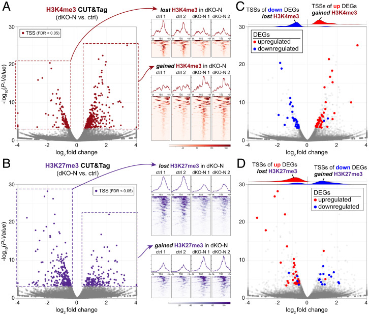
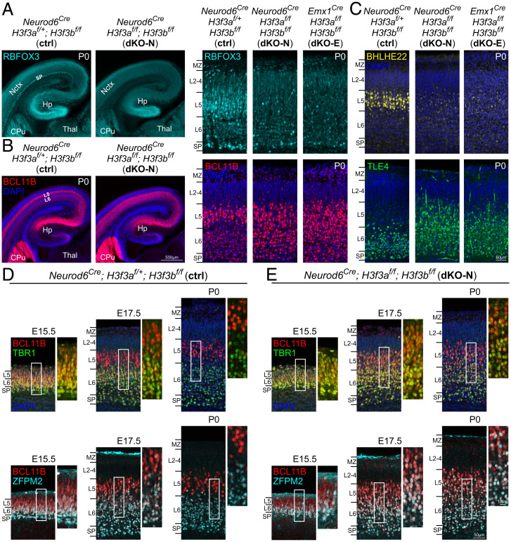
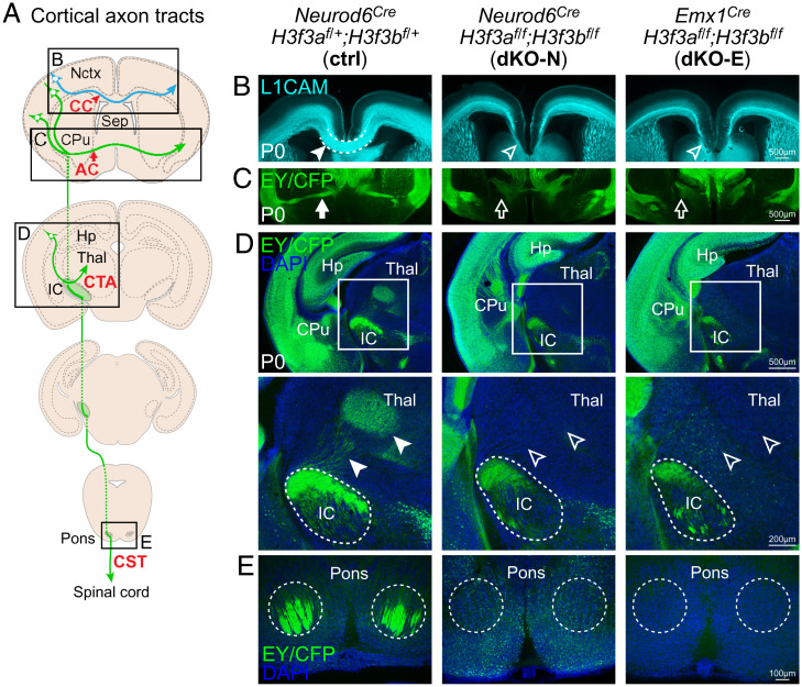
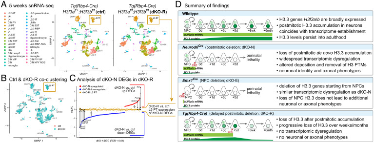
Histone variants, which can be expressed outside of S-phase and deposited DNA synthesis-independently, provide long-term histone replacement in postmitotic cells, including neurons. Beyond replenishment, histone variants also play active roles in gene regulation by modulating chromatin states or enabling nucleosome turnover. Here, we uncover crucial roles for the histone H3 variant H3.3 in neuronal development. We find that newborn cortical excitatory neurons, which have only just completed replication-coupled deposition of canonical H3.1 and H3.2, substantially accumulate H3.3 immediately postmitosis. Codeletion of H3.3-encoding genes H3f3a and H3f3b from newly postmitotic neurons abrogates H3.3 accumulation, markedly alters the histone posttranslational modification landscape, and causes widespread disruptions to the establishment of the neuronal transcriptome. These changes coincide with developmental phenotypes in neuronal identities and axon projections. Thus, preexisting, replication-dependent histones are insufficient for establishing neuronal chromatin and transcriptome; de novo H3.3 is required. Stage-dependent deletion of H3f3a and H3f3b from 1) cycling neural progenitor cells, 2) neurons immediately postmitosis, or 3) several days later, reveals the first postmitotic days to be a critical window for de novo H3.3. After H3.3 accumulation within this developmental window, codeletion of H3f3a and H3f3b does not lead to immediate H3.3 loss, but causes progressive H3.3 depletion over several months without widespread transcriptional disruptions or cellular phenotypes. Our study thus uncovers key developmental roles for de novo H3.3 in establishing neuronal chromatin, transcriptome, identity, and connectivity immediately postmitosis that are distinct from its role in maintaining total histone H3 levels over the neuronal lifespan.

Keywords: cerebral cortex; chromatin; development; histone modification; neuronal identity.


Conflict of interest statement

The authors declare no competing interest.

Figures

Fig. 1.
Fig. 1.
Postmitotic accumulation of H3.3 in cortical neurons and genetic manipulation of H3f3a and H3f3b. (A) snRNA-seq of wild-type E14.5 cortex visualized by Uniform Manifold Approximation and Projection (UMAP). H3f3a and H3f3b mRNA expression was found in each of the 19 identified cell clusters and in 98.0% and 98.2% of cells, respectively. (B) Analysis of H3.3 and pan-H3 protein in cortical development by immunostaining. On E14.5 and E16.5, low levels of H3.3 (green) were present in the VZ and SVZ, composed largely of cycling NPCs. High levels of H3.3 were present in the CP, composed largely of postmitotic neurons. In E16.5 CP and P0 cortex, higher levels of H3.3 were present in the early-born neurons of the SP and deep layers, compared to the late-born neurons of the upper layers (open arrowheads). Pan-H3 (red) was broadly present and showed no preference for early-born neurons; high levels were present in late-born upper-layer neurons (solid arrowhead). (C) Temporal analysis of H3.3 accumulation. Neurons born on E13.5 were labeled by a single pulse of EdU. EdU-labeled neurons (magenta, arrowheads) were analyzed by H3.3 immunostaining (green). (D) Quantitative analysis of immunofluorescence revealed a progressive increase in H3.3 levels over the first postmitotic days (one-way ANOVA with Tukey’s post hoc test, n.s., not significant; *P < 0.01; ***P < 0.0001). (E) A schematic of H3.1/H3.2 and H3.3 accumulation, and genetic manipulation of H3.3-encoding genes H3f3a and H3f3b. (F) Analysis of Cre expression (yellow) by immunostaining confirmed the published specificities of Neurod6Cre in newly postmitotic neurons and of Emx1Cre in NPCs at E14.5. (G) Analysis of H3 proteins in P0 control (ctrl) and conditional H3.3 gene double knockouts, Neurod6Cre;H3f3af/f;H3f3bf/f (dKO-N) and Emx1Cre;H3f3af/f;H3f3bf/f (dKO-E). In both dKO-N and dKO-E, H3.3 (green) was largely lost from neocortex (Nctx) but preserved in caudate putamen (CPu) and septum (Sep). The levels of pan-H3 (red) were unaffected in dKO-N or dKO-E. CIN, cortical interneuron; CR, Cajal-Retzius cell; CPN, cortical plate neuron; dCP, deep cortical plate; IPC, intermediate progenitor cell; IZ, intermediate zone; Ln, layer n; MZ, marginal zone; RGC, radial glial cell; SPN, subplate neuron; uCP, upper cortical plate.
Fig. 2.
Fig. 2.
Disrupted developmental acquisition of the neuronal transcriptome following H3f3a and H3f3b codeletion. (A) Volcano plot of UMI RNA-seq comparing P0 cortex of dKO-N to control. Of 948 DEGs (FDR < 0.01), 579 were down-regulated (blue) and 369 were up-regulated (red). (B) The developmental expression trajectories of dKO-N DEGs and 3,128 unchanged genes based on ENCODE RNA-seq data of mouse forebrain and normalized to E10.5 levels. Genes down-regulated in dKO-N are characterized by a progressive increase in expression across forebrain development. (C) Genes were categorized based on developmental trajectory using logistic regression on the ENCODE forebrain RNA-seq data. Genes that normally increase in expression over embryonic development (tangerine) were significantly down-regulated in dKO-N. (D) Intersectional analysis of DEGs with genes selectively expressed in CP neurons (magenta) or VZ NPCs (green) at E14.5. NPC-selective genes were overrepresented in up-regulated DEGs and neuron-selective genes were overrepresented in down-regulated DEGs. (E) Immunostaining validation of expression changes in the P0 cortex. In ctrl, NR4A2 (NURR1, cyan) immunostaining labeled deep-layer neurons. In dKO-N, NR4A2 staining was lost. In ctrl, SATB2 (yellow) immunostaining labeled L2-L5 neurons. In dKO-N, SATB2 staining was decreased, especially in the deep layers. (F and G) Volcano plots comparing dKO-E to control (F) and dKO-N to dKO-E (G). DEGs are indicated as in A. (H) Experimental overlap of dKO-N and dKO-E DEGs compared to random overlap. (I) Heatmap of dKO-N and dKO-E DEGs based on log2FC. dKO-N and dKO-E DEGs showed congruent expression-change directionality. dev., developmental; reg., regulated; TPM, transcripts per million.
Fig. 3.
Fig. 3.
Disrupted histone modifications following H3f3a and H3f3b codeletion. (A) Volcano plot of H3K4me3 CUT&Tag signal at annotated TSSs comparing the P0 cortex of dKO-N to control, and normalized H3K4me3 CUT&Tag profiles 5 kb upstream and downstream of TSSs. At FDR < 0.05, 300 TSSs showed significantly altered H3K4me3 in dKO-N (burgundy), 220 gained H3K4me3, and 80 lost H3K4me3. (B) Volcano plot of H3K27me3 CUT&Tag signal at TSSs comparing dKO-N to control, and normalized H3K27me3 CUT&Tag profiles: 275 TSSs showed significantly altered H3K27me3 in dKO-N (purple), 105 gained H3K27me3, and 170 lost H3K27me3. (C and D) Intersectional analysis of significantly altered TSSs and DEGs in dKO-N. Down-regulated DEGs in dKO-N (blue) largely lost the activating mark H3K4me3 (C) at their TSS but gained the repressive mark H3K27me3 (D). Conversely, up-regulated DEGs (red) largely gained the activating mark H3K4me3 at their TSSs but lost the repressive mark H3K27me3. DEGs directionally correlated with PTM changes in dKO-N.
Fig. 4.
Fig. 4.
Disrupted establishment of layer-dependent neuronal identities following H3f3a and H3f3b codeletion. (A) In the P0 control cortex, RBFOX3 (NEUN, cyan) immunostaining was present in all cortical layers and intensely labeled L5 and SP neurons. In dKO-N and dKO-E cortex, RBFOX3 expression was decreased and intense staining of L5 and SP neurons was absent. (B) In P0 control, BCL11B (CTIP2, red) showed intense staining in L5 neurons and moderate staining in L6 neurons. In dKO-N and dKO-E, differential BCL11B staining between L5 and L6 was lost. (C) In P0 control, BHLHE22 (yellow) immunostaining intensely labeled L5 neurons, and TLE4 (green) immunostaining was restricted to L6 neurons. In dKO-N and dKO-E, intense L5 staining of BHLHE22 was lost, and TLE4 staining was aberrantly present in L5. (D and E) Postmitotic refinement of deep-layer neuronal identities was analyzed in E15.5, E17.5, and P0 cortex by layer marker costaining. In control (D), TBR1 (green)- or ZFPM2 (FOG2, cyan)-labeled L6 neurons abundantly coexpressed BCL11B (red) at E15.5. TBR1- or ZFPM2-labeled L6 neurons have begun to down-regulate BCL11B at E17.5 and largely did not express high levels of BCL11B by P0. In dKO-N (E), BCL11B was coexpressed with TBR1 or ZFPM2 at E15.5 in a manner similar to control. Abundant marker coexpression, however, persisted at E17.5 and P0. Deep-layer neurons maintained a developmentally immature, mixed L5/L6 molecular identity in dKO-N until P0. Hp, hippocampus; Thal, thalamus.
Fig. 5.
Fig. 5.
Defective axon tract development following H3f3a and H3f3b codeletion. (A) A schematic of major cortical axon tracts. (B) L1CAM (cyan) staining of P0 cortex. dKO-N and dKO-E were characterized by loss of white matter thickness and agenesis of the CC (arrowheads). (C–E) Cre-dependent fluorescent reporters expressed from the H3f3a and H3f3b floxed loci were detected by anti-EGFP immunostaining of EYFP and ECFP residues. (C) EY/CFP (green) reporter staining revealed failed midline crossing of the AC (arrows) and misrouting of AC axons to the hypothalamus in dKO-N and dKO-E. (D) In dKO-N and dKO-E, corticofugal axons reached internal capsule (IC), but corticothalamic tract axons (CTA, arrowheads) failed to innervate thalamus (Thal). (E) Analysis of the CST revealed an absence of axons that reached the level of the pons in dKO-N and dKO-E.
Fig. 6.
Fig. 6.
H3f3a and H3f3b codeletion after initial H3.3 accumulation in new neurons. (A) Analysis of Tg(Rbp4-Cre)–mediated recombination by Cre-dependent reporter ROSA-tdTomato (red). Tg(Rbp4-Cre)–mediated recombination was present in BCL11B-labeled (green) L5 neurons starting at ∼E18.5, ∼5 d after terminal mitosis. (B) A schematic of H3.3 accumulation and genetic manipulation of H3f3a and H3f3b by Tg(Rbp4-Cre) in Tg(Rbp4-Cre);H3f3af/f;H3f3bf/f (dKO-R). (C) At P7, tdTomato-labeled axons (red) arising from L5 neurons abundantly innervated CST and reached the medullary pyramids (arrowheads) in dKO-R. (D) Analysis of BHLHE22 (green) and BCL11B (blue) by immunostaining revealed normal BHLHE22/BCL11B coexpression in tdTomato-labeled L5 neurons (red) in dKO-R. (E) Temporal analysis of H3.3 levels in tdTomato-labeled L5 neurons (red, arrowheads) by immunostaining. In the control cortex, H3.3 (green) is maintained in tdTomato-labeled L5 neurons at each analyzed age. In dKO-R, H3.3 levels were unaffected at P0 but showed a progressive loss from L5 neurons over weeks and months. (F) Quantitative analysis of immunofluorescence (unpaired t test with Welch’s correction, n.s., not significant; *P < 0.01; **P < 0.001). (G) At 6 mo postnatal, pan-H3 and H3 carrying PTMs H3K4me3 or H3K27me3 (green) were assessed in tdTomato-labeled L5 neurons (red, arrowheads) by immunostaining. Despite loss of H3.3, pan-H3 and H3 carrying PTMs were not observably different in dKO-R compared to control.
Fig. 7.
Fig. 7.
Transcriptome maintenance following H3f3a and H3f3b codeletion after initial H3.3 accumulation. (A and B) snRNA-seq of control and dKO-R cortex at postnatal 5 wk visualized by UMAP. Clustering of ctrl (salmon) and dKO-R (turquoise) cells showed the same 34 cell clusters in UMAP space (A) with substantial overlap of each cluster (B), including the L5 PT neurons (gold boxes) in which Tg(Rbp4-Cre) is active. (C) DEGs from dKO-N were compared based on log2FC in dKO-N and in dKO-R L5 PT neurons. Both up (red) and down (blue) regulated dKO-N DEGs showed normalization toward log2FC of 0 (no change) in dKO-R L5 PT neurons (gold). (D) Summary of key findings. Cortical neurons undergo substantial de novo H3.3 accumulation postmitosis. Postmitotic H3.3 is required for developmental establishment of neuronal chromatin and transcriptome, acquisition of distinct neuronal identities, and formation of axon tracts. The first few postmitotic days are a critical window for de novo H3.3 in neurons. This early H3.3 accumulation differs in timescale from long-term H3.3 turnover, which occurs over weeks to months.

Similar articles

Cited by

References

    1. Henikoff S., Smith M. M., Histone variants and epigenetics. Cold Spring Harb. Perspect. Biol. 7, a019364 (2015). - PMC - PubMed
    1. Venkatesh S., Workman J. L., Histone exchange, chromatin structure and the regulation of transcription. Nat. Rev. Mol. Cell Biol. 16, 178–189 (2015). - PubMed
    1. Rando O. J., Chang H. Y., Genome-wide views of chromatin structure. Annu. Rev. Biochem. 78, 245–271 (2009). - PMC - PubMed
    1. Elsaesser S. J., Goldberg A. D., Allis C. D., New functions for an old variant: No substitute for histone H3.3. Curr. Opin. Genet. Dev. 20, 110–117 (2010). - PMC - PubMed
    1. Gallegos D. A., Chan U., Chen L. F., West A. E., Chromatin regulation of neuronal maturation and plasticity. Trends Neurosci. 41, 311–324 (2018). - PMC - PubMed

Publication types