. 2022 Dec 8;140(23):2477-2489.
doi: 10.1182/blood.2022015568.

MAPK-interacting kinase 1 regulates platelet production, activation, and thrombosis

Affiliations

MAPK-interacting kinase 1 regulates platelet production, activation, and thrombosis

Bhanu Kanth Manne et al. Blood. .

Erratum in

Abstract

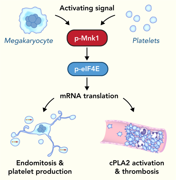
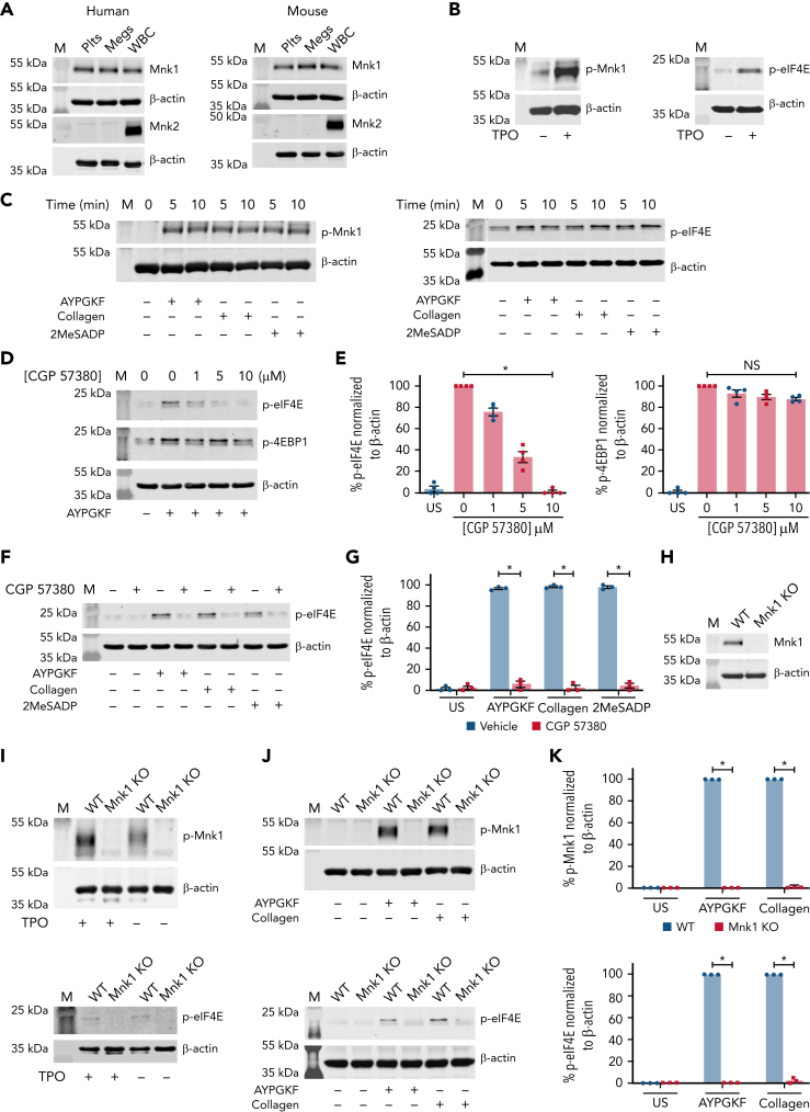
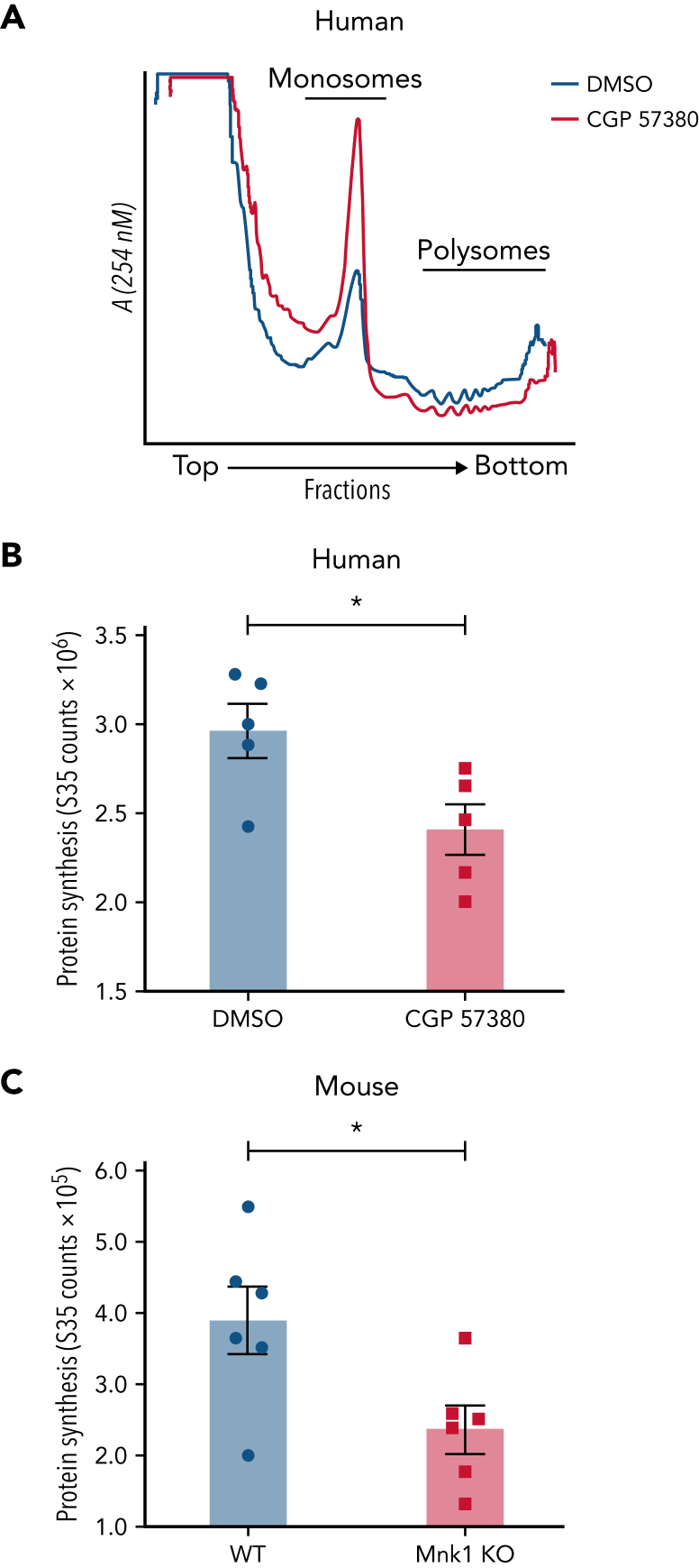
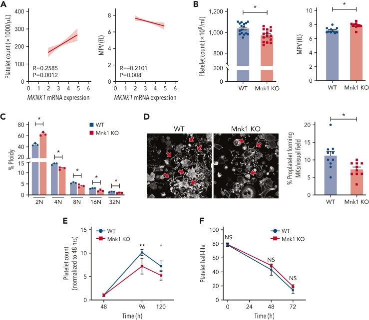
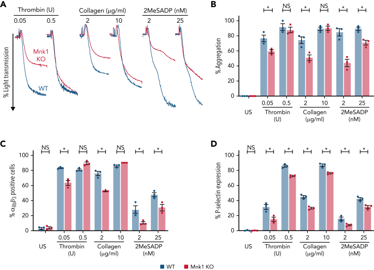
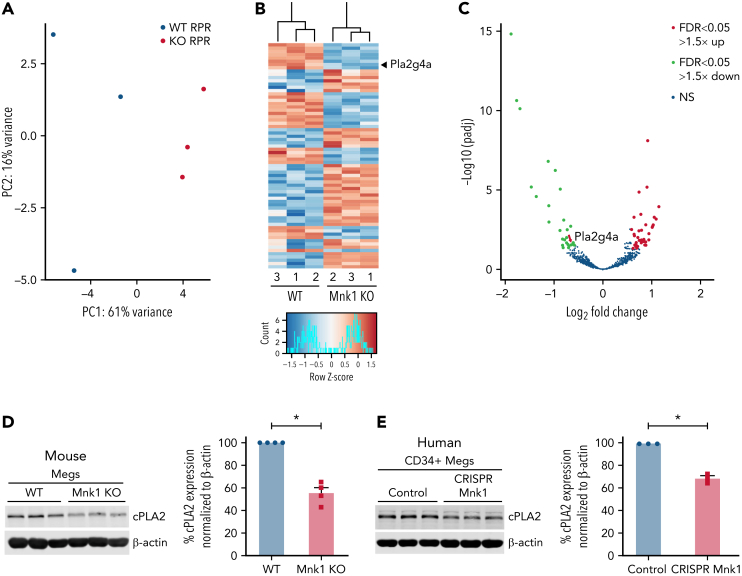
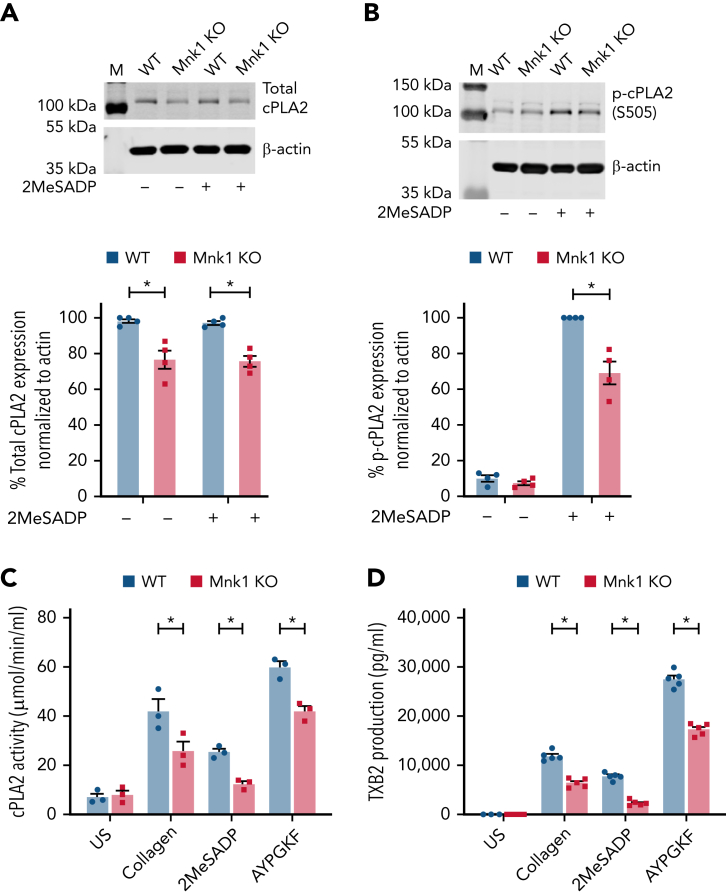
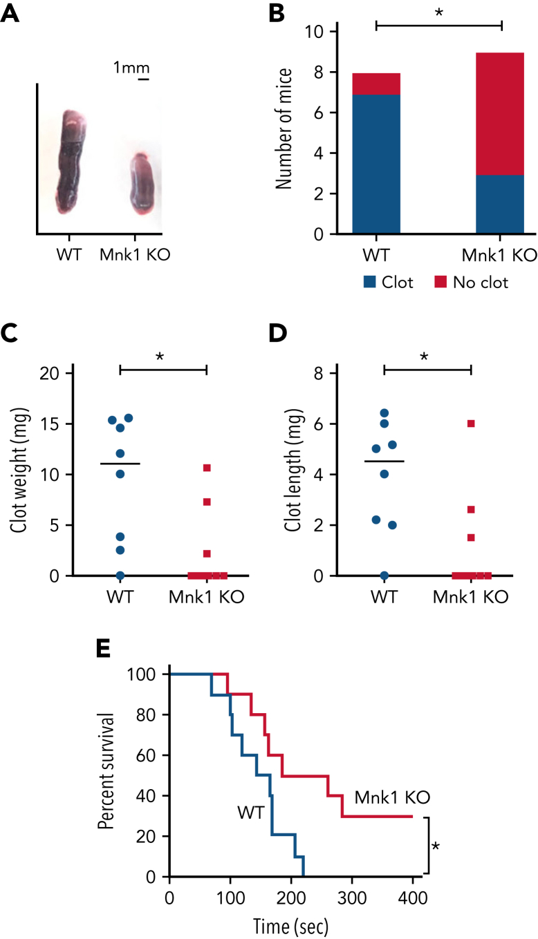
The MAPK-interacting kinase (Mnk) family includes Mnk1 and Mnk2, which are phosphorylated and activated in response to extracellular stimuli. Mnk1 contributes to cellular responses by regulating messenger RNA (mRNA) translation, and mRNA translation influences platelet production and function. However, the role of Mnk1 in megakaryocytes and platelets has not previously been studied. The present study investigated Mnk1 in megakaryocytes and platelets using both pharmacological and genetic approaches. We demonstrate that Mnk1, but not Mnk2, is expressed and active in human and murine megakaryocytes and platelets. Stimulating human and murine megakaryocytes and platelets induced Mnk1 activation and phosphorylation of eIF4E, a downstream target of activated Mnk1 that triggers mRNA translation. Mnk1 inhibition or deletion significantly diminished protein synthesis in megakaryocytes as measured by polysome profiling and [35S]-methionine incorporation assays. Depletion of Mnk1 also reduced megakaryocyte ploidy and proplatelet forming megakaryocytes in vitro and resulted in thrombocytopenia. However, Mnk1 deletion did not affect the half-life of circulating platelets. Platelets from Mnk1 knockout mice exhibited reduced platelet aggregation, α granule secretion, and integrin αIIbβ3 activation. Ribosomal footprint sequencing indicated that Mnk1 regulates the translation of Pla2g4a mRNA (which encodes cPLA2) in megakaryocytes. Consistent with this, Mnk1 ablation reduced cPLA2 activity and thromboxane generation in platelets and megakaryocytes. In vivo, Mnk1 ablation protected against platelet-dependent thromboembolism. These results provide previously unrecognized evidence that Mnk1 regulates mRNA translation and cellular activation in platelets and megakaryocytes, endomitosis and thrombopoiesis, and thrombosis.


Conflict of interest statement

Conflict-of-interest disclosure: M.T.R. and J.W.R. disclose that they are coinventors on a patent using platelet transcriptomics. The remaining authors declare no competing financial interests.

Figures

None
Graphical abstract
Figure 1.
Figure 1.
Megakaryocytes and platelets express Mnk1 protein. (A) Washed platelets (Plts), megakaryocytes (Megs), and white blood cells (as a control) from both healthy human donors and WT C57Bl/6 mice were lysed and analyzed for total Mnk1 and Mnk2 protein expression by western blotting. β-Actin was used as a loading control. Images representative of n ≥ 3 independent experiments. (B) CD34+ megakaryocytes derived and cultured from human cord blood were lysed after stimulation with or without TPO (100 ng/mL) on culture day 13 for 15 minutes. Lysates were analyzed for phosphorylated Mnk1 (p-Mnk1, left) and phosphorylated eIF4E (p-eIF4E, right) by western blotting. β-Actin was used as a loading control. Images representative of n ≥ 3 independent experiments. (C) Washed human platelets were left alone (0 minutes) or stimulated with AYPGKF (100 μM), collagen (5 μg/mL), or 2MeSADP (50 nM) for 5 and 10 minutes. Samples were analyzed for p-Mnk1 (left) and p-EIF4E (right) by western blotting. β-Actin was used as a loading control. Images representative of n ≥ 3 independent experiments. (D-E) Washed human platelets were left unstimulated (US) in DMSO vehicle control or treated with different concentrations of the Mnk1 inhibitor CGP 57380 for 5 minutes. Then, platelets were stimulated with AYPGKF (100 μM, 5 minutes). Cells were then lysed and analyzed for p-eIF4E and p-4EBP1 by western blotting. β-Actin was used as a loading control. Quantitative analysis of p-eIF4E and p-4EBP1 normalized to β-actin are shown in the bar graphs to the right. Western blot images are representative of n ≥ 3 independent experiments. (F-G) Washed human platelets were left US or treated with CGP 57380 (10 μM) for 5 minutes. The cells were then left alone in DMSO (Veh) or stimulated with AYPGKF (100 μM), collagen (5 μg/mL), or 2MeSADP (50 nM) for 5 minutes and probed for p-eIF4E by western blotting. β-Actin was used as a loading control. Quantitative analysis of p-eIF4E normalized to β-actin is shown in the bar graph to the right (∗P < .05). Western blot images are representative of n ≥ 3 independent experiments. (H) The expression of total Mnk1 protein in platelets from either WT or Mnk1 KO mice was analyzed by western blotting. β-Actin was used as a loading control. Western blot images are representative of n ≥ 3 independent experiments. (I) Cultured bone marrow megakaryocytes from either WT or Mnk1 KO mice were left alone or stimulated with TPO (100 μM) for 15 minutes and then analyzed for p-Mnk1 (top) and p-eIF4E (bottom) by western blotting. β-Actin was used as a loading control. Western blot images are representative of n ≥ 3 independent experiments. (J) Washed platelets from either WT or Mnk1 KO mice were left unstimulated or stimulated with AYPGKF (100 μM) or collagen (5 μg/mL) for 5 and 10 minutes. Platelets were analyzed for p-Mnk1 (top) and p-eIF4E (bottom) by western blotting. β-Actin was used as a loading control. Images are representative of n ≥ 3 independent experiments. (K) Quantitative analyses of p-Mnk1 (top) and p-EIF4E (bottom) normalized to β-actin (∗P < .05). M, marker; NS, not significant.
Figure 2.
Figure 2.
Mnk1 regulates mRNA translation and de novo protein synthesis in human and murine megakaryocytes. (A) Human, cord blood–derived, CD34+ megakaryocytes were left alone with vehicle control (DMSO, blue line) or treated with CGP 57380 (10 μM, red line) on culture day 13 and then allowed to adhere on fibrinogen-coated plates for 2 hours. Megakaryocytes were then lysed and sedimented by centrifugation on a 5% to 50% sucrose gradient. Isolated monosome and polysome fractions are indicated. Graphs are representative of n = 3 independent experiments. (B) Human, cord blood–derived, CD34+ megakaryocytes were cultured in the presence of CGP 57380 (10 μM) or vehicle control (DMSO) on culture day 13. Megakaryocytes were then resuspended in [35S]-methionine media and allowed to adhere on fibrinogen-coated plates for 2 hours. Protein synthesis was quantified using a scintillation counter (∗P < .05; n = 5 independent experiments). (C) Bone marrow–derived megakaryocytes from either WT or Mnk1 KO mice were resuspended in [35S]-methionine media and allowed to adhere on fibrinogen-coated plates for 2 hours. Protein synthesis was quantified using a scintillation counter (∗P < .05; n = 6 independent experiments).
Figure 3.
Figure 3.
Mnk1 regulates megakaryocyte endomitosis and platelet production. (A) Pearson correlation analyses of the association between platelet counts (left) and MPV (right) with platelet MKNK1 mRNA expression (n = 154 healthy human donors). Light gray lines represent 95% confidence intervals. (B) Platelet counts (left) and mean platelet volume (right) in WT or Mnk1 KO mice (n > 10 mice per group, ∗P < .05). (C) Bone marrow–derived megakaryocytes from WT or Mnk1 KO mice were collected at culture day 5, and ploidy distribution of megakaryocytes was quantified (n = 3 independent experiments), ∗PANOVA < .05). (D) Bone marrow–derived megakaryocytes from WT or Mnk1 KO mice were collected at culture day 5. Megakaryocytes were then allowed to adhere to fibrinogen-coated plates and incubated overnight at 37°C. Megakaryocytes were then stained with phalloidin, and proplatelet formation was assessed by confocal microscopy. The right bar graph shows the number of proplatelet forming megakaryocytes (∗P < .05, n = 3 independent experiments). (E-F) Platelets were depleted from WT or Mnk1 KO mice by a single IV injection of an anti-GPIbα antibody (Emfret Analytics), leading to near-complete platelet clearance by 48 hours. Platelet counts were measured over 120 hours (eg, 5 days) by Hemavet. The (E) percentage of platelet count recovery was calculated from the platelet nadir at 48 hours, and (F) platelet clearance was calculated from the time of injection of the anti-GPIbα antibody (n = 5 mice per group, ∗P < .05; ∗∗P < .01). ANOVA, analysis of variance; NS, not significant.
Figure 4.
Figure 4.
Mnk1 deficiency reduces platelet activation. (A-B) Washed platelets from WT and Mnk1 KO mice were left unstimulated (US) or were stimulated with thrombin (0.05 and 0.5 U), collagen (2 and 10 μg/mL), and 2MeSADP (2 and 25 nM). The curves are representative of n = 3 independent experiments. The bar graphs on the right show quantitation of platelet aggregation (n = 3 mice per group; ∗P < .05). (C-D) Flow cytometric analyses of JON/A binding (for activated integrin αIIbβ3) or P-selectin surface expression on washed platelets from WT and Mnk1 KO mice following stimulation with thrombin (0.05 and 0.5 U), collagen (2 and 10 μg/mL), and 2MeSADP (2 and 25 nM). Graphs show data from n = 3 mice per group (∗P < .05). NS, not significant.
Figure 5.
Figure 5.
Mnk1 regulates the translation of mRNAs, including cPLA2, in megakaryocytes. Bone marrow megakaryocytes were isolated from WT (n = 3) or Mnk1 KO (n = 3) mice and cultured for 5 days. Ribosomal footprint profiling was performed to identify RNAs with ≥1 ribosomes attached (RPRs), suggestive of mRNAs being actively translated. (A-B) Principal component analysis and heat map showing mRNAs with differentially abundant RPRs. The gene Pla2g4a is enlarged and highlighted with a black arrowhead. (C) Volcano plot showing significantly (false discovery rate < 0.05) upregulated (log2 fold change > 1.5, red) and downregulated (log2 fold change < 1.5, green) RPRs in megakaryocytes from WT or Mnk1 KO mice. Blue circles represent RPRs that were not significantly changed in megakaryocytes between WT and Mnk1 KO mice. The gene Pla2g4a is enlarged and highlighted with a red arrow. (D) Bone marrow–derived megakaryocytes from WT and Mnk1 KO mice analyzed for cPLA2 protein expression. β-Actin was used as a loading control. Bar graph shows quantification of total cPLA2 protein in Mnk1 KO megakaryocytes compared with WT megakaryocytes as assessed by densitometry (n = 4 per group, ∗P < .05). (E) Immunoblot of cPLA2 protein after Mnk1 CRISPR-Cas9 (CRISPR Mnk1)-based knockdown in day 13 human CD34+-derived cultured megakaryocytes. Guide RNAs not targeting known genes were used as a negative control (Control). Bar graph shows quantification of total cPLA2 protein expression as assessed by densitometry (n = 3 per group, ∗P < .05). NS, not significant.
Figure 6.
Figure 6.
Mnk1 regulates cPLA2 activity and thromboxane production by platelets. (A-B) Total (A) and phosphorylated (B) cPLA2 protein expression was assessed by immunoblot in unstimulated or 2MeSADP-stimulated (10 nM, 5 minutes) platelets from WT and Mnk1 KO mice. β-Actin was used as a loading control. Immunoblots are representative of n = 3 independent experiments. (C-D) Washed platelets from WT and Mnk1 KO mice were left unstimulated (US) or stimulated with collagen (2 μg/ml), 2MeSADP (2 nM), or AYPGKF (150 μM) 10 minutes. Then, platelet cPLA2 activity (C) and TXB2 production (D) were assessed (n = 3-5 mice per group, ∗P < .05). M, marker.
Figure 7.
Figure 7.
Mnk1 regulates thrombus formation in vitro and in vivo. (A-D) WT (n = 8) or Mnk1 KO (n = 9) mice were subject to a venous stasis model of thrombosis. (A) Representative clots from WT or Mnk1 KO mice. (B) Number of mice in each group with thrombosis (∗P < .05). (C-D) Clots were removed, measured, and weighed (∗P < .05). (E) Survival proportions in WT and Mnk1 KO mice after induction of pulmonary thromboembolism by injecting a collagen/epinephrine mixture through retro-orbital injection (n = 10 mice per group, ∗P < .05).

Comment in

Similar articles

Cited by

References

    1. Machlus KR, Italiano JE., Jr. The incredible journey: from megakaryocyte development to platelet formation. J Cell Biol. 2013;201(6):785–796. - PMC - PubMed
    1. Hartwig J, Italiano J., Jr. The birth of the platelet. J Thromb Haemost. 2003;1(7):1580–1586. - PubMed
    1. Hartwig JH, Italiano JE., Jr. Cytoskeletal mechanisms for platelet production. Blood Cells Mol Dis. 2006;36(2):99–103. - PubMed
    1. Thon JN, Italiano JE. Platelet formation. Semin Hematol. 2010;47(3):220–226. - PMC - PubMed
    1. Hvas AM. Platelet function in thrombosis and hemostasis. Semin Thromb Hemost. 2016;42(3):183–184. - PubMed

Publication types