. 2022 Sep:154:405-420.
doi: 10.1016/j.cortex.2022.02.018. Epub 2022 Jun 28.

Enhanced positive emotional reactivity in frontotemporal dementia reflects left-lateralized atrophy in the temporal and frontal lobes

Affiliations

Enhanced positive emotional reactivity in frontotemporal dementia reflects left-lateralized atrophy in the temporal and frontal lobes

Suzanne M Shdo et al. Cortex. 2022 Sep.

Abstract

In frontotemporal dementia (FTD), left-lateralized atrophy patterns have been associated with elevations in certain positive emotions. Here, we investigated whether positive emotional reactivity was enhanced in semantic variant primary progressive aphasia (svPPA), an FTD syndrome that targets the left anterior temporal lobe. Sixty-one participants (16 people with svPPA, 24 people with behavioral variant FTD, and 21 healthy controls) viewed six 90-sec trials that were comprised of a series of photographs; each trial was designed to elicit a specific positive emotion, negative emotion, or no emotion. Participants rated their positive emotional experience after each trial, and their smiling behavior was coded with the Facial Action Coding System. Results indicated that positive emotional experience and smiling were elevated in svPPA in response to numerous affective and non-affective stimuli. Voxel-based morphometry analyses revealed that greater positive emotional experience and greater smiling in the patients were both associated with smaller gray matter volume in the left superior temporal gyrus (pFWE < .05), among other left-lateralized frontotemporal regions. Whereas enhanced positive emotional experience related to atrophy in middle superior temporal gyrus and structures that promote cognitive control and emotion regulation, heightened smiling related to atrophy in posterior superior temporal gyrus and structures that support motor control. Our results suggest positive emotional reactivity is elevated in svPPA and offer new evidence that atrophy in left-lateralized emotion-relevant systems relates to enhanced positive emotions in FTD.

Keywords: Elation; Euphoria; Mood; Neurodegeneration; Semantic dementia.


Conflict of interest statement

Declaration of competing interest The authors report no competing interests.

Figures

Figure 1.
Figure 1.. Mean positive emotional experience across trials in each diagnostic group
*= p<.05, **=p<.001. Separate ANCOVAs (controlling for age and sex) were run for each trial to decompose the significant diagnosis by trial interaction that was detected in our primary analysis on total positive emotional experience. Tukey-adjusted pairwise comparisons were conducted to correct for multiple comparisons. Error bars represent the standard error. These analyses revealed that people with svPPA reported significantly higher levels of total positive emotional experience during the awe (p<.001), love (p<.05), and neutral (p<.05) trials than the bvFTD and healthy control groups. People with svPPA also reported significantly higher levels of positive emotional experience during the fear trial than healthy controls (p<.05).
Figure 2.
Figure 2.. Mean smiling behavior across trials in each diagnostic group.
*=p<.05. Separate ANCOVAs (controlling for age and sex) were run for each trial to decompose the significant diagnosis by trial interaction that was detected in our primary analysis on smiling behavior. Tukey-adjusted pairwise comparisons were used to correct for multiple comparisons. People with svPPA had a higher total smiling score during the fear (p<.05) and sadness (p<.05) trials than those with bvFTD. People with bvFTD had a greater total smiling score during the amusement trial than the healthy controls (p<.05).
Figure 3.
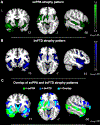
Figure 3.. Distinct and overlapping atrophy patterns in svPPA and bvFTD.
Whole-brain voxel-based morphometry analyses (controlling for age, sex, and TIV) confirmed that the A) svPPA and (B) bvFTD groups had smaller gray matter volume than the healthy controls in frontotemporal and subcortical regions typically atrophied in these syndromes (pFWE<.05). (C) In svPPA and bvFTD, there was some overlapping atrophy, but the people with svPPA tended to have more left lateralized atrophy than those with bvFTD, whose atrophy was more bilateral (pFWE<.05). Statistical maps are superimposed on the Montreal Neurological Institute template.
Figure 4.
Figure 4.. Left-lateralized atrophy related to greater total positive emotional experience and greater total smiling behavior.
Voxel-based morphometry analyses (controlling for age, sex, diagnosis, MMSE, and TIV) revealed that (A) smaller gray matter volume in left superior temporal gyrus was associated with greater total positive emotional experience (pFWE<.05) and greater total smiling behavior (pFWE<.05). (B) Greater total positive emotional experience was associated with smaller gray matter volume in the left superior temporal gyrus, left dorsal anterior insula, and left orbitofrontal cortex (p<.001), and (C) greater total smiling behavior was associated with smaller gray matter volume in the left temporal lobe and middle frontal gyrus. Color bars represent T-scores at p<.001. Statistical maps are superimposed on the Montreal Neurological Institute template.
Figure 5.
Figure 5.. Linear association between total positive emotional experience and gray matter volume across the clinical groups.
A regression model (controlling for age, sex, diagnosis, TIV, and MMSE) confirmed that total positive emotional experience had a linear association across the clinical groups with gray matter volume in the left superior temporal gyrus and left dorsal anterior insula clusters that emerged in the VBM analyses. These results suggest the neuroimaging results reflected linear brain-emotion associations and were not driven by one diagnostic group.

Similar articles

Cited by

References

    1. Adolphs R (2003). Cognitive neuroscience: Cognitive neuroscience of human social behaviour. Nature Reviews Neuroscience, 4(3), 165–178. 10.1038/nrn1056 - DOI - PubMed
    1. Agosta F, Galantucci S, Canu E, Cappa SF, Magnani G, Franceschi M, Falini A, Comi G, & Filippi M (2013). Disruption of structural connectivity along the dorsal and ventral language pathways in patients with nonfluent and semantic variant primary progressive aphasia: A DT MRI study and a literature review. Brain and Language, 127(2), 157–166. 10.1016/j.bandl.2013.06.003 - DOI - PubMed
    1. Aron AR, Robbins TW, & Poldrack RA (2004). Inhibition and the right inferior frontal cortex. In Trends in Cognitive Sciences (Vol. 8, Issue 4, pp. 170–177). 10.1016/j.tics.2004.02.010 - DOI - PubMed
    1. Balota DA, Yap MJ, Cortese MJ, Hutchison KA, Kessler B, Loftis B, Neely JH, Nelson DL, Simpson GB, & Treiman R (2007). The english lexicon project. In Behavior Research Methods (Vol. 39, Issue 3, pp. 445–459). Springer; New York LLC. 10.3758/BF03193014 - DOI - PubMed
    1. Beauchamp MS, Lee KE, Argall BD, & Martin A (2004). Integration of auditory and visual information about objects in superior temporal sulcus. Neuron, 41(5), 809–823. 10.1016/S0896-6273(04)00070-4 - DOI - PubMed

Publication types