Review
. 2022 Oct:90:78-89.
doi: 10.1016/j.clinimag.2022.07.009. Epub 2022 Jul 29.

Advanced cardiac imaging in the spectrum of COVID-19 related cardiovascular involvement

Affiliations
Review

Advanced cardiac imaging in the spectrum of COVID-19 related cardiovascular involvement

Anna Palmisano et al. Clin Imaging. 2022 Oct.

Abstract

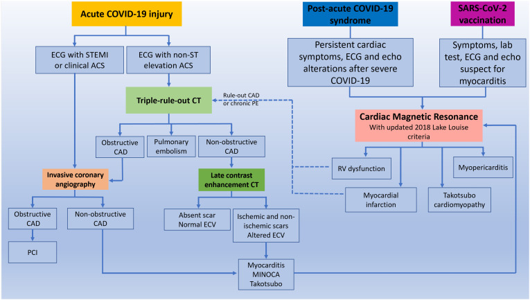
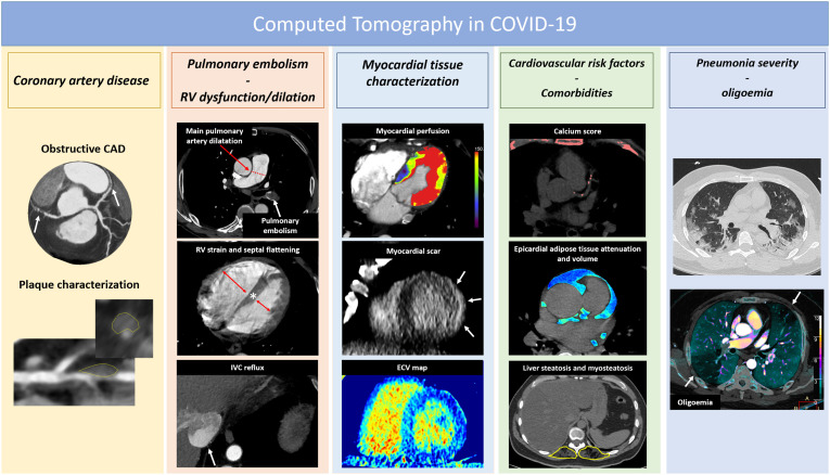
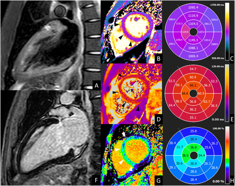
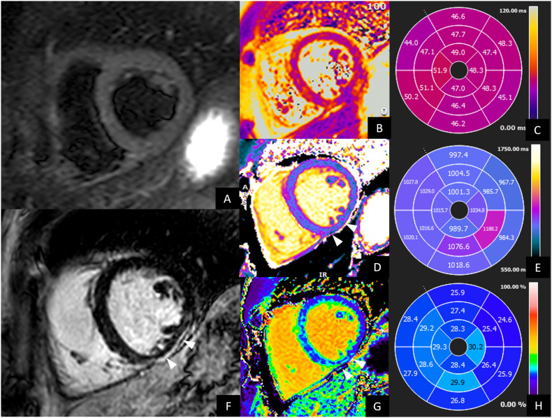
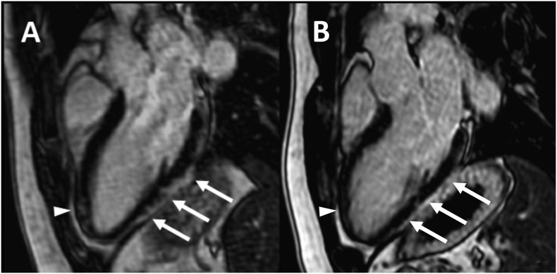
Cardiovascular involvement is a common complication of COVID-19 infection and is associated to increased risk of unfavorable outcome. Advanced imaging modalities (coronary CT angiography and Cardiac Magnetic Resonance) play a crucial role in the diagnosis, follow-up and risk stratification of patients affected by COVID-19 pneumonia with suspected cardiovascular involvement. In the present manuscript we firstly review current knowledge on the mechanisms by which SARS-CoV-2 can trigger endothelial and myocardial damage. Secondly, the implications of the cardiovascular damage on patient's prognosis are presented. Finally, we provide an overview of the main findings at advanced cardiac imaging characterizing COVID-19 in the acute setting, in the post-acute syndrome, and after vaccination, emphasizing the potentiality of CT and CMR, the indication and their clinical implications.

Keywords: COVID-19; Cardiac Magnetic Resonance; Coronary CT angiography; Myocarditis; Pulmonary embolism; Vaccine.


Figures

Fig. 1
Fig. 1
Role of CT and CMR in the diagnostic algorithm of COVID-19 related cardiac complication. After clinical evaluation, patients with chest pain, ST elevation ACS, high pretest probability of CAD and high risk of mortality should be referred to emergent invasive coronary angiography (ICA). Patients with NSTEMI, atypical symptoms and ECG abnormality should be referred to CT. A triple rule-out protocol should be preferred for the simultaneous exclusion of pulmonary embolism (PE) and coronary artery disease (CAD). Patients with obstructive CAD should be referred to ICA for percutaneous intervention (PCI), while patients with non-obstructive CAD to tissue characterization. This could be obtained directly from CT, and in presence of scar and ECV alteration according to multidisciplinary evaluation a diagnostic confirmation with CMR can be performed. In patients with suspected long COVID-19 syndrome and post-vaccination symptoms, CMR is the first level examination. CT can have a role subsequently in order to exclude chronic PE or obstructive CAD in patients with long COVID-19 syndrome.
Fig. 2
Fig. 2
The spectrum of CT potentialities in the setting of COVID-19.
Fig. 3
Fig. 3
CMR of acute left ventricle dysfunction during COVID-19. A 39-year-old male presented to the emergency department for fever, caught and dyspnea. Nasopharyngeal swab was positive for SARS-CoV 2 infection. Laboratory tests showed increased troponin T level (42,6 ng/mL, normal value <14 ng/mL) and a moderate depression of left ventricle systolic function (ejection fraction <40%) was documented at echocardiography. CMR was performed 8 days later and showed a slight diffuse hypokinesia of left ventricle (LV ejection fraction 51%) with absent focal edema on short-tau inversion recovery images (A) and absent LGE (B), but diffuse alteration of T2 values (B) (56 ms, normal value ≤ 50 ms; C), of native T1 (D) (1084 ms, normal value ≤ 1045 ms E) and of extracellular volume fraction (G) (28%, normal value ≤ 27%; H) with higher values in mid-apical septum and mid-apical anterior wall (arrows in B, D and G). These findings were suggestive for acute myocarditis according to 2018 Lake Louise criteria. Endomyocardial biopsy confirmed these findings, showing diffuse edema and macrophage infiltrate. After 1 month, the patient was discharged with complete resolution of cardiac alteration.
Fig. 4
Fig. 4
CMR of a 33-year-old male with persistent palpitation and tachycardia especially during physical activity at 1 year after COVID-19 recovery. Holter ECG documented frequent ectopic ventricular beats. Hence, CMR was performed. CMR showed preserved left and right ventricle ejection fraction, without wall motion alteration. No edema was evident on short-tau inversion recovery images (A) neither on T2 maps (B and C). LGE images (F) showed a thin subepicardial scar on the inferior mid-ventricular wall, associated to increased native T1 (arrows in D, values in E) and ECV values (arrows in H, values in G). These findings were suggestive for post-myocarditis scar.
Fig. 5
Fig. 5
CMR of a 30-year-old male with COVID-19 vaccine-related myocarditis. LGE imaging performed along three-chambers view 5 days after the onset of patient's symptoms (A) shows subepicardial enhancement along the infero-lateral myocardial segments (arrows) with minimal involvement of the anterior wall in the apical region (arrowhead). Cardiac MRI performed 3-months later (B) shows almost complete resolution of myocardial LGE in the same segments.

References

    1. Esposito A., Palmisano A., Cao R., Rancoita P., Landoni G., Grippaldi D., et al. Quantitative assessment of lung involvement on chest CT at admission: impact on hypoxia and outcome in COVID-19 patients. Clin Imaging. 2021;77:194–201. doi: 10.1016/J.CLINIMAG.2021.04.033. - DOI - PMC - PubMed
    1. Giustino G., Croft L.B., Stefanini G.G., Bragato R., Silbiger J.J., Vicenzi M., et al. Characterization of myocardial injury in patients with COVID-19. J Am Coll Cardiol. 2020;76:2043–2055. doi: 10.1016/J.JACC.2020.08.069. - DOI - PMC - PubMed
    1. Chung M.K., Zidar D.A., Bristow M.R., Cameron S.J., Chan T., Harding C.V., et al. COVID-19 and cardiovascular disease: from bench to bedside. Circ Res. 2021;128:1214–1236. doi: 10.1161/CIRCRESAHA.121.317997. - DOI - PMC - PubMed
    1. Bansal M. Cardiovascular disease and COVID-19. Diabetes Metab Syndr Clin Res Rev. 2020;14:247–250. doi: 10.1016/J.DSX.2020.03.013. - DOI - PMC - PubMed
    1. Lala A., Johnson K.W., Januzzi J.L., Russak A.J., Paranjpe I., Richter F., et al. Prevalence and impact of myocardial injury in patients hospitalized with COVID-19 infection. J Am Coll Cardiol. 2020;76:533–546. doi: 10.1016/J.JACC.2020.06.007. - DOI - PMC - PubMed