

High endothelial venules as potential gateways for therapeutics
- PMID: 35931612
- PMCID: PMC10804419
- DOI: 10.1016/j.it.2022.07.002
High endothelial venules as potential gateways for therapeutics
Abstract
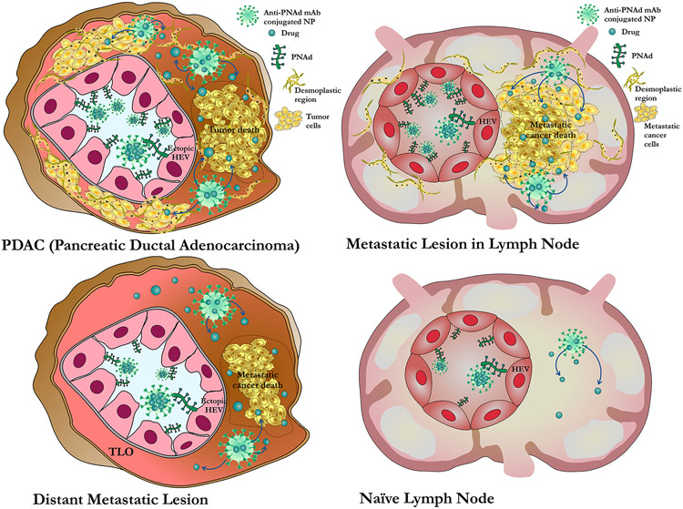
High endothelial venules (HEVs) are specialized blood vessels that support the migration of lymphocytes from the bloodstream into lymph nodes (LNs). They are also formed ectopically in mammalian organs affected by chronic inflammation and cancer. The recent arrival of immunotherapy at the forefront of many cancer treatment regimens could boost a crucial role for HEVs as gateways for the treatment of cancer. In this review, we describe the microanatomical and biochemical characteristics of HEVs, mechanisms of formation of newly made HEVs, immunotherapies potentially dependent on HEV-mediated T cell homing to tumors, and finally, how HEV-targeted therapies might be used as a complementary approach to potentially shape the therapeutic landscape for the treatment of cancer and immune-mediated diseases.
Keywords: cancer immunology; high endothelial venule; lymph node; nanomedicine; nanoparticles; transplantation.
Copyright © 2022. Published by Elsevier Ltd.
Conflict of interest statement
Declaration of interests The authors have no interests to declare.
Figures

References
-
- Singh SK and Singh R (2022) Nanotherapy: targeting the tumour microenvironment. Nat Rev Cancer 22 (5), 258. - PubMed
-
- Drayton DL et al. (2006) Lymphoid organ development: from ontogeny to neogenesis. Nat Immunol 7 (4), 344–53. - PubMed
-
- Ager A. (1998) High Endothelial Venules. In Encyclopedia of Immunology (Second Edition) (Delves PJ ed), pp. 1093–1101, Elsevier.
Publication types
MeSH terms
Grants and funding
LinkOut - more resources
Full Text Sources
Medical
