

Differential diagnosis and prognosis of small renal masses: association with collateral vessels detected using contrast-enhanced computed tomography
- PMID: 35932010
- PMCID: PMC9354334
- DOI: 10.1186/s12885-022-09971-w
Differential diagnosis and prognosis of small renal masses: association with collateral vessels detected using contrast-enhanced computed tomography
Abstract
Background: Active surveillance (AS) is one of the treatment methods for patients with small renal masses (SRMs; < 4 cm), including renal cell carcinomas (RCCs). However, some small RCCs may exhibit aggressive neoplastic behaviors and metastasize. Little is known about imaging biomarkers capable of identifying potentially aggressive small RCCs. Contrast-enhanced computed tomography (CECT) often detects collateral vessels arising from neoplastic angiogenesis in RCCs. Therefore, this study aimed to evaluate the association between SRM differential diagnoses and prognoses, and the detection of collateral vessels using CECT.
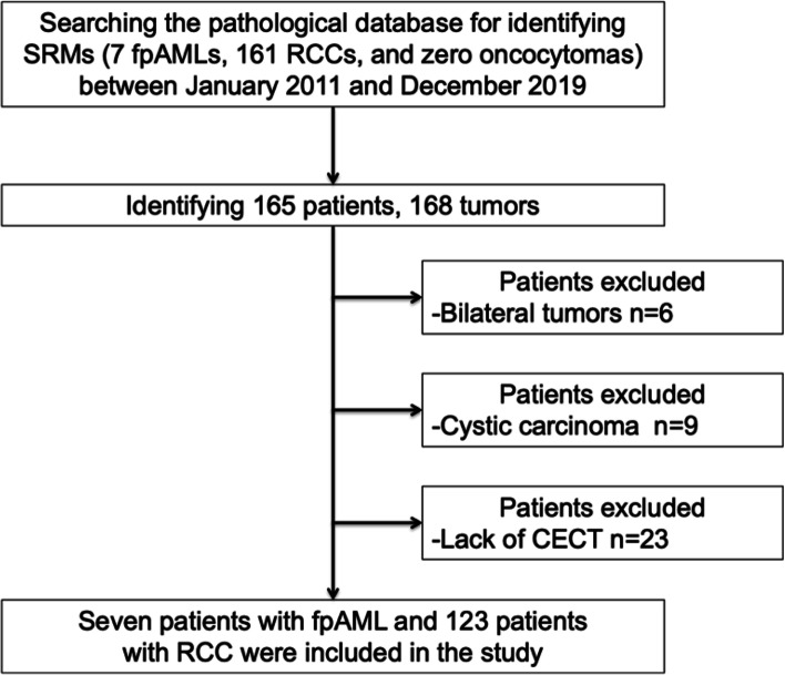
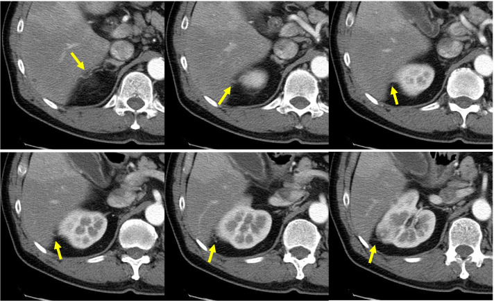
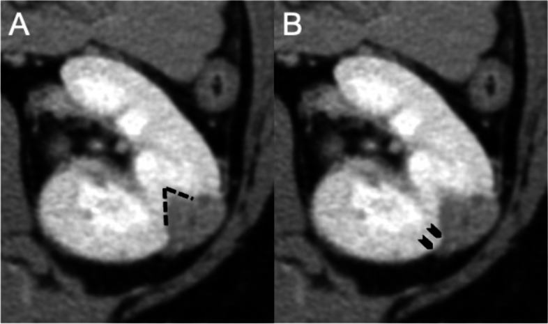
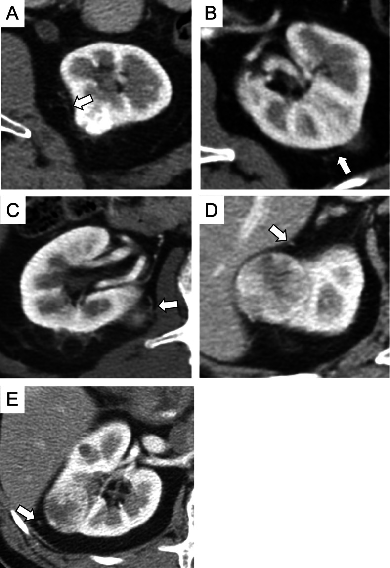
Methods: A total of 130 consecutive patients with pathologically confirmed non-metastatic SRMs (fat-poor angiomyolipomas [fpAMLs; n = 7] and RCCs [n = 123]) were retrospectively enrolled. Between 2011 and 2019, SRM diagnoses in these patients were confirmed after biopsy or surgical resection. All RCCs were surgically resected. Regardless of diameter, a collateral vessel (CV) was defined as any blood vessel connecting the tumor from around the kidney using CECT. First, we analyzed the role of CV-detection in differentiating between fpAML and RCC. Then, we evaluated the sensitivity, specificity, positive predictive value (PPV), negative predictive value (NPV), and accuracy of RCC diagnosis based on CV-detection using CECT. We also assessed the prognostic value of CV-detection using the Fisher exact test, and Kaplan-Meier method and the log-rank test.
Results: The sensitivity, specificity, PPV, NPV, and accuracy of CV-detection for the diagnosis of small RCCs was 48.5, 45.5, 100, 100, and 9.5% respectively. Five of 123 (4.1%) patients with RCC experienced recurrence. CV-detection using CECT was the only significant factor associated with recurrence (p = 0.0177). Recurrence-free survival (RFS) was significantly lower in patients with CV compared with in those without CV (5-year RFS 92.4% versus 100%, respectively; p = 0.005). In addition, critical review of the CT images revealed the CVs to be continuous with the venous vessels around the kidney.
Conclusions: The detection of CVs using CECT is useful for differentiating between small fpAMLs and RCCs. CV-detection may also be applied as a predictive parameter for small RCCs prone to recurrence after surgical resection. Moreover, AS could be suitable for small RCCs without CVs.
Keywords: Collateral vessel; Contrast-enhanced computed tomography; Diagnostic accuracy; Fat-poor angiomyolipoma; Renal cell carcinoma; Small renal masses.
© 2022. The Author(s).
Conflict of interest statement
The authors declare that they have no competing interests.
Figures

Similar articles
-
Histogram analysis of small solid renal masses: differentiating minimal fat angiomyolipoma from renal cell carcinoma.AJR Am J Roentgenol. 2012 Feb;198(2):377-83. doi: 10.2214/AJR.11.6887. AJR Am J Roentgenol. 2012. PMID: 22268181
-
Contrast-enhanced ultrasound for differentiating benign from malignant solid small renal masses: comparison with contrast-enhanced CT.Abdom Radiol (NY). 2017 Aug;42(8):2135-2145. doi: 10.1007/s00261-017-1111-x. Abdom Radiol (NY). 2017. PMID: 28331942
-
Morphologic analysis with computed tomography may help differentiate fat-poor angiomyolipoma from renal cell carcinoma: a retrospective study with 602 patients.Abdom Radiol (NY). 2018 Mar;43(3):647-654. doi: 10.1007/s00261-017-1244-y. Abdom Radiol (NY). 2018. PMID: 28677004
-
CT radiomics for differentiating fat poor angiomyolipoma from clear cell renal cell carcinoma: Systematic review and meta-analysis.PLoS One. 2023 Jul 27;18(7):e0287299. doi: 10.1371/journal.pone.0287299. eCollection 2023. PLoS One. 2023. PMID: 37498830 Free PMC article.
-
[Epidemiology and diagnostic assessment of small renal masses].Urologe A. 2018 Mar;57(3):274-279. doi: 10.1007/s00120-018-0585-7. Urologe A. 2018. PMID: 29460170 Review. German.
Cited by
-
CT features based preoperative predictors of aggressive pathology for clinical T1 solid renal cell carcinoma and the development of nomogram model.BMC Cancer. 2024 Jan 30;24(1):148. doi: 10.1186/s12885-024-11870-1. BMC Cancer. 2024. PMID: 38291357 Free PMC article.
-
Role of collateral vessels on contrast-enhanced computed tomography in predicting metastatic potential for small renal cell carcinoma.Discov Oncol. 2024 Oct 4;15(1):523. doi: 10.1007/s12672-024-01409-y. Discov Oncol. 2024. PMID: 39365374 Free PMC article.
-
Recent nanotheranostic approaches in cancer research.Clin Exp Med. 2024 Jan 19;24(1):8. doi: 10.1007/s10238-023-01262-3. Clin Exp Med. 2024. PMID: 38240834 Free PMC article. Review.
-
Clinical Application Value of Contrast-Enhanced Ultrasound in the Diagnosis of Renal Space-Occupying Lesions.Int J Nephrol Renovasc Dis. 2023 Dec 5;16:253-259. doi: 10.2147/IJNRD.S432436. eCollection 2023. Int J Nephrol Renovasc Dis. 2023. PMID: 38075192 Free PMC article.
References
-
- Fujii Y, Komai Y, Saito K, Iimura Y, Yonese J, Kawakami S, et al. Incidence of benign pathologic lesions at partial nephrectomy for presumed RCC renal masses: Japanese dual-center experience with 176 consecutive patients. Urology. 2008;72(3):598–602. doi: 10.1016/j.urology.2008.04.054. - DOI - PubMed
MeSH terms
Substances
LinkOut - more resources
Full Text Sources
Medical
Research Materials
