. 2022 Nov;28(11):1718-1732.
doi: 10.1111/cns.13907. Epub 2022 Aug 6.

Treatment with FRAX486 rescues neurobehavioral and metabolic alterations in a female mouse model of CDKL5 deficiency disorder

Affiliations

Treatment with FRAX486 rescues neurobehavioral and metabolic alterations in a female mouse model of CDKL5 deficiency disorder

Claudia Fuchs et al. CNS Neurosci Ther. 2022 Nov.

Abstract

Introduction: CDKL5 deficiency disorder (CDD) is a rare neurodevelopmental condition, primarily affecting girls for which no cure currently exists. Neuronal morphogenesis and plasticity impairments as well as metabolic dysfunctions occur in CDD patients. The present study explored the potential therapeutic value for CDD of FRAX486, a brain-penetrant molecule that was reported to selectively inhibit group I p21-activated kinases (PAKs), serine/threonine kinases critically involved in the regulation of neuronal morphology and glucose homeostasis.

Methods: The effects of treatment with FRAX486 on CDD-related alterations were assessed in vitro (100 nM for 48 h) on primary hippocampal cultures from Cdkl5-knockout male mice (Cdkl5-KO) and in vivo (20 mg/Kg, s.c. for 5 days) on Cdkl5-KO heterozygous females (Cdkl5-Het).

Results: The in vitro treatment with FRAX486 completely rescued the abnormal neuronal maturation and the number of PSD95-positive puncta in Cdkl5-KO mouse neurons. In vivo, FRAX486 normalized the general health status, the hyperactive profile and the fear learning defects of fully symptomatic Cdkl5-Het mice. Systemically, FRAX486 treatment normalized the levels of reactive oxidizing species in the whole blood and the fasting-induced hypoglycemia displayed by Cdkl5-Het mice. In the hippocampus of Cdkl5-Het mice, treatment with FRAX486 rescued spine maturation and PSD95 expression and restored the abnormal PAKs phosphorylation at sites which are critical for their activation (P-PAK-Ser144/141/139) or for the control cytoskeleton remodeling (P-PAK1-Thr212).

Conclusions: Present results provide evidence that PAKs may represent innovative therapeutic targets for CDD.

Keywords: CDKL5 deficiency disorder; animal model; behavior; cytoskeleton; therapeutic approach; transfection.


Conflict of interest statement

The authors declare no conflict of interest.

Figures

FIGURE 1
FIGURE 1
Treatment with FRAX486 normalizes neurite outgrowth in an in vitro model of CDD. Primary hippocampal neurons were prepared from 1‐day‐old (P1) wild‐type (wt) and Cdkl5 knockout (Cdkl5‐KO) mice. On the 7th day, in vitro (DIV7) differentiated hippocampal cultures were treated with 100 nM FRAX486 or control solution (ctrl) and fixed 48 h later (DIV9) for immunofluorescence analysis. (A) Quantification of neurite outgrowth of microtubule‐associated protein 2 (MAP2)‐positive (1:100; Merck Millipore) in hippocampal neurons was done using the image analysis system Image‐Pro Plus. FRAX486 treatment rescued the abnormal dendritic development in Cdkl5‐KO neurons. (B) The degree of synaptic innervation was evaluated by counting the number of PSD95+ puncta (1:200; Abcam) on proximal dendrites and expressed as the number of PSD95+ puncta per 20 μm of length. FRAX486 treatment rescued the reduced density of PSD95+ puncta in Cdkl5‐KO neurons. (C and D) Representative fluorescence images of differentiated MAP2‐positive hippocampal neurons (C; scale bar: 10 μm) and proximal dendrites immunopositive for PSD95 (D; scale bar: 1 μm) of wt and Cdkl5‐KO mice. (E) Quantification of dendritic branching represented as the total number of intersections. FRAX486 normalized the number of intersections in Cdkl5‐KO neurons. Statistical significance was assessed using one‐way ANOVA (main effect of experimental group) followed by Dunnett's post hoc test (setting Cdkl5‐KO, ctrl as control group). Data are mean ± SEM. **p < 0.01, ***p < 0.001; N = 3–4 per group
FIGURE 2
FIGURE 2
FRAX486 treatment improves behavioral abnormalities, general health status and oxidative stress levels in the blood of Cdkl5‐Het female mice at an advanced stage of the disease, a validated CDD mouse model. Cdkl5 heterozygous (Cdkl5‐Het) female mice received subcutaneous (s.c) injections with FRAX486 (20 mg/kg, daily) or control (ctrl) solution for 5 days. Wild‐type (wt) littermates were treated only with ctrl. A battery of tests was performed to assess drug effects on behavior and whole blood was collected to measure the levels of reactive oxidizing species (ROS). (A) The general health status was qualitatively evaluated by a trained observer blind to mouse genotype and treatment. Briefly, mice received a score (ranging from 0 = normal appearance to 4 = highly compromised) for each of the following symptoms: gait, mobility, breathing, kyphosis, fur, hindlimb clasping, tremors, seizures occurence and general condition. The individual scores for each category were subsequently averaged to obtain a semiquantitative measure of symptom status, called general health score (measured in average units, AU). FRAX486 treatment normalized the general health status of Cdkl5‐Het female mice. (B) Time spent freezing during the baseline (BL) and the testing phase (Test) of the fear conditioning task was automatically monitored (Any Maze v.6.3, Stoelting). FRAX486 treatment improved the fear memory of Cdkl5‐Het female mice. (C) Distance traveled during a 15‐min‐long open field task was measured by the means of a tracking software (Any Maze v.6.3, Stoelting) and considered as a measure of locomotor activity in a novel environment. FRAX486 treatment rescued the hyperactive profile of Cdkl5‐Het female mice. (D) Nitroxyl 3‐carboxy‐proxyl radical (CP) intensity was measured in whole blood by electron paramagnetic resonance (EPR) and considered as an index of ROS production; data are normalized to wt, ctrl values. FRAX486 treatment rescued the pro‐oxidant status that occurs in the blood of Cdkl5‐Het mice. Statistical significance was assessed using one‐way ANOVA (main effect of experimental group) followed by Dunnett's post hoc test (setting Cdkl5‐Het, ctrl as control group). Repeated measures ANOVA (setting experimental group as between factor and repeated measurements as within factor) followed by Tukey's post hoc test was also performed. In case of deviation from normality and homoschedasticity, data were analyzed by means of nonparametric Kruskal–Wallis test followed by Dunn's post hoc test. Bar plots represent mean ± SEM (B, C) while boxplots represent the 10th, 25th, 50th (median), 75th, and 90th percentiles (A, D). *p < 0.05, **p < 0.01, ***p < 0.001; N = 12–15 per group (A–C) or N = 6–13 per group (D)
FIGURE 3
FIGURE 3
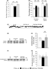
Treatment with FRAX486 rescues the aberrant blood glucose homeostasis displayed by symptomatic Cdkl5‐Het mice. (A) Glucose tolerance test (GTT). Blood glucose levels (glycemia) in Cdkl5 heterozygous (Cdkl5‐Het) female mice and wild‐type (wt) littermates were measured using a commercial glucometer (Menarini diagnostic) from tail vein blood collected at 0 (baseline), 30, 60, 120 and 180 min following intraperitoneal (i.p.) injection with 2 g/kg body weight d‐glucose (10% D glucose solution; Sigma Aldrich) after overnight fasting. No significant genotype differences were found in the GTT. (B) Insulin sensitivity test (IST). One week later, mice were i.p. injected with human recombinant insulin solution (0.4 U/kg body weight, Humulin, Eli‐Lilly) after being food‐deprived for 5 h. Blood was collected from tail vein at 0 (baseline) and 15 min after insulin administration and glycemia was measured as in (a). Cdkl5‐Het mice showed fasting‐induced hypoglycemia. (C) Cdkl5‐Het and wt mice from a separate cohort were treated for 5 consecutive days via s.c. injections of ctrl and, on the 5th day of treatment, glycemia was measured through tail vein incision under both basal conditions and after 5 h of fasting (1° week). The following week (2° week), Cdkl5‐Het mice received once daily, for 5 consecutive days, s.c. injections of 20 mg/kg of FRAX486, while wt mice received ctrl solution, and glycemia was measured as in the first week. A 5‐day‐long treatment with FRAX486 completely rescued the fasting‐induced hypoglycemia of Cdkl5‐Het mice, restoring wt‐like glycemic levels. Statistical significance was assessed using repeated measures ANOVA (setting genotype as between factor and repeated measurements as within factor) followed by Tukey's post hoc test. Data are mean ± SEM. *p < 0.05, **p < 0.01; N = 4–5 per group (A, B) or N = 7–10 per group (C)
FIGURE 4
FIGURE 4
Treatment with FRAX486 rescues spine maturation and PSD95 expression in the hippocampus of Cdkl5‐Het mice. Symptomatic Cdkl5 heterozygous (Cdkl5‐Het) female mice received daily subcutaneous (s.c.) injections of FRAX486 (20 mg/kg, daily) or control (ctrl) solution for 5 consecutive days. Wild‐type (wt) littermates received only ctrl. Six hours after the last injection hippocampal tissue was dissected for histological analyses. (A) Dendritic spine density was measured on Golgi‐stained brain coronal sections by manually counting the number of dendritic spines on apical dendrites of CA1‐pyramidal neurons. In each mouse, 10–15 dendritic segments (segment length, 10–30 μm) were analyzed and the linear spine density was calculated by dividing the total number of counted spines by the length of the sampled segment. FRAX486 treatment increased the defective spine density in Cdkl5‐Het female mice. (B) Based on their morphology, dendritic spines can be divided into 5 different classes (immature spines: Filopodium‐like, thin‐shaped, and stubby‐shaped; mature spines: Mushroom‐ and cup‐shaped), which also reflect their state of maturation. The number of spines belonging to each class was counted and expressed as a percentage in relation to the total number of protrusions in neurons. FRAX486 treatment rescued spine imbalance in CA1 pyramidal neurons of Cdkl5‐Het mice. (C) Examples of Golgi‐stained dendritic branches of CA1 pyramidal neurons of one animal from each experimental group. Scale bar: 1 μm. (D, E) PSD95 levels were normalized to total GAPDH contents and expressed as a percentage of those of wt, ctrl mice. The following primary antibodies were used: Rabbit polyclonal anti‐PSD95 (1:1000; Abcam), rabbit polyclonal anti‐GAPDH (1:5000; Sigma‐Aldrich). Densitometric analysis of digitized images was performed using Chemidoc XRS Imaging Systems and Image Lab™ Software (Bio‐Rad). FRAX486 treatment increased the reduced PSD95 levels in hippocampal extracts of Cdkl5‐Het mice. Immunoblots were assembled to show examples from two animals of each experimental group. (F, G) Synaptophysin 1 levels were normalized to total β‐Actin contents and expressed as a percentage of those of wt, ctrl mice. The following primary antibodies were used: Rabbit polyclonal anti‐synaptophysin 1 (1:1000, SYSY) and mouse monoclonal anti‐β‐actin (1:5000, Sigma‐Adrich). Densitometric analysis of digitized images was performed using Chemidoc XRS Imaging Systems and Image Lab™ Software (Bio‐Rad). No genotype or treatment effects were found in synaptophysin 1 levels in hippocampal extracts of experimental mice. Immunoblots were assembled to show examples from two animals of each experimental group. Statistical significance was assessed using one‐way ANOVA (main effect of experimental group) followed by Dunnett's post hoc test (setting Cdkl5‐Het, ctrl as control group). Data are mean ± SEM. **p < 0.01; N = 3–4 per group (A, B), N = 4–7 per group (E), N = 8–12 per group (G)
FIGURE 5
FIGURE 5
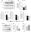
FRAX486 treatment rescues the abnormal phosphorylation levels of PAKs in Cdkl5‐Het mouse hippocampus. Fully symptomatic Cdkl5 heterozygous (Cdkl5‐Het) female mice received daily subcutaneous (s.c.) injections of FRAX486 (20 mg/kg, daily) or control (ctrl) for 5 consecutive days. Wild‐type (wt) littermates received only ctrl solution. Six hours after the last injection hippocampal tissue was dissected for western blot analyses of phospho‐PAK1/2/3 (P‐PAKs) normalized to total PAK1/2/3 content (PAKs), phospho‐PAK1 threonine‐212 (P‐PAK1 Thr212), and phospho‐PAK1 serine‐204 (P‐PAK1 Ser204) levels in hippocampal homogenates. P‐PAK1 levels were normalized to total PAK1 contents, while total PAK1 levels were normalized to GAPDH levels in hippocampal extracts. (A) Immunoblots were assembled to show examples from two animals of each experimental group. (B) FRAX486 normalized the reduced P‐PAKs/PAKs ratio in hippocampal extracts from Cdkl5‐Het mice at wt‐like levels. (C) FRAX486 rescued the increased levels of P‐PAK1 at Thr212 in hippocampal extracts from Cdkl5‐Het mice. (D and E) No significant difference was found in the analysis of P‐PAK1 at Ser204 or in total PAK1 contents. (F–H) To substantiate the suggested link between disrupted Cdkl5 functionality and aberrant phosphorylation of phospho‐PAK1 on threonine‐212 (P‐PAK1Thr‐212), 293 T cells were transfected with pCMV14‐3xFLAG‐hCDKL5 carrying the CDKL5_1 isoform that generates a protein of 960 amino acids (107 kDa) and pCMV14‐3xFLAG empty backbone as a control (ctrl, Sigma Aldrich) using Metafectene Easy Plus (Biontex). Twenty‐four hours later, transfection cells were processed for western blotting. (F) Immunoblots from technical replicates of P‐PAK1 (Thr212, Ser204) levels in 293 T total protein extracts from control and CDKL5‐transfected cells. (G) Western blot analysis revealed a reduction of P‐PAK1 at Thr212 in the presence of the overexpression of CDKL5. (H) No significant difference was found in the analysis of phospho‐PAK1 serine‐204 (P‐PAK1 at Ser 204). P‐PAK1 levels were normalized to total protein contents in cells. Data in B–E are expressed as a percentage of the values of wt, ctrl while in G,H as a percentage of those of cells transfected with the empty vector (ctrl). Statistical significance was assessed using one‐way ANOVA (main effect of experimental group) followed by Dunnett's post hoc test (setting Cdkl5‐Het, ctrl as control group) for B–E and with Student's t‐test for G,H. Data are mean ± SEM. *p < 0.05, **p < 0.01, ***p < 0.001; N = 7–11 per group (B–E) or N = 3 (G, H)

References

    1. Olson HE, Demarest ST, Pestana‐Knight EM, et al. Cyclin‐dependent kinase‐like 5 deficiency disorder: clinical review. Pediatr Neurol. 2019;97:18‐25. - PMC - PubMed
    1. Bahi‐Buisson N, Nectoux J, Rosas‐Vargas H, et al. Key clinical features to identify girls with CDKL5 mutations. Brain J Neurol. 2008;131(Pt 10):2647‐2661. - PubMed
    1. Fehr S, Downs J, Ho G, et al. Functional abilities in children and adults with the CDKL5 disorder. Am J Med Genet A. 2016;170(11):2860‐2869. - PubMed
    1. Kalscheuer VM, Tao J, Donnelly A, et al. Disruption of the serine/threonine kinase 9 gene causes severe X‐linked infantile spasms and mental retardation. Am J Hum Genet. 2003;72(6):1401‐1411. - PMC - PubMed
    1. Zhu YC, Xiong ZQ. Molecular and synaptic bases of CDKL5 disorder. Dev Neurobiol. 2019;79(1):8‐19. - PubMed

Publication types

Supplementary concepts