Review
. 2022 Oct;15(5):411-426.
doi: 10.1007/s12178-022-09780-5. Epub 2022 Aug 6.

Radiographic Evaluation of Pediatric Patients with Patellofemoral Instability

Affiliations
Review

Radiographic Evaluation of Pediatric Patients with Patellofemoral Instability

Kevin J Orellana et al. Curr Rev Musculoskelet Med. 2022 Oct.

Abstract

Purpose of review: The purpose of this review is to highlight the radiographic assessments of utility in the evaluation of a pediatric patient with patellofemoral instability to facilitate a thorough work-up. Understanding of these measures is useful in understanding evolving research in this field, providing accurate patient risk assessment, and appropriately directing surgical decision-making.

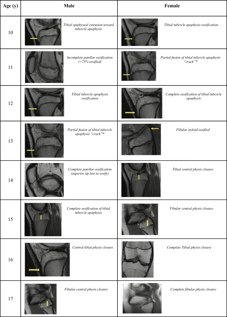
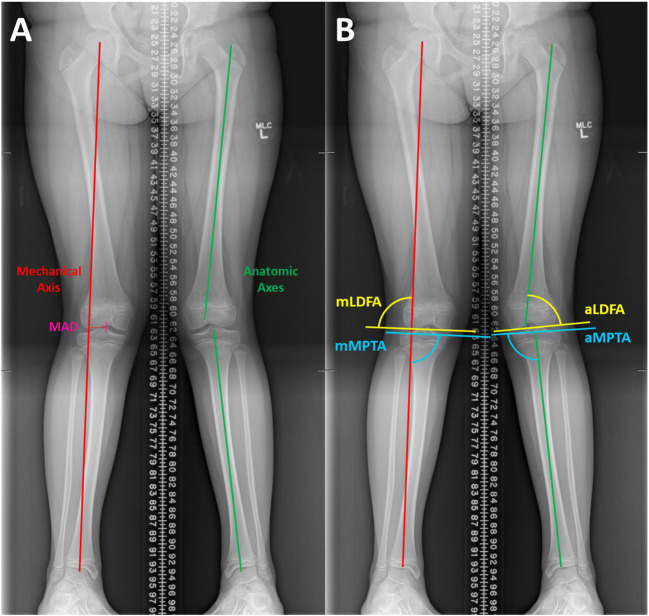
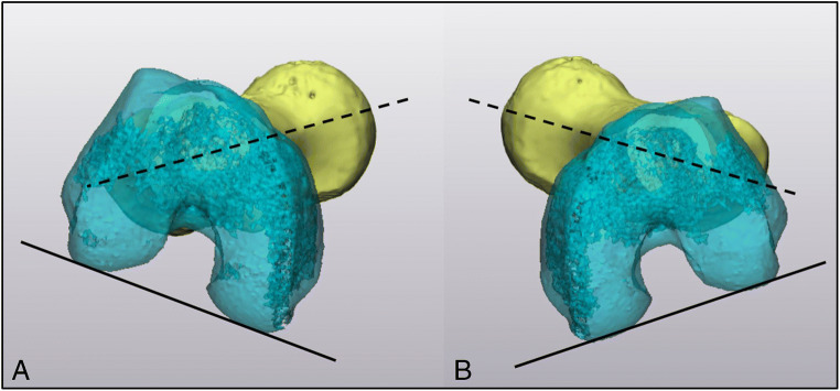
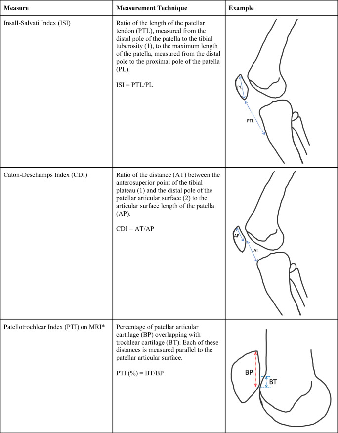
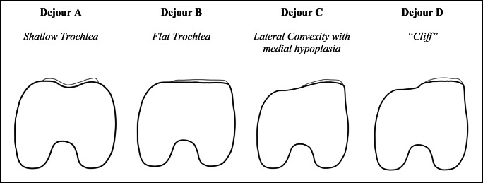
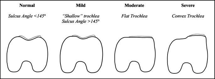
Recent findings: Recent literature has broadened the radiographic characterization of the pediatric patellar instability and its anatomic risk factors. Knee MRI can inform the assessment of skeletal maturity and novel axial alignment measurements may enhance our identification of patients at increased risk of recurrent instability. Additional improvements have been made in the objective measurement and classification of trochlear dysplasia. Knee MRI-based skeletal age assessments may obviate the need for hand bone age assessments in growing children with patellofemoral instability. Novel objective measures exist in the evaluation of pediatric patellar instability both in the assessment of axial alignment and trochlear dysplasia. Future work should focus on how these measures can aid in guiding surgical decision-making.

Keywords: Axial alignment; Coronal alignment; Pediatric knee morphology; Pediatric patellofemoral instability; Radiographic evaluation in pediatrics; Trochlear dysplasia.


Conflict of interest statement

Kevin J. Orellana, Morgan G. Batley, Jie C. Nguyen, and Brendan A. Williams declare that they have no conflict of interest. J. Todd R. Lawrence is a board or committee member of the American Academy of Pediatrics and has received IP royalties from Sawbones/Pacific Research Laboratories.

Figures

Fig. 1
Fig. 1
Reproducible regional physeal changes of the knee visualized on sagittal intermediate-weighted non-fat saturated images in males and females aged 10 to 17 years old. *Crack represents incomplete fusion and should cross entire apophysis. Figure adapted from Meza et al. [••]
Fig. 2
Fig. 2
Measures for assessment of coronal plane angular deformity using standing alignment film. a Illustrates the mechanical axis, mechanical axis deviation (MAD), and anatomic axes of the femur and tibia. b Illustrates angles of interest relative to the mechanical and anatomical axes: lateral distal femoral angle (LDFA) and medial proximal tibial angle (MPTA)
Fig. 3
Fig. 3
CT version study with 3D reconstruction of the right (a) and left (b) legs in a patient with patellar instability and femoral anteversion. Versional assessment is determined by finding the angle made by the femoral neck (dashed line) and the posterior condylar axis of the femur (solid line). In this patient, femoral anteversion was 36 degrees in the right femur and 35 degrees in the left femur
Fig. 4
Fig. 4
Measurement techniques for sagittal alignment ratios of ISI, CDI, and PTI. *PTI measured on the midline sagittal section MRI through the patella with the thickest articular cartilage and maximal length of the patella
Fig. 5
Fig. 5
Measurement techniques for axial alignment assessments including TT-TG, PT-LTR, and LTR-TT are described and presented with representative drawings
Fig. 6
Fig. 6
The Dejour classification of trochlear dysplasia is described along with representative figures depicting expected findings on axial MRI (MRI Dejour [•]) at the proximal-most aspect of the trochlear cartilage for each subtype are presented
Fig. 7
Fig. 7
The Oswestry-Bristol classification of trochlear dysplasia is described and presented along with representative axial MRI findings at the proximal-most aspect of the trochlear cartilage
Fig. 8
Fig. 8
Objective trochlear measurements to identify and characterize trochlear dysplasia on axial MRI of the knee are presented. Measurement techniques for SA, LTI, TDI, and MCTO are described with representative drawings

References

    1. Fithian DC, Paxton EW, Stone ML, Silva P, Davis DK, Elias DA, et al. Epidemiology and natural history of acute patellar dislocation. Am J Sports Med. 2004;32:1114–1121. doi: 10.1177/0363546503260788. - DOI - PubMed
    1. Sanders TL, Pareek A, Hewett TE, Stuart MJ, Dahm DL, Krych AJ. Incidence of first-time lateral patellar dislocation: a 21-year population-based study. Sports Health. 2018;10:146–151. doi: 10.1177/1941738117725055. - DOI - PMC - PubMed
    1. Sillanpää P, Mattila VM, Iivonen T, Visuri T, Pihlajamäki H. Incidence and risk factors of acute traumatic primary patellar dislocation. Med Sci Sports Exerc. 2008;40:606–611. doi: 10.1249/MSS.0b013e318160740f. - DOI - PubMed
    1. Nietosvaara Y, Aalto K, Kallio PE. Acute patellar dislocation in children: incidence and associated osteochondral fractures. J Pediatr Orthop. 1994;14:513–515. doi: 10.1097/01241398-199407000-00018. - DOI - PubMed
    1. Buchner M, Baudendistel B, Sabo D, Schmitt H. Acute traumatic primary patellar dislocation: long-term results comparing conservative and surgical treatment. Clin J Sport Med. 2005;15:62–66. doi: 10.1097/01.jsm.0000157315.10756.14. - DOI - PubMed