. 2022 Oct 15:1793:148040.
doi: 10.1016/j.brainres.2022.148040. Epub 2022 Aug 4.

Eyeblink tract tracing with two strains of herpes simplex virus 1

Affiliations

Eyeblink tract tracing with two strains of herpes simplex virus 1

Deidre E O'Dell et al. Brain Res. .

Abstract

Background: Neuroinvasive herpes simplex-1 (HSV-1) isolates including H129 and McIntyre cross at or near synapses labeling higher-order neurons directly connected to infected cells. H129 spreads predominately in the anterograde direction while McIntyre strains spread only in the retrograde direction. However, it is unknown if neurons are functional once infected with derivatives of H129 or McIntyre.

New method: We describe a previously unpublished HSV-1 recombinant derived from H129 (HSV-373) expressing mCherry fluorescent reporters and one new McIntyre recombinant (HSV-780) expressing the mCherry fluorophore and demonstrate how infections affect neuron viability.

Results and comparison with existing methods: Each recombinant virus behaved similarly and spread to the target 4 days post-infection. We tested H129 recombinant infected neurons for neurodegeneration using Fluoro-jade C and found them to be necrotic as a result of viral infection. We performed dual inoculations with both HSV-772 and HSV-780 to identify cells comprising both the anterograde pathway and the retrograde pathway, respectively, of our circuit of study. We examined the presence of postsynaptic marker PSD-95, which plays a role in synaptic plasticity, in HSV-772 infected and in dual-infected rats (HSV-772 and HSV-780). PSD-95 reactivity decreased in HSV-772-infected neurons and dual-infected tissue had no PSD-95 reactivity.

Conclusions: Infection by these new recombinant viruses traced the circuit of interest but functional studies of the cells comprising the pathway were not possible because viral-infected neurons died as a result of necrosis or were stripped of PSD-95 by the time the viral labels reached the target.

Keywords: Eyeblink Conditioning Circuit; H129; McIntyre; Neuroinvasive Viruses; Transsynaptic Tracers.


Conflict of interest statement

Declaration of Competing Interest

The authors declare that they have no known competing financial interests or personal relationships that could have appeared to influence the work reported in this paper.

Figures

Fig. 1.
Fig. 1.
A schematic showing the rat eyeblink conditioning circuit. The anterior interpositus nucleus is shown receiving several excitatory and inhibitory inputs, suggesting this region is involved in cerebellar learning and memory.
Fig. 2.
Fig. 2.
Experimental Timeline. The figure shows the overall timeline used to carry out our experiments.
Fig. 3.
Fig. 3.
HSV-772 Reactivity in the AIN appears at 4 days post infection. A.) HSV-772 at 3 days post infection shows little to no HSV-772 reactivity. Coordinates: Bregma:10.80 mm, Interaural: −1.80 mm. B.) HSV-772 has some reactivity in the brainstem but none in any of the DCN at 3.5 days post infection. Coordinates: Bregma: 11.28 mm, Interaural: −2.28 mm. C.) Shows minor reactivity of HSV-772 at 4 days post infection at the earliest. Coordinates: Bregma: 11.16 mm, Interaural: −2.16 mm. C’.) Shows a zoomed in image of the AIN. There are at least 3 HSV-772 positive cells in this region at 4 days post infection. Same coordinates as in C. D.) Shows HSV-772 is present not only at the AIN but also in the cerebellar cortex. The circle outlines the location of the AIN. Coordinates: Bregma: 10.68 mm, Interaural: −1.68 mm. D’.) Shows a zoomed in image of the AIN. A large group of HSV-772–positive cells is visible. Same coordinates as in D. AIN: Anterior Interpositus Nucleus, DCN: Deep Cerebellar Nuclei. Coordinates are taken from Paxinos and Watson Rat Atlas. Magnification 4x.
Fig. 4.
Fig. 4.
HSV-780 reactivity appears in the AIN 4 days post infection. Discrete labeling of HSV-780 is found in the AIN 4 days following infection. The circle identifies where the AIN is located. There is also extensive infection throughout the brainstem in areas associated with the facial nerve and the red nucleus, which are identified by the black rectangle. Coordinates: Bregma: 11.04 mm, Interaural: −2.04 mm. AIN: Anterior Interpositus Nucleus. Coordinates are taken from Paxinos and Watson Rat Atlas. Magnification 4x.
Fig. 5.
Fig. 5.
HSV-373 infected cells of the AIN. 3.5 days post infection appear necrotic. The figure shows the Fluoro-jade C (upper right panel) is present in cells that are also infected with HSV-373 mCherry (upper left panel). Magnification 63x.
Fig. 6.
Fig. 6.
HSV-373 infected cells in the AIN 4 days post infection. Neurons infected with HSV-373 have amorphous shape and abnormal cell bodies with displaced nuclei. Neurons with H129 recombinant infection are also green with fluoro-jade C reactivity (upper right panel). Magnification 63x.
Fig. 7.
Fig. 7.
The AIN of uninfected rats lacks Flour-Jade C positive cells. There are no cells positive for Fluoro-Jade C staining in the AIN of a control uninfected rat. Magnification 63x.
Fig. 8.
Fig. 8.
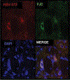
Neurons with dual infection in the AIN lack PSD-95. 4 days post infection with HSV-780 and HSV-772 leads to synaptic stripping of infected neurons. There is no visible PSD-95 in the AIN of dual inoculated rats. AIN: Anterior Interpositus Nucleus. Magnification 63x.
Fig. 9.
Fig. 9.
Neurons with HSV-772 infection in the AIN have diminished PSD-95 reactivity. PSD-95 reactivity is minimal in the AIN of a rat euthanized 4 days after HSV-772 infection. AIN: Anterior Interpositus Nucleus. Magnification 63x.
Fig. 10.
Fig. 10.
PSD-95 reactivity is extensive in an uninfected rat AIN. Control rats without H129 or McIntyre have PSD-95 in their AIN. AIN: Anterior Interpositus Nucleus. Magnification 63x.

References

    1. Almeida CG, Tampellini D, Takahashi RH, Greengard P, Lin MT, Snyder EM, Gouras GK, 2005. Beta-amyloid accumulation in APP mutant neurons reduces PSD-95 and GluR1 in synapses. Neurobiol. Dis. 20, 187–198. 10.1016/j.nbd.2005.02.008. - DOI - PubMed
    1. Bagnall MW, Zingg B, Sakatos A, Moghadam SH, Zeilhofer HU, du Lac S, 2009. Glycinergic Projection Neurons of the Cerebellum. J. Neurosci. 29, 10104–10110. 10.1523/JNEUROSCI.2087-09.2009. - DOI - PMC - PubMed
    1. Banfield BW, Kaufman JD, Randall JA, Pickard GE, 2003. Development of pseudorabies virus strains expressing red fluorescent proteins: new tools for multisynaptic labeling applications. J. Virol. 77 (18), 10106–10112. - PMC - PubMed
    1. Barnett E, Cassell M, Perlman S, 1993. 2 Neurotropic Viruses, Herpes-Simplex Virus Type-1 and Mouse Hepatitis-Virus, Spread Along Different Neural Pathways from the Main Olfactory-Bulb. Neuroscience 57, 1007–1025. 10.1016/0306-4522(93)90045-H. - DOI - PMC - PubMed
    1. Barnett EM, Evans GD, Sun N, Perlman S, Cassell MD, 1995. Anterograde tracing of trigeminal afferent pathways from the murine tooth pulp to cortex using herpes simplex virus type 1. J. Neurosci. 15 (4), 2972–2984. - PMC - PubMed

Publication types