. 2022 Sep:148:103814.
doi: 10.1016/j.ibmb.2022.103814. Epub 2022 Aug 3.

Characterization of two kdr mutations at predicted pyrethroid receptor site 2 in the sodium channels of Aedes aegypti and Nilaparvata lugens

Affiliations

Characterization of two kdr mutations at predicted pyrethroid receptor site 2 in the sodium channels of Aedes aegypti and Nilaparvata lugens

Huahua Sun et al. Insect Biochem Mol Biol. 2022 Sep.

Abstract

Pyrethroid insecticides prolong the opening of insect sodium channels by binding to two predicted pyrethroid receptor sites (PyR), PyR1 and PyR2. Many naturally-occurring sodium channel mutations that confer pyrethroid resistance (known as knockdown resistance, kdr) are located at PyR1. Recent studies identified two new mutations, V253F and T267A, at PyR2, which co-exist with two well-known mutations F1534C or M918T, at PyR1, in pyrethroid-resistant populations of Aedes aegypti and Nilaparvata lugens, respectively. However, the role of the V253F and T267A mutations in pyrethroid resistance has not been functionally examined. Here we report functional characterization of the V253F and T267A mutations in the Ae. aegypti sodium channel AaNav2-1 and the N. lugens sodium channel NlNav1 expressed in Xenopus oocytes. Both mutations alone reduced channel sensitivity to pyrethroids, including etofenprox. We docked etofenprox in a homology model of the pore module of the NlNav1 channel based on the crystal structure of an open prokaryotic sodium channel NavMs. In the low-energy binding pose etofenprox formed contacts with V253, T267 and a previously identified L1014 within PyR2. Combining of V253F or T267A with F1534C or M918T results in a higher level of pyrethroid insensitivity. Furthermore, both V253F and T267A mutations altered channel gating properties. However, V253F- and T267A-induced gating modifications was not observed in the double mutant channels. Our findings highlight the first example in which naturally-found combinational mutations in PyR1 and PyR2 not only confer higher level pyrethroid insensitivity, but also reduce potential fitness tradeoff in pyrethroid-resistant mosquitoes caused by kdr mutation-induced sodium channel gating modifications.

Keywords: Knockdown resistance; Pyrethroid insecticides; Pyrethroid receptor site 2; Sodium channel.


Figures

Figure 1.
Figure 1.
(A) Sodium channel topology. Positions of T267A, M918T and L1014F in N. lugens and V253F and F1534C in Ae. aegypti are shown with residues designated by numbers in the house fly sodium channel (GenBank accession number: X96668). (B) Sequence alignment of helices S4-S5, S5 and S6 in sodium channels and potassium channel Kv1.2. We use a residue-labeling scheme that is universal for P-loop channels (Du et al., 2013; Zhorov and Tikhonov, 2004). Position of a residue is designated by a label that contains domain number, a symbol “k”, “o”, or “i” representing, respectively, the S4-S5 linker, the outer helix, and the inner helix, and a relative position of the residue in the segment. (Zhorov and Tikhonov, 2004).
Figure 2.
Figure 2.
Voltage dependence of activation, fast inactivation and slow inactivation of NlNav1 and mutant channels T267A. (A) Voltage dependence of activation. (B) Voltage dependence of fast inactivation. (C) Voltage dependence of slow inactivation. Voltage step protocols used to generate the G-V curves are indicated above the panels. The error bars represent SEM from at least 8 oocytes.
Figure 3.
Figure 3.
Effect of NlNav1 channel mutations T267A on channel sensitivity to four pyrethroids. (A-D) Tail currents in wild-type and mutant channel T267A induced by individual pyrethroids. (E) Percentage of channel modification by 1 μM pyrethroid from oocytes expressing wild-type or mutant channel T267A. Mark (*) indicates significant differences between the wild-type and the mutant channels as determined by non-parametric Mann-Whitney test (p < 0.05). The error bars represent SEM from at least 5 oocytes.
Figure 4.
Figure 4.
Voltage dependence of activation, fast inactivation and slow inactivation of AaNav2-1 and its mutant V253F. (A) Voltage dependence of activation. (B) Voltage dependence of fast inactivation. (C) Voltage dependence of slow inactivation. Voltage step protocols used to generate the G-V or I-V curves are indicated above each panel. The error bars represent SEM from at least 7 oocytes.
Figure 5.
Figure 5.
Modification of the AaNav2-1 channel and mutant channels V253F, F1534C or V253F+F1534C by pyrethroids (A) bifenthrin, (B) etofenprox, (C) deltamethrin, and (D) λ-cyhalothrin. Top row: Structural formulae of pyrethroids. Middle row: Tail currents induced by different concentrations of the pyrethroids in oocytes expressing the AaNav2-1 channel. Bottom row: Percentage of channel modification by 1 μM pyrethroids in oocytes expressing AaNav2-1 and mutant channels. One-way ANOVA Tukey’s test was applied for the statistical analysis, with p < 0.05 indicating a significant difference. The bars are labeled by the same letter when the difference between pyrethroid effects on the channels is not statistically significant (p > 0.05). The error bars represent SEM from at least 10 oocytes.
Figure 6.
Figure 6.
Effects of mutations on sodium channel sensitivity to DDT. (A) Representative traces from AaNav2-1, V253F, F1534C, and V253F+F1534C mutant channels in the absence of DDT (top row), and after incubation with DDT (100 μM) (bottom row). (B) Percentages of channel inactivation inhibited by DDT (100 μM). One-way ANOVA Tukey’s test was applied for the statistical analysis, with p < 0.05 indicating a significant difference. The bars are labeled by the same letter when the difference between DDT effects on the channels is not statistically significant (p > 0.05). The error bars represent SEM from at least 11 oocytes.
Figure 7.
Figure 7.
Intracellular (A) and intra-membrane (B) views at the pore module of superimposed cryo-EM structures of sodium channels NavPaS (red, PDB ID: 5×0m) (Shen et al., 2017), rNav1.5 (cyan, PDB ID: 6uz3) (Jiang et al., 2020) and crystal structure of prokaryotic sodium channel NavMs with completely open pore module (green, PDB ID: 5hvx) (Sula et al., 2017). The structures are superimposed by minimizing root mean square deviations of Cα atoms in the four P1 helices, whose mutual disposition is well conserved in P-loop channels, against the reference crystal structure of chimeric potassium channel Kv1.2/Kv2.1 (PDB ID: 2R9R), the first eukaryotic P-loop channel obtained at resolution below 2.5 Å (Long et al., 2007). The general folding of the three channels is similar. However, the diameter of cytoplasmic half of the inner pore in the non-functional channel NavPaS is much smaller than that in the rNav1.5 or NavMs channels. The NavMs structure with the fully open symmetric pore module appears a better template for modeling insect sodium channels with pyrethroids, which binds to open channels, than asymmetric structure of the Nav1.5 channel in partially inactivated state.
Figure 8.
Figure 8.
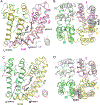
Etofenprox in the NavMs-based models of insect sodium channels. Domains I, II, III and IV in the pore module are pink, yellow, green and gray, respectively. Etofenprox is shown by thick bonds in left panels and by spherical atoms in right panels. Residues within 4 Å from etofenprox are shown as thin bonds. Residues whose substitutions are explored in this study are indicated by one-letter code with sequential numbers according to the house fly sodium channel and labels according to a universal labeling system for P-loop channels. (A, B) Intra-membrane (A) and extracellular (B) views at etofenprox in the PyR2 site of N. lugens NlNav1sodium channel. (C, D) Intra-membrane and extracellular views at the pore module of bumble bee BiNav1 (GenBank accession number: KY123916) sodium channel. The image is produced using coordinates of an earlier model (Wu et al., 2017).

References

    1. Ahern CA, Payandeh J, Bosmans F, Chanda B, 2016. The hitchhiker’s guide to the voltage-gated sodium channel galaxy. The Journal of general physiology 147, 1–24. - PMC - PubMed
    1. Auteri M, La Russa F, Blanda V, Torina A, 2018. Insecticide resistance associated with kdr mutations in Aedes albopictus: an update on worldwide evidences. BioMed research internationa, 3098575. - PMC - PubMed
    1. Catterall WA, 2010. Ion channel voltage sensors: structure, function, and pathophysiology. Neuron 67, 915–928. - PMC - PubMed
    1. Catterall WA, 2014. Structure and function of voltage-gated sodium channels at atomic resolution. Experimental physiology 99, 35–51. - PMC - PubMed
    1. Catterall WA, 2017. Forty years of sodium channels: structure, function, pharmacology, and epilepsy. Neurochemical research 42, 2495–2504. - PMC - PubMed

Publication types

LinkOut - more resources