

Corneal endothelial cell density and its correlation with birth weight, anthropometric parameters, and ocular biometric parameters in Chinese school children
- PMID: 35933331
- PMCID: PMC9356483
- DOI: 10.1186/s12886-022-02561-1
Corneal endothelial cell density and its correlation with birth weight, anthropometric parameters, and ocular biometric parameters in Chinese school children
Abstract
Background: To describe the distribution of corneal endothelial cell density (ECD), and to explore its correlation with birth weight (BW), anthropometric parameters, and ocular biometric parameters in Chinese school children.
Methods: In the population-based cross-sectional Nanjing Eye Study, children were measured for anthropometric information, for ECD by the noncontact specular microscope and for ocular biometric parameters by the optic low-coherent reflectometer. Data from right eyes were analyzed to illustrate the distribution of ECD and for determining correlated factors with ECD using univariate and multiple linear regression analysis. Comparisons among three different BW groups were performed using a one-way ANOVA analysis followed by the Bonferroni correction for pairwise comparisons.
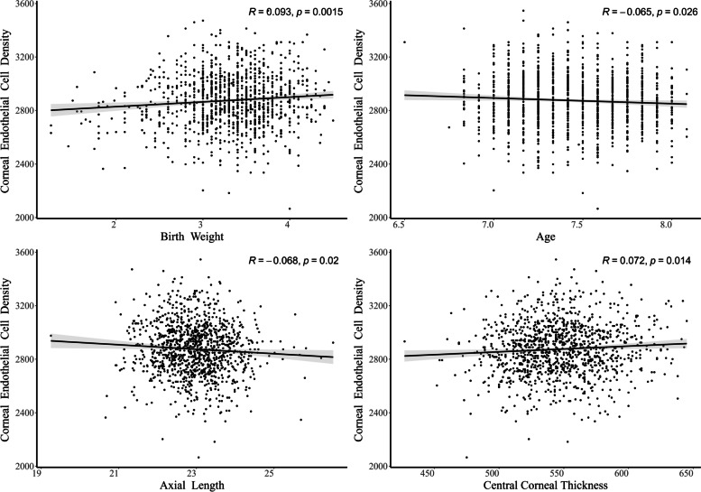
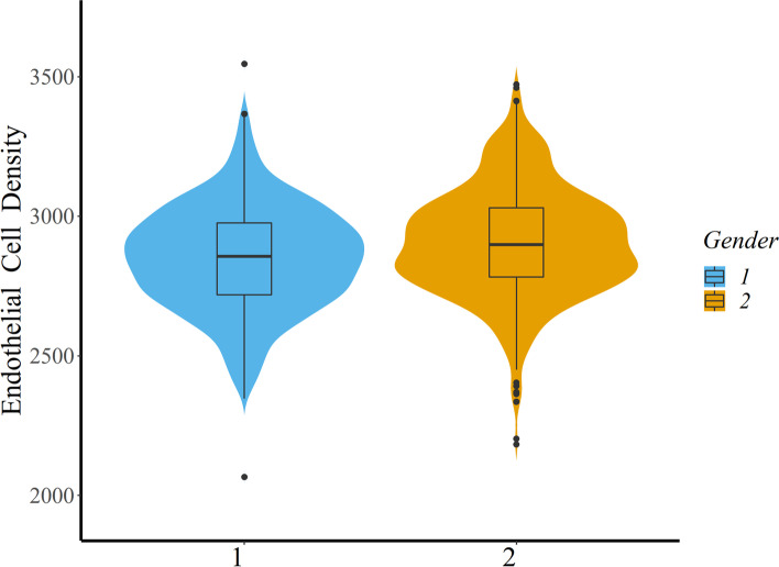
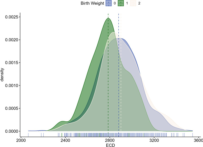
Results: Of 1171 children, the mean (± standard deviation) ECD was 2875.34 ± 195.00 cells/mm2. In the Multiple Linear Regression analysis, BW, gender and central corneal thickness were significantly associated with ECD. The ECD increased by 36.16 cells/mm2 with BW increasing by 1 kg (P = 0.001) and increased by 0.44 cells/mm2 for every additional 1 mm in central corneal thickness (P = 0.01). The ECD of girls was 54.41 cells/mm2 higher than boys (P < 0.001). Children born with low BW presented significantly lower ECD than those born with normal BW (P < 0.05) and high BW (P < 0.05). Age and axial length were not significantly associated with ECD (P = 0.06 and P = 0.21, respectively).
Conclusions: In Chinese school children aged 82 to 94 months, the ECD is positively correlated with BW and central corneal thickness, in which BW is a newly identified associated factor. It is like that gender plays an important role in ECD distribution while girls have relatively greater ECD than boys.
Keywords: Birth weight; Body mass index; Corneal endothelial cell density; Epidemiology; Ocular parameters.
© 2022. The Author(s).
Conflict of interest statement
The authors declare that they have no competing interests.
Figures
Similar articles
-
Corneal thickness and endothelial cell density measured by non-contact specular microscopy and pachymetry in Rhesus macaques (Macaca mulatta) with laser-induced ocular hypertension.Exp Eye Res. 2003 Jun;76(6):671-7. doi: 10.1016/s0014-4835(03)00055-1. Exp Eye Res. 2003. PMID: 12742349
-
Corneal endothelial morphology of healthy myopic Malaysian children of Chinese ethnicity aged 8-9 years and its association with axial length.F1000Res. 2022 Mar 21;11:339. doi: 10.12688/f1000research.110560.2. eCollection 2022. F1000Res. 2022. PMID: 36111215 Free PMC article.
-
Corneal Endothelial Morphology and Ocular Biometric Indexes in Premature Children With and Without Retinopathy of Prematurity.Invest Ophthalmol Vis Sci. 2024 May 1;65(5):37. doi: 10.1167/iovs.65.5.37. Invest Ophthalmol Vis Sci. 2024. PMID: 38780946 Free PMC article.
-
Overestimation of corneal endothelial cell density by automated method in glaucomatous eyes with impaired corneal endothelial cells.Int Ophthalmol. 2022 Jan;42(1):133-145. doi: 10.1007/s10792-021-02008-4. Epub 2021 Sep 5. Int Ophthalmol. 2022. PMID: 34482487 Free PMC article.
-
Corneal thickness and endothelial morphology in Normal Thai eyes.BMC Ophthalmol. 2020 Apr 28;20(1):167. doi: 10.1186/s12886-020-01385-1. BMC Ophthalmol. 2020. PMID: 32345246 Free PMC article.
Cited by
-
Secondary Trifocal Intraocular Lens Implantation in Dense Cataracts: A Promising Alternative to One-Step Surgery.Ophthalmol Ther. 2025 Aug 20. doi: 10.1007/s40123-025-01202-2. Online ahead of print. Ophthalmol Ther. 2025. PMID: 40833456
References
-
- Murphy C, Alvarado J, Juster R, Maglio M. Prenatal and postnatal cellularity of the human corneal endothelium. A quantitative histologic study. Invest Ophthalmol Vis Sci. 1984;25(3):312–22. - PubMed
-
- Bourne WM, Nelson LR, Hodge DO. Central corneal endothelial cell changes over a ten-year period. Invest Ophthalmol Vis Sci. 1997;38(3):779–782. - PubMed
MeSH terms
LinkOut - more resources
Full Text Sources
