. 2022 Aug 6;19(1):201.
doi: 10.1186/s12974-022-02563-2.

Microglia-specific deletion of histone deacetylase 3 promotes inflammation resolution, white matter integrity, and functional recovery in a mouse model of traumatic brain injury

Affiliations

Microglia-specific deletion of histone deacetylase 3 promotes inflammation resolution, white matter integrity, and functional recovery in a mouse model of traumatic brain injury

Yongfang Zhao et al. J Neuroinflammation. .

Abstract

Background: Histone deacetylases (HDACs) are believed to exacerbate traumatic brain injury (TBI) based on studies using pan-HDAC inhibitors. However, the HDAC isoform responsible for the detrimental effects and the cell types involved remain unknown, which may hinder the development of specific targeting strategies that boost therapeutic efficacy while minimizing side effects. Microglia are important mediators of post-TBI neuroinflammation and critically impact TBI outcome. HDAC3 was reported to be essential to the inflammatory program of in vitro cultured macrophages, but its role in microglia and in the post-TBI brain has not been investigated in vivo.

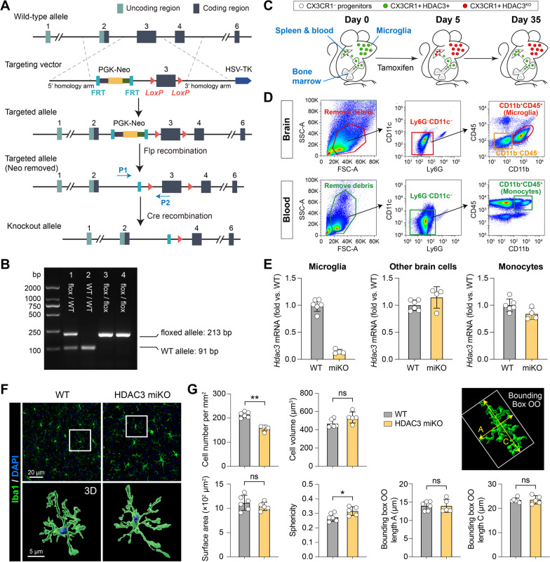
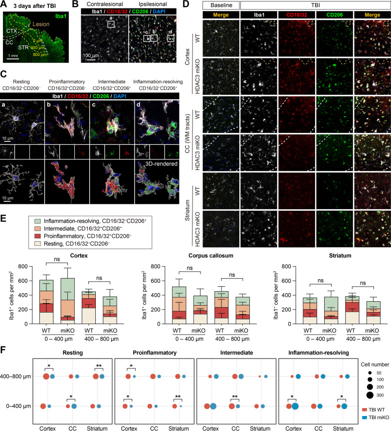
Methods: We generated HDAC3LoxP mice and crossed them with CX3CR1CreER mice, enabling in vivo conditional deletion of HDAC3. Microglia-specific HDAC3 knockout (HDAC3 miKO) was induced in CX3CR1CreER:HDAC3LoxP mice with 5 days of tamoxifen treatment followed by a 30-day development interval. The effects of HDAC3 miKO on microglial phenotype and neuroinflammation were examined 3-5 days after TBI induced by controlled cortical impact. Neurological deficits and the integrity of white matter were assessed for 6 weeks after TBI by neurobehavioral tests, immunohistochemistry, electron microscopy, and electrophysiology.

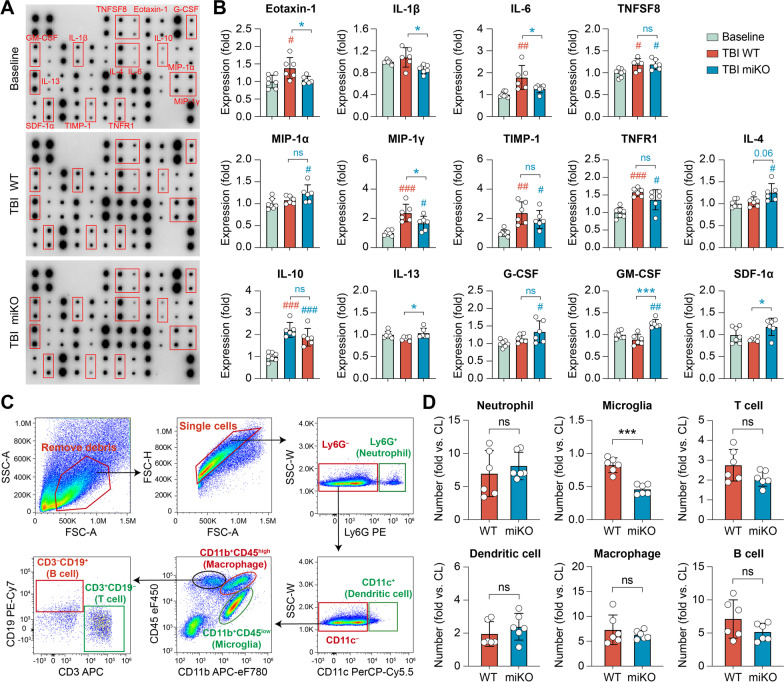
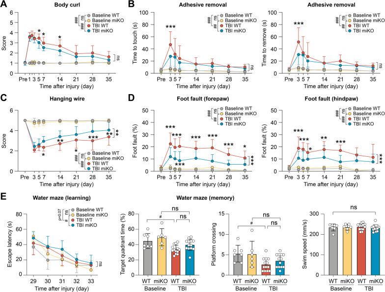
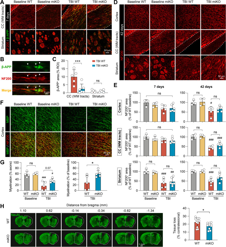
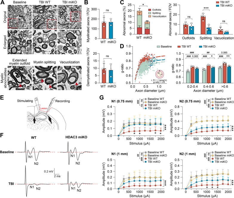
Results: HDAC3 miKO mice harbored specific deletion of HDAC3 in microglia but not in peripheral monocytes. HDAC3 miKO reduced the number of microglia by 26%, but did not alter the inflammation level in the homeostatic brain. After TBI, proinflammatory microglial responses and brain inflammation were markedly alleviated by HDAC3 miKO, whereas the infiltration of blood immune cells was unchanged, suggesting a primary effect of HDAC3 miKO on modulating microglial phenotype. Importantly, HDAC3 miKO was sufficient to facilitate functional recovery for 6 weeks after TBI. TBI-induced injury to axons and myelin was ameliorated, and signal conduction by white matter fiber tracts was significantly enhanced in HDAC3 miKO mice.

Conclusion: Using a novel microglia-specific conditional knockout mouse model, we delineated for the first time the role of microglial HDAC3 after TBI in vivo. HDAC3 miKO not only reduced proinflammatory microglial responses, but also elicited long-lasting improvement of white matter integrity and functional recovery after TBI. Microglial HDAC3 is therefore a promising therapeutic target to improve long-term outcomes after TBI.

Keywords: Conditional gene knockout; Controlled cortical impact; HDAC3; Neuroinflammation.


Conflict of interest statement

The authors declare that they have no competing interests.

Figures

Fig. 1
Fig. 1
Generation and characterization of microglia-specific HDAC3 knockout mice. A Targeting strategy to generate HDAC3LoxP mice with LoxP-flanked exon 3 of the Hdac3 gene. In the presence of Cre, the LoxP sites recombine and delete the floxed region of Hdac3. P1 and P2, genotyping primers. B PCR genotyping demonstrating generation of heterozygous mice (lane 1) and subsequent breeding to obtain homozygous mice containing the LoxP sites on both alleles (lanes 3 and 4). Using primers flanking the 5′ LoxP site, the floxed allele runs at a weight of 213 bp, whereas the wild-type (WT) allele has a weight of 91 bp. C Pulse knockout strategy to restrict tamoxifen-induced HDAC3 KO to microglia. Peripheral CX3CR1+ cells are continuously replaced from CX3CR1 progenitor cells in the bone marrow. HDAC3 is deleted 5 days after tamoxifen treatment in both microglia and peripheral CX3CR1+ cells. After another 30 days, peripheral CX3CR1+ cells are replenished from bone marrow progenitors by new CX3CR1+ cells that have not undergone recombination. Therefore, only microglia are HDAC3 KO, and most peripheral monocytes express HDAC3. D FACS gating strategy for brain microglia (CD11b+CD45+), other brain cells (CD11bCD45), and blood monocytes (CD11b+CD45+). E Quantitative PCR was performed 30 days after tamoxifen treatment on FACS-sorted cells to verify the deletion of HDAC3 in microglia but not in other brain cells or blood monocytes. n = 6 (WT) and 4 (HDAC3 miKO) mice. F, G The number and morphology of microglia were assessed in the brain of HDAC3 miKO mice and WT control mice after Iba1 immunostaining and 3D rendering of images. n = 6 mice (237 cells) for WT. n = 5 mice (140 cells) for HDAC3 miKO. Shown are the mean ± SD. *p < 0.05, **p < 0.01. ns no significant difference
Fig. 2
Fig. 2
HDAC3 miKO enhances inflammation-resolving responses of microglia after TBI. HDAC3 miKO mice and WT control mice were subjected to TBI induced by controlled cortical impact. The phenotype of microglia was examined 3 days after TBI by triple-label immunostaining of CD16/32, CD206, and Iba1. A Iba1 immunofluorescence in the ipsilesional brain hemisphere illustrates the boundary of the TBI lesion and the peri-lesion areas in the cortex (CTX), corpus callosum (CC), and striatum (STR) where images in BD were taken from. B Triple-label immunosignal of Iba1, CD16/32, and CD206 in the peri-lesion striatum and in the corresponding region in the non-injured contralesional cortex. Rectangles, areas enlarged in C. C Images taken under high magnification demonstrate 4 typical phenotypes of microglia based on their expression of CD16/32 and CD206: resting (a), proinflammatory (b), intermediate (c), and inflammation-resolving (d). Lower panels: images 3D-rendered by Imaris. D Representative images taken from the peri-lesion cortex, CC white matter (WM) tracts, and striatum 3 days after TBI or taken from the corresponding regions in baseline control brains. See Additional file 1: Fig. S2 for images of baseline controls under individual color channel. E The number of Iba1+ cells under each of the four phenotypic categories was counted in the peri-lesion cortex, CC and striatum, 0–400 μm and 400–800 μm from the lesion boundary. ns no significant difference in total Iba1+ cells between HDAC3 miKO and WT mice. Shown are the mean ± SD. F The numbers of Iba1+ cells in the 4 phenotype categories were illustrated in dot plots, where the area of a dot represents the number of cells. n = 6 (WT) and 5 (HDAC3 miKO) mice. *p < 0.05, **p < 0.01 HDAC3 miKO vs. WT
Fig. 3
Fig. 3
HDAC3 miKO alleviates neuroinflammation after TBI without altering infiltrating blood immune cells. HDAC3 miKO mice and WT control mice were subjected to TBI induced by CCI, and brain inflammation profiles were examined 5 days after TBI. A, B A panel of 40 inflammatory cytokines was measured in the ipsilesional brain hemisphere using an antibody array. A Representative blots with significantly altered markers labeled. B Summarized data showing the mean expression levels of 14 markers that were significantly different among groups. Non-injured baseline controls were pooled from both WT and HDAC3 miKO mice, between which there was no significant difference (see Additional file 1: Fig. S1). C, D Infiltration of peripheral immune cells into the post-TBI brain was assessed using flow cytometry 5 days after TBI. C Flow cytometry gating strategy for various immune cells in the brain. D Summarized data showing the numbers of immune cells in the ipsilesional brain hemisphere, expressed as fold changes over the non-injured contralesional side. Shown are the mean ± SD. n = 6–7 mice per group. #p < 0.05, ##p < 0.01, ###p < 0.001 TBI vs. baseline control. *p < 0.05, ***p < 0.001 HDAC3 miKO vs. WT after TBI. ns no significant difference
Fig. 4
Fig. 4
HDAC3 miKO promotes long-term functional recovery after TBI. Adult male HDAC3 miKO mice and age- and sex-matched WT control mice were subjected to TBI induced by CCI or non-injury control procedures (baseline control). AD Sensorimotor deficits were assessed in male mice before (Pre) and up to 35 days after injury by the body curl (A), adhesive removal (B), hanging wire (C), and foot fault (D) tests. n = 9 (baseline) or 13 (TBI) mice per group. E The Morris water maze test was performed to assess spatial learning and spatial memory in male mice at 29–34 days after injury. Mice in all groups had comparable swim speeds, suggesting similar gross locomotor functions. n = 6–7 (baseline) or 13 (TBI) mice per group. Shown are the mean ± SD. #p < 0.05, ##p < 0.01, ###p < 0.001 TBI vs. baseline. *p < 0.05, **p < 0.01, ***p < 0.001 HDAC3 miKO vs. WT after TBI. ns no significant difference
Fig. 5
Fig. 5
HDAC3 miKO improves short-term and long-term integrity of white matter after TBI. Immunofluorescence staining was performed at 7 and 42 days after TBI or non-injury control procedures (baseline controls) to assess brain injury and white matter integrity in HDAC3 miKO and WT mice. AC Axonal injury was assessed 7 days after TBI using NF200 and β-APP double-label immunostaining. A Representative images taken from the white matter-enriched corpus callosum (CC) and striatum in the ipsilesional brain hemisphere. Dashed line, the boundary of CC. B β-APP immunosignal in the shape of classic axonal bulbs and varicosities (arrows) was observed after TBI, suggesting axonal damage. C Summarized data on β-APP-immunopositive areas. D Axonal integrity was assessed 42 days after TBI by NF200 immunostaining. E Summarized data on NF200-immunopositive areas in the ipsilesional cortex, CC and striatum 7 and 42 days after TBI, expressed as percentages of the WT baseline group. F Myelin integrity in the peri-lesion cortex was assessed 42 days after TBI using MBP and NF200 double-immunostaining. G Summarized data on the degree of myelination (areas immunopositive for both MBP and NF200), expressed as percentages of myelinated axons to total axons (left panel), or as percentages to baseline controls (right panel). n = 4 (baseline) or 5–6 (TBI) per group. H The volume of chronic brain tissue loss was measured 42 days after TBI on coronal brain sections immunostained for the neuronal marker NeuN. Dashed line, the relative area of the contralesional hemisphere to illustrate ipsilesional tissue loss. n = 12 mice per group. Shown are the mean ± SD. #p < 0.05, ##p < 0.01, ###p < 0.001 TBI vs. baseline. *p < 0.05, ***p < 0.001 HDAC3 miKO vs. WT. ns no significant difference
Fig. 6
Fig. 6
HDAC3 miKO improves nerve signal conduction in the white matter after TBI. AD White matter ultrastructure was examined in the ipsilesional CC 42 days after TBI using transmission electron microscopy (TEM). A Representative TEM images. TBI induced demyelination and abnormal myelin morphology in myelinated axons (lower panel), such as extended myelin outfold, myelin splitting, and vacuolization (arrow). B, C The numbers of myelinated axons, demyelinated axons, and myelinated axons with abnormal myelin morphology were counted in post-TBI HDAC3 miKO mice and control WT mice. Data were expressed as the number of axons per field of view (FOV; 200 μm2) or percentage of total myelinated axons. D Scatter plot (left panel) and summarized data (right panel) showing the g-ratio of all axons counted from 3 to 4 mice in each group. Baseline control group was pooled from both WT and HDAC3 miKO mice. EG Axonal conduction at the ipsilesional CC/external capsule was assessed 42 days after TBI by measuring the evoked compound action potentials (CAPs). E Diagram showing the location of the stimulating and recording electrodes at the external capsule. Dashed line: the boundary of the lesion. F Representative traces of the evoked CAPs at 2000 µA stimulation to show the N1 (through myelinated axons) and N2 (through unmyelinated axons) components, recorded 1 mm from the stimulation point. G Quantification of the N1 and N2 amplitudes after various stimulation current intensities, recorded 0.75 mm and 1 mm from the stimulating point. n = 5–8 mice per group. Shown are the mean ± SD. ##p < 0.01, ###p < 0.001 TBI vs. baseline. *p < 0.05, ***p < 0.001 HDAC3 miKO vs. WT after TBI. ns no significant difference

Similar articles

Cited by

References

    1. Haberland M, Montgomery RL, Olson EN. The many roles of histone deacetylases in development and physiology: implications for disease and therapy. Nat Rev Genet. 2009;10(1):32–42. doi: 10.1038/nrg2485. - DOI - PMC - PubMed
    1. Shein NA, Grigoriadis N, Alexandrovich AG, Simeonidou C, Lourbopoulos A, Polyzoidou E, et al. Histone deacetylase inhibitor ITF2357 is neuroprotective, improves functional recovery, and induces glial apoptosis following experimental traumatic brain injury. FASEB J. 2009;23(12):4266–4275. doi: 10.1096/fj.09-134700. - DOI - PMC - PubMed
    1. Wang G, Jiang X, Pu H, Zhang W, An C, Hu X, et al. Scriptaid, a novel histone deacetylase inhibitor, protects against traumatic brain injury via modulation of PTEN and AKT pathway: scriptaid protects against TBI via AKT. Neurotherapeutics. 2013;10(1):124–142. doi: 10.1007/s13311-012-0157-2. - DOI - PMC - PubMed
    1. Wang G, Shi Y, Jiang X, Leak RK, Hu X, Wu Y, et al. HDAC inhibition prevents white matter injury by modulating microglia/macrophage polarization through the GSK3beta/PTEN/Akt axis. Proc Natl Acad Sci USA. 2015;112(9):2853–2858. doi: 10.1073/pnas.1501441112. - DOI - PMC - PubMed
    1. Chen X, Wang H, Zhou M, Li X, Fang Z, Gao H, et al. Valproic acid attenuates traumatic brain injury-induced inflammation in vivo: involvement of autophagy and the Nrf2/ARE signaling pathway. Front Mol Neurosci. 2018;11:117. doi: 10.3389/fnmol.2018.00117. - DOI - PMC - PubMed