

Neuropathology in chronic traumatic encephalopathy: a systematic review of comparative post-mortem histology literature
- PMID: 35933388
- PMCID: PMC9356428
- DOI: 10.1186/s40478-022-01413-9
Neuropathology in chronic traumatic encephalopathy: a systematic review of comparative post-mortem histology literature
Abstract
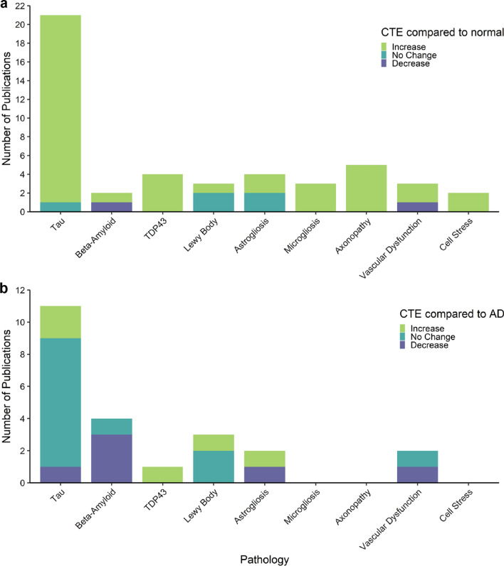
Chronic traumatic encephalopathy (CTE) is a neurodegenerative disease associated with repetitive head trauma and is characterised by the perivascular accumulation of hyperphosphorylated tau (p-tau) in the depths of cortical sulci. CTE can only be diagnosed postmortem and the cellular mechanisms of disease causation remain to be elucidated. Understanding the full scope of the pathological changes currently identified in CTE is necessary to identify areas requiring further research. This systematic review summarises the current literature on CTE pathology from postmortem human tissue histology studies published until 31 December 2021. Publications were included if they quantitively or qualitatively compared postmortem human tissue pathology in CTE to neuropathologically normal cases or other neurodegenerative diseases such as Alzheimer's disease (AD). Pathological entities investigated included p-tau, beta-amyloid, TDP-43, Lewy bodies, astrogliosis, microgliosis, axonopathy, vascular dysfunction, and cell stress. Of these pathologies, p-tau was the most frequently investigated, with limited reports on other pathological features such as vascular dysfunction, astrogliosis, and microgliosis. Consistent increases in p-tau, TDP-43, microgliosis, axonopathy, and cell stress were reported in CTE cases compared to neuropathologically normal cases. However, there was no clear consensus on how these pathologies compared to AD. The CTE cases used for these studies were predominantly from the VA-BU-CLF brain bank, with American football and boxing as the most frequent sources of repetitive head injury exposure. Overall, this systematic review highlights gaps in the literature and proposes three priorities for future research including: 1. The need for studies of CTE cases with more diverse head injury exposure profiles to understand the consistency of pathology changes between different populations. 2. The need for more studies that compare CTE with normal ageing and AD to further clarify the pathological signature of CTE for diagnostic purposes and to understand the disease process. 3. Further research on non-aggregate pathologies in CTE, such as vascular dysfunction and neuroinflammation. These are some of the least investigated features of CTE pathology despite being implicated in the acute phase response following traumatic head injury.
Keywords: Chronic traumatic encephalopathy; Dementia pugilistica; Neuropathology; Systematic review.
© 2022. The Author(s).
Conflict of interest statement
The authors declare no competing interests.
Figures
Similar articles
-
Perivascular glial reactivity is a feature of phosphorylated tau lesions in chronic traumatic encephalopathy.Acta Neuropathol. 2025 Feb 8;149(1):16. doi: 10.1007/s00401-025-02854-x. Acta Neuropathol. 2025. PMID: 39921702 Free PMC article.
-
CCL2 is associated with microglia and macrophage recruitment in chronic traumatic encephalopathy.J Neuroinflammation. 2020 Dec 5;17(1):370. doi: 10.1186/s12974-020-02036-4. J Neuroinflammation. 2020. PMID: 33278887 Free PMC article.
-
Home treatment for mental health problems: a systematic review.Health Technol Assess. 2001;5(15):1-139. doi: 10.3310/hta5150. Health Technol Assess. 2001. PMID: 11532236
-
Signs and symptoms to determine if a patient presenting in primary care or hospital outpatient settings has COVID-19.Cochrane Database Syst Rev. 2022 May 20;5(5):CD013665. doi: 10.1002/14651858.CD013665.pub3. Cochrane Database Syst Rev. 2022. PMID: 35593186 Free PMC article.
-
Amyloid PET across the cognitive spectrum in former professional and college American football players: findings from the DIAGNOSE CTE Research Project.Alzheimers Res Ther. 2023 Oct 5;15(1):166. doi: 10.1186/s13195-023-01315-5. Alzheimers Res Ther. 2023. PMID: 37798671 Free PMC article.
Cited by
-
Neurobiology and Impact of Chronic Traumatic Encephalopathy in Athletes: A Focused Review.Cureus. 2025 Jun 19;17(6):e86367. doi: 10.7759/cureus.86367. eCollection 2025 Jun. Cureus. 2025. PMID: 40688873 Free PMC article. Review.
-
Intimate Partner Violence-Related Brain Injury: Unmasking and Addressing the Gaps.J Neurotrauma. 2024 Oct;41(19-20):2219-2237. doi: 10.1089/neu.2023.0543. Epub 2024 Apr 5. J Neurotrauma. 2024. PMID: 38323539 Review.
-
Blood-Based Biomarkers in the Diagnosis of Chronic Traumatic Encephalopathy: Research to Date and Future Directions.Int J Mol Sci. 2023 Aug 8;24(16):12556. doi: 10.3390/ijms241612556. Int J Mol Sci. 2023. PMID: 37628736 Free PMC article. Review.
-
Chronic traumatic encephalopathy-neuropathologic change in a routine neuropathology service: 7-year follow-up.J Neuropathol Exp Neurol. 2023 Oct 20;82(11):948-957. doi: 10.1093/jnen/nlad079. J Neuropathol Exp Neurol. 2023. PMID: 37846159 Free PMC article.
-
Mild traumatic brain injury increases cortical iron: evidence from individual susceptibility mapping.Brain Commun. 2025 Mar 12;7(2):fcaf110. doi: 10.1093/braincomms/fcaf110. eCollection 2025. Brain Commun. 2025. PMID: 40161218 Free PMC article.
References
-
- Adams JW, Alvarez VE, Mez J, Huber BR, Tripodis Y, Xia W, Meng G, Kubilus CA, Cormier K, Kiernan PT, Daneshvar DH, Chua AS, Svirsky S, Nicks R, Abdolmohammadi B, Evers L, Solomon TM, Cherry JD, Aytan N, Mahar I, Devine S, Auerbach S, Alosco ML, Nowinski CJ, Kowall NW, Goldstein LE, Dwyer B, Katz DI, Cantu RC, Stern RA, Au R, McKee AC, Stein TD. Lewy body pathology and chronic traumatic encephalopathy associated with contact sports. J Neuropathol Exp Neurol. 2018;77:757–768. doi: 10.1093/jnen/nly065. - DOI - PMC - PubMed
-
- Alosco ML, Stein TD, Tripodis Y, Chua AS, Kowall NW, Huber BR, Goldstein LE, Cantu RC, Katz DI, Palmisano JN, Martin B, Cherry JD, Mahar I, Killiany RJ, McClean MD, Au R, Alvarez V, Stern RA, Mez J, McKee AC. Association of white matter rarefaction, arteriolosclerosis, and tau with dementia in chronic traumatic encephalopathy. JAMA Neurol. 2019;76:1298–1308. doi: 10.1001/jamaneurol.2019.2244. - DOI - PMC - PubMed
-
- Ameen-Ali KE, Bretzin A, Lee EB, Folkerth R, Hazrati L-N, Iacono D, Keene CD, Kofler J, Kovacs GG, Nolan A, Perl DP, Priemer DS, Smith DH, Wiebe DJ, Stewart W, Al-Sarraj S, Cortes E, Crary J, Dams-O’Connor K, Diaz-Arrastia R, Dollé J-P, Edlow B, Fischl B, Hinds CS, Johnson VE, Manley G, Meaney D, Okonkwo D, Schneider ALC, Schneider J, Troakes C, Trojanowski JQ, van der Kouwe A, Yaffe K. Detection of astrocytic tau pathology facilitates recognition of chronic traumatic encephalopathy neuropathologic change. Acta Neuropathol Commun. 2022;10(1):1–11. doi: 10.1186/S40478-022-01353-4. - DOI - PMC - PubMed
-
- Anderson EN, Morera AA, Kour S, Cherry JD, Ramesh N, Gleixner A, Schwartz JC, Ebmeier C, Old W, Donnelly CJ, Cheng JP, Kline AE, Kofler J, Stein TD, Pandey UB. Traumatic injury compromises nucleocytoplasmic transport and leads to TDP-43 pathology. Elife. 2021 doi: 10.7554/eLife.67587. - DOI - PMC - PubMed
Publication types
MeSH terms
Substances
LinkOut - more resources
Full Text Sources
Medical
