. 2022 Aug 6;12(1):123.
doi: 10.1186/s13578-022-00860-0.

GREM1/PPP2R3A expression in heterogeneous fibroblasts initiates pulmonary fibrosis

Affiliations

GREM1/PPP2R3A expression in heterogeneous fibroblasts initiates pulmonary fibrosis

Xiaoni Shi et al. Cell Biosci. .

Abstract

Background: Fibroblasts have important roles in the synthesis and remodeling of extracellular matrix (ECM) proteins during pulmonary fibrosis. However, the spatiotemporal distribution of heterogeneous fibroblasts during disease progression remains unknown.

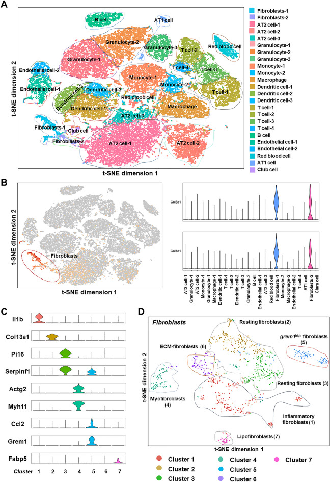
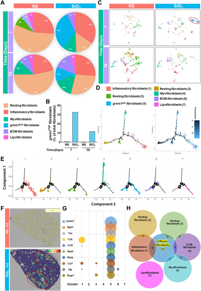
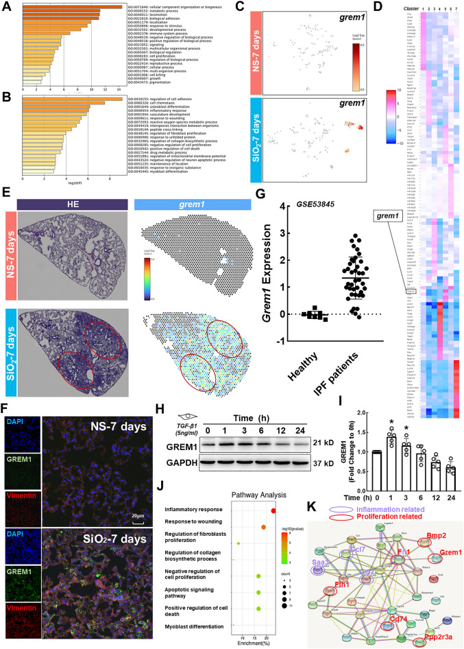
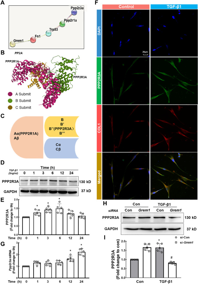
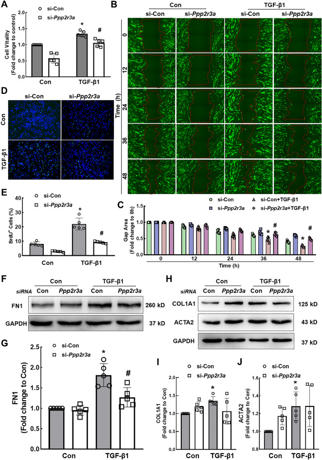
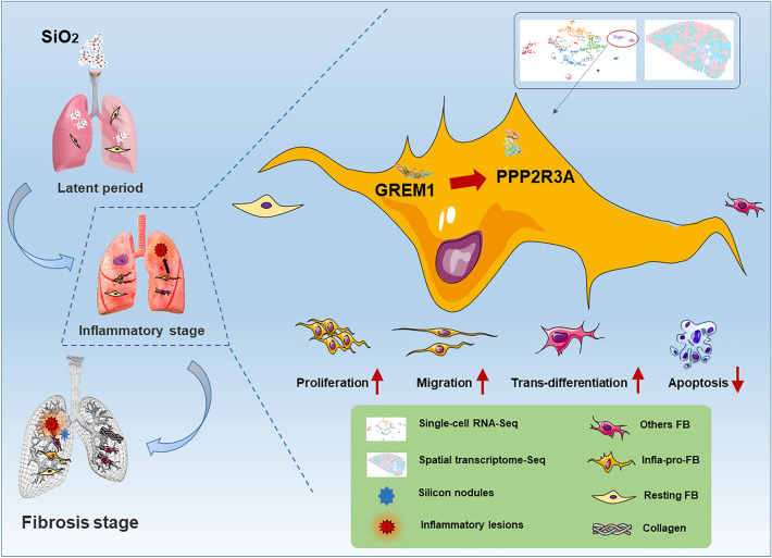
Results: In the current study, silica was used to generate a mouse model of pathological changes in the lung, and single-cell sequencing, spatial transcriptome sequencing and an analysis of markers of cell subtypes were performed to identify fibroblast subtypes. A group of heterogeneous fibroblasts that play an important role at the early pathological stage were identified, characterized based on the expression of inflammatory and proliferation genes (termed inflammatory-proliferative fibroblasts) and found to be concentrated in the lesion area. The expression of GREM1/protein phosphatase 2 regulatory subunit B''alpha (PPP2R3A) in inflammatory-proliferative fibroblasts was found to initiate early pulmonary pathological changes by increasing the viability, proliferation and migration of cells.

Conclusions: Inflammatory-proliferative fibroblasts play a key role in the early pathological changes that occur in silicosis, and during this process, GREM1 is the driving factor that targets PPP2R3A and initiates the inflammatory response, which is followed by irreversible fibrosis induced by SiO2. The GREM1/PPP2R3A pathway may be a potential target in the early treatment of silicosis.

Keywords: Heterogeneous fibroblasts; Pulmonary fibrosis; Single-cell transcriptomics; Spatial transcriptomics.


Conflict of interest statement

The authors have no conflicts of interest to declare.

Figures

Fig. 1
Fig. 1
Classification of fibroblasts using single-cell transcriptome sequencing. A Single-cell sequencing divided all the cells in lung tissue into 24 subtypes. B Fibroblasts were separated based on Col1a1 and Col3a1 expression. C A marker gene used for the subtyping of fibroblasts. D Fibroblasts were subdivided into seven subtypes
Fig. 2
Fig. 2
Correlation analysis of fibroblast subtypes. A Percentage of each fibroblast subtype among the total fibroblasts in different groups (saline and silica groups) at different time points (7 and 56 days). B Proportion of grem1high fibroblasts in different groups at different time points. C Distribution of each subtype of fibroblasts in the saline and silica groups at 7 and 56 days. D The quasi-chronological analysis shows the status of the whole fibroblast population at each node of transdifferentiation. E Status of each subtype of fibroblasts at the node of transdifferentiation. F The lesion area contains a large number of grem1high fibroblasts. G Based on the expression of the top 10 genes with multiple changes in Cluster 5 compared with other subtypes, the numbers 1–10 correspond to the 10 genes in the left column, and the bubble size represents the size of multiple changes. grem1 is only expressed in Cluster 5. H Venn diagram showing the number of identical genes expressed in the different subtypes
Fig. 3
Fig. 3
Results of biochemical analyses and validation of the scRNA-Seq analysis of inflammatory-proliferative fibroblasts. A GO enrichment analysis of the molecular functions of the top 50 genes in inflammatory-proliferative fibroblasts. B GO enrichment analysis of signaling pathways related to the top 50 genes in inflammatory-proliferative fibroblasts. C Expression of grem1 in seven subtypes. D Gene heatmap of the seven subtypes. E The spatial transcriptomic sequencing results show that grem1 is expressed at high levels in the lesion area. F Immunohistochemical staining showing that GREM1 is expressed on fibroblasts. The expression level of the experimental group was higher than that of the control group; the scale bar represents 20 μm. G In the GEO database, the expression of Grem1 in the lung tissue of patients with IPF was significantly higher than that found in the healthy group (*p < 0.05). H, I In HPF-a cells, GREM1 expression first increased and then decreased over time, and the expression levels at 1 and 3 h were significantly different from those at 0 h (*p < 0.05). J The bubble chart shows the pathways of interest among the related pathways identified in inflammatory-proliferative fibroblasts. K Interaction map between proteins enriched in signaling pathways examined in this study
Fig. 4
Fig. 4
Effect of TGF-β1 on PPP2R3A expression in HPF-a cells and the relationship between GREM1 and PPP2R3A. A Protein interaction map of the main components investigated in our research. B Three-dimensional structure of the PP2A protein. C Location of each subunit of PP2A. D, E In HPF-a cells, the expression of PPP2R3A first increased and then decreased over time. The expression levels at 1, 3, and 6 h were significantly different from those at 0 h (*p < 0.05). F Representative images of immunofluorescence staining show that TGF-β1 treatment increased the expression of PPP2R3A protein in HPF-a cells; scale bar = 20 μm. G The Ppp2r3a mRNA level increased over time, and the expression levels at 12 and 24 h were significantly different from those at 0 h (*p < 0.05). H, I Representative Western blot results showing that Grem1 knockdown partially reversed the upregulation of PPP2R3A induced by TGF-β1. *p < 0.05 indicates that the expression level of PPP2R3A in the si-Con group was higher than that in the control group after TGF-β1 treatment, showing successful establishment of the cell model. #p < 0.05 indicates that PPP2R3A was expressed at a lower level in the si-Grem1 group than in the si-Con group after TGF-β1 treatment
Fig. 5
Fig. 5
PPP2R3A mediates TGF-β1 signaling to induce the proliferation and activation of HPF-a cells. A CCK-8 assays show that Ppp2r3a knockdown partially reversed the increase in the viability of HPF-a cells induced by TGF-β1. *p < 0.05 indicates that the cell viability of the si-Con group after TGF-β1 treatment was higher than that in the control group and that the model was therefore successfully established. #p < 0.05 indicates that the cell viability of the si-Ppp2r3a group was lower than that of the si-Con group after TGF-β1 treatment. B, C Wound healing experiments showed that the downregulation of Ppp2r3a expression attenuated cell migration induced by TGF-β1. *p < 0.05 indicates that the cell migration in the si-Con group after TGF-β1 treatment was higher than that in the control group and that the model was successfully established. #p < 0.05 indicates that the cell migration of the si-Ppp2r3a group was lower than that of the si-Con group after TGF-β1 treatment. D The combined immunofluorescence images of BrdU (green) and DAPI (blue) show that the downregulation of Ppp2r3a expression attenuated cell proliferation induced by TGF-β1. E Percentage of BrdU-positive cells in five independent experiments. *p < 0.05 indicates that the cell proliferation of the si-Con group after TGF-β1 treatment was higher than that of the control group, which showed that the model was successfully established. #p < 0.05 indicates that the cell proliferation of the si-Ppp2r3a group was lower than that of the si-Con group after TGF-β1 treatment. F, G Downregulation of Ppp2r3a expression partially reversed the increase in FN1 expression induced by TGF-β1. *p < 0.05 indicates that the expression of FN1 in the si-Con group after TGF-β1 treatment was higher than that in the control group and thus that the model was successfully established. #p < 0.05 indicates that the expression of FN1 in the si-Ppp2r3a group was lower than that in the si-Con group after TGF-β1 treatment. H Downregulation of Ppp2r3a expression had little effect on the increase in Col1 and α-SMA expression induced by TGF-β1. I, J *p < 0.05 indicates that the expression of Col1 and α-SMA in the si-Con group after TGF-β1 treatment was higher than that in the control group, which revealed that the model was successfully established
Fig. 6
Fig. 6
Mouse model of the early pathology of lung silicosis and expression of PPP2R3A in mouse lung tissue. A Sirius Red staining shows significantly greater collagen deposition in the lung tissue of the silica group than that of the saline group, which revealed that the model was successfully established. B Representative WB results showing higher PPP2R3A expression in the lung tissue of the silica group than that of the saline group. C *p < 0.05 indicates that the difference between the two groups is significant. D Immunohistochemical staining showing that PPP2R3A is expressed on fibroblasts. The expression level in the experimental group was higher than that in the control group. E The scRNA-Seq results show increased ppp2r3a expression in inflammatory-proliferative fibroblasts. F The scRNA-Seq results show that vimentin expression was increased in inflammatory-proliferative fibroblasts
Fig. 7
Fig. 7
PPP2R3A affects the function and mechanism of heterogeneous fibroblasts (GREM1) during early pathological changes in the lung

Similar articles

Cited by

References

    1. Barnes H, Goh NSL, Leong TL, Hoy R. Silica-associated lung disease: an old-world exposure in modern industries. Respirology. 2019;24(12):1165–1175. doi: 10.1111/resp.13695. - DOI - PubMed
    1. Takenouchi Y, Kitakaze K, Tsuboi K, Okamoto Y. Growth differentiation factor 15 facilitates lung fibrosis by activating macrophages and fibroblasts. Exp Cell Res. 2020;391(2):112010. doi: 10.1016/j.yexcr.2020.112010. - DOI - PubMed
    1. Alvarez RF, Gonzalez CM, Martinez AQ, Perez JJB, Fernandez LC, Fernandez AP. Guidelines for the diagnosis and monitoring of silicosis. Arch Bronconeumol. 2015;51(2):86–93. - PubMed
    1. Barron L, Gharib SA, Duffield JS. Lung pericytes and resident fibroblasts: busy multitaskers. Am J Pathol. 2016;186(10):2519–2531. doi: 10.1016/j.ajpath.2016.07.004. - DOI - PMC - PubMed
    1. Lynch MD, Watt FM. Fibroblast heterogeneity: Implications for human disease. J Clin Invest. 2018;128(1):26–35. doi: 10.1172/JCI93555. - DOI - PMC - PubMed

LinkOut - more resources