. 2022 Aug 6;13(8):684.
doi: 10.1038/s41419-022-05124-w.

USP22 controls type III interferon signaling and SARS-CoV-2 infection through activation of STING

Affiliations

USP22 controls type III interferon signaling and SARS-CoV-2 infection through activation of STING

Rebekka Karlowitz et al. Cell Death Dis. .

Abstract

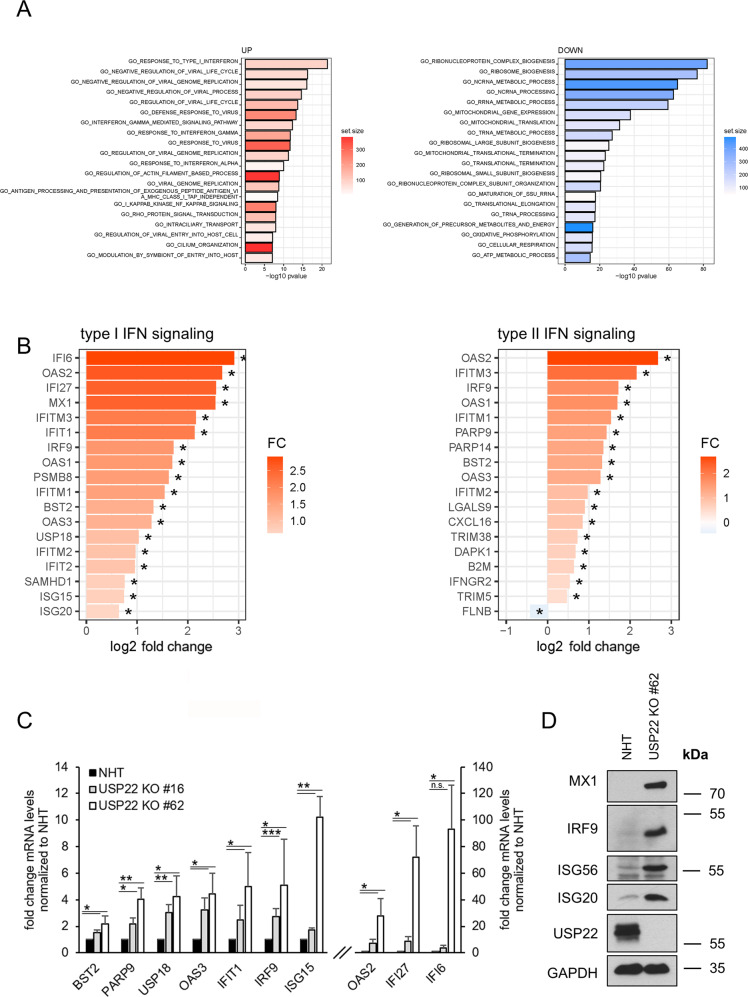
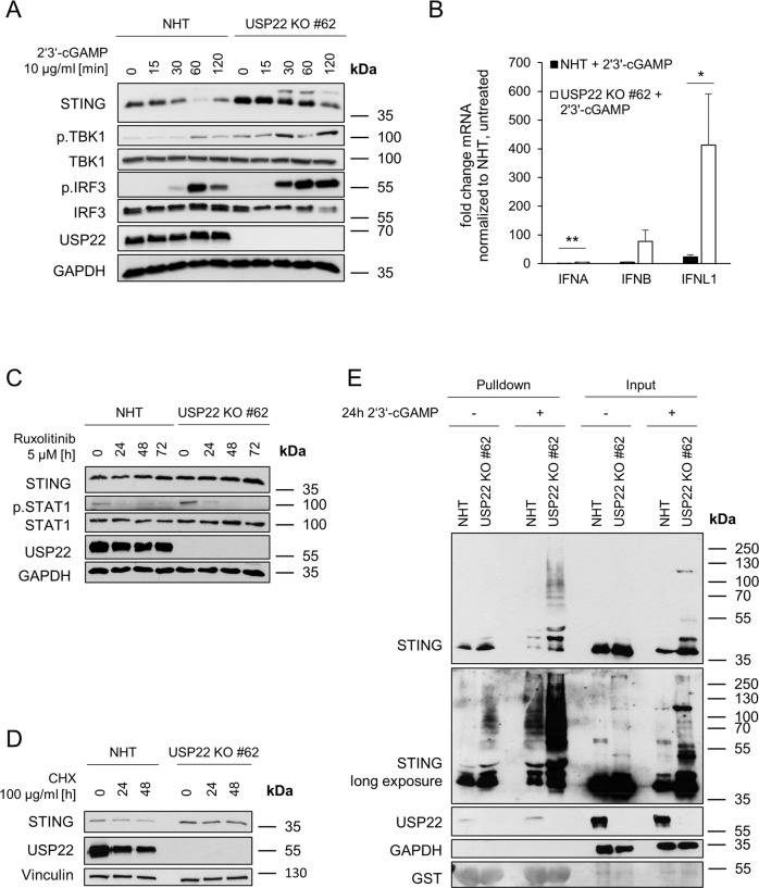
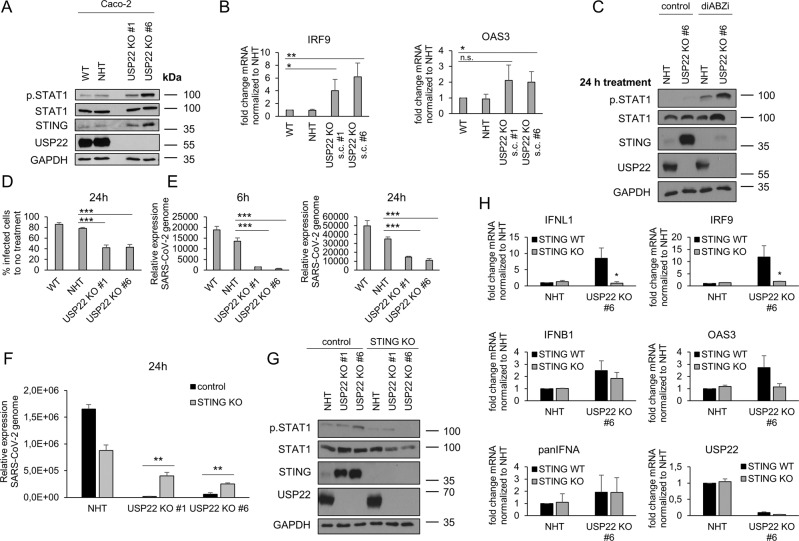
Pattern recognition receptors (PRRs) and interferons (IFNs) serve as essential antiviral defense against SARS-CoV-2, the causative agent of the COVID-19 pandemic. Type III IFNs (IFN-λ) exhibit cell-type specific and long-lasting functions in auto-inflammation, tumorigenesis, and antiviral defense. Here, we identify the deubiquitinating enzyme USP22 as central regulator of basal IFN-λ secretion and SARS-CoV-2 infections in human intestinal epithelial cells (hIECs). USP22-deficient hIECs strongly upregulate genes involved in IFN signaling and viral defense, including numerous IFN-stimulated genes (ISGs), with increased secretion of IFN-λ and enhanced STAT1 signaling, even in the absence of exogenous IFNs or viral infection. Interestingly, USP22 controls basal and 2'3'-cGAMP-induced STING activation and loss of STING reversed STAT activation and ISG and IFN-λ expression. Intriguingly, USP22-deficient hIECs are protected against SARS-CoV-2 infection, viral replication, and the formation of de novo infectious particles, in a STING-dependent manner. These findings reveal USP22 as central host regulator of STING and type III IFN signaling, with important implications for SARS-CoV-2 infection and antiviral defense.


Conflict of interest statement

The authors declare no competing interests.

Figures

Fig. 1
Fig. 1. Profiling USP22-mediated gene expression in HT-29 hIECs.
A. Volcano plot showing the differential gene expression patterns of two independent single-cell HT-29 USP22 CRISPR/Cas9 KO clones (#16 and #62) compared to CRISPR/Cas9 control (NHT) HT-29 cells. Color code represents the log2 fold change compared to NHT. B Heatmap of the top-50 differentially regulated genes between HT-29 USP22 KO single clones #16 and #62 and the NHT control. Color coding represents the row-wise scaled (Z-score) RNA intensities. Genes are sorted according to their log2 fold change, compared to NHT. C Basal mRNA expression levels of the indicated genes were determined in control and two independent USP22 KO HT-29 single clones using qRT-PCR. Gene expression was normalized against 28S mRNA and is presented as x-fold mRNA expression compared to NHT. Mean and SD of three independent experiments in triplicate are shown. *P < 0.05; **P < 0.01, ***P < 0.001.
Fig. 2
Fig. 2. Loss of USP22 specifically enriches for genes involved in IFN signaling and response to viral infection.
A Bar plot showing the top-20 regulated GO terms in two independent single-cell HT-29 USP22 CRISPR/Cas9 KO clones (#16 and #62) compared to control (NHT) HT-29 cells. Color code represents the number of annotated genes within each gene set. B Heatmap of differentially expressed genes contributing to the GO terms response to type I IFN (left) and type II signaling (right). Color code represents the log2 fold change compared to NHT. Note that due to lack of annotation and overlapping ISGs between type I/II and type III IFNs, response to type III IFN as GO term was not included. C Basal mRNA expression levels of GO- enriched genes related to IFN signaling in control (NHT) and two independent USP22 KO HT-29 single clones using qRT-PCR. Gene expression was normalized against 28S mRNA and is presented as x-fold mRNA expression compared to NHT. Mean and SD of three independent experiments in triplicate are shown. *P < 0.05; **P < 0.01, ***P < 0.001, n.s. not significant. D Western blot analysis of basal MX1, IRF9, ISG56, ISG20, and USP22 expression levels in control and USP22 KO HT-29 cells (clone USP22 KO #62). GAPDH served as loading control. Representative blots of at least two different independent experiments are shown.
Fig. 3
Fig. 3. USP22 negatively regulates STAT1 signaling and IFN-λ1 expression.
A. Basal mRNA expression levels of total IFNA (panIFNA) and IFNB1 in control (NHT) and the CRISPR/Cas9-generated USP22 KO HT-29 single clone (USP22 KO #62). Gene expression was normalized against 28 S mRNA and is presented as x-fold mRNA expression compared to NHT. Mean and SD of three independent experiments in triplicate are shown. *P < 0.05; **P < 0.01. B Western blot analysis of basal phosphorylated and total levels of STAT1 and USP22 in control and USP22 KO HT-29 cells (USP22 KO #62). GAPDH served as loading control. Representative blots of at least two different independent experiments are shown. C FACS-based analysis of the indicated basal secretion patterns of the viral defense cytokine panel in supernatants of control and USP22 KO HT-29 cells (USP22 KO #62). Data are presented as absolute levels of cytokines (in pg/ml). Samples below lower detection limit were set to zero, values above upper detection limit were set to detection limit. Mean and SD of three independent experiments in triplicate are shown. *P < 0.05; n.s. not significant. D Basal mRNA expression levels of IFNL1 in control and USP22 KO HT-29 single clone (USP22 KO #62). Gene expression was normalized against 28 S mRNA and is presented as x-fold mRNA expression compared to NHT. Mean and SD of three independent experiments in triplicate are shown. *P < 0.05. E Western blot analysis of phosphorylated and total levels of STAT1 as well as USP22 in control and USP22 KO HT-29 cells (USP22 KO #62) after 24 h incubation with 1 µg/ml IFNAR2 blocking antibody. As positive control, cells were pre-treated for 1 h with IFNAR2 blocking antibody, then treated 1 h with 0.05 ng/ml IFN-β. Vinculin served as loading control. Representative blots of at least two different independent experiments are shown.
Fig. 4
Fig. 4. USP22 regulates type III IFN signaling via STING.
A. Western blot analysis of basal RIG-I, MDA5, TLR3, and USP22 expression levels in control (NHT) and the CRISPR/Cas9-generated USP22 KO HT-29 single clone (USP22 KO #62). GAPDH served as loading control. Representative blots of at least two different independent experiments are shown. B Western blot analysis of basal RIG-I, phosphorylated and total STAT1, ISG56 and USP22 expression levels in control, USP22 KO HT-29 cells (USP22 KO #62) as well as two NHT-control and one USP22-DDX58 dKO HT-29 single clones. GAPDH served as loading control. Representative blots of at least two different independent experiments are shown. C Idem as (B), one MDA5/IFIH1 KO single clone instead of RIG-I/DDX58. D Idem as (B), three TLR3 KO single clones instead of RIG-I/DDX58. E Western blot analysis of phosphorylated and total STAT1, RIG-I, STING, and USP22 expression levels in control and USP22 KO HT-29 cells (USP22 KO #62) subjected to transfection with transfection reagent (control) alone or in the presence of ISD and poly(I:C) (2 µg/well) for 24 h. Vinculin served as loading control. Representative blots of at least two different independent experiments are shown. F mRNA expression levels of OAS3 (left) and IRF9 (right) in control and USP22 KO HT-29 cells (USP22 KO #62) subjected to transfection with ISD and poly(I: C) (each 2 µg/well) for 24 h. Gene expression was normalized against 28 S mRNA and is presented as x-fold mRNA expression compared to NHT. Mean and SD of three independent experiments in triplicate are shown. *P < 0.05; **P < 0.01; n.s. not significant. G Western blot analysis of phosphorylated and total STAT1, STING, and USP22 expression levels in control NHT and USP22 KO HT-29 cells (USP22 KO #62) as well as in the indicated NHT- and USP22-STING dKO cells. GAPDH served as loading control. Representative blots of at least two different independent experiments are shown. H Basal mRNA expression levels of the indicated genes in control NHT and USP22 KO HT-29 cells (USP22 KO #62) as well as in the indicated NHT and USP22 STING dKO cells. Gene expression was normalized against 28 S mRNA and is presented as x-fold mRNA expression compared to NHT. Mean and SD of three independent experiments in triplicate are shown. **P < 0.01; ***P < 0.001. I Basal mRNA expression levels of IFNA, IFNB, and IFNL1 in control NHT and USP22 KO HT-29 cells (USP22 KO #62) as well as in the indicated NHT- and USP22-STING dKO cells. Gene expression was normalized against 28 S mRNA and is presented as x-fold mRNA expression compared to NHT. Mean and SD of three independent experiments in triplicate are shown. **P < 0.01; ***P < 0.001.
Fig. 5
Fig. 5. USP22 negatively regulates STING activation and ubiquitination.
A Western blot analysis of STING, phosphorylated and total TBK1, phosphorylated and total IRF3 and USP22 expression levels in control (NHT) and CRISPR/Cas9-generated USP22 KO HT-29 single clone (USP22 KO #62) subjected to 2’3’-cGAMP (10 µg/ml) for the indicated time points. GAPDH served as loading control. Representative blots of at least two different independent experiments are shown. B mRNA expression levels of IFNA, IFNB, and IFNL1 in control and USP22 KO HT-29 cells (USP22 KO #62) subjected to 2’3’-cGAMP (10 µg/ml) for 3 h. Gene expression was normalized against 28 S mRNA and is presented as x-fold mRNA expression compared to NHT. Mean and SD of three independent experiments in triplicate are shown. *P < 0.05; **P < 0.01. C Western blot analysis of STING, phosphorylated and total STAT1 and USP22 expression levels in control and USP22 KO HT-29 cells (USP22 KO #62) subjected to the JAK/STAT inhibitor ruxolitinib (5 µM) for the indicated time points. GAPDH served as loading control. Representative blots of at least two different independent experiments are shown. D Western blot analysis of STING and USP22 expression levels in control and USP22 KO HT-29 cells (USP22 KO #62) subjected to CHX (100 µg/ml) for the indicated time points. Vinculin served as loading control. Representative blots of at least two different independent experiments are shown. E Western blot analysis of TUBE-enriched ubiquitin-modified STING from control and USP22 KO HT-29 cells (USP22 KO #62) subjected to 2’3’-cGAMP (10 µg/ml) for 24 h. GAPDH served as loading control and Ponceau S staining confirms equal loading of GST-TUBE beads. Representative blots of at least two different independent experiments are shown.
Fig. 6
Fig. 6. Loss of USP22 protects against SARS-CoV-2 infection, replication and de novo infectious virus production in a STING-dependent manner.
A Western blot analysis of phosphorylated and total STAT1, STING, and USP22 expression levels in WT, control (NHT), and two CRISPR/Cas9-generated USP22 KO Caco-2 single clones (USP22 KO #1 and #6). GAPDH served as loading control. Representative blots of at least two different independent experiments are shown. B Basal mRNA expression levels of IRF9 and OAS3 in WT, control, and USP22 KO Caco-2 cells (USP22 KO #1 and #6). Gene expression was normalized against 28 S mRNA and is presented as x-fold mRNA expression compared to NHT. Mean and SD of three independent experiments in triplicate are shown. *P < 0.05; **P < 0.01, n.s. not significant. C Western blot analysis of phosphorylated and total STAT1 and total STING expression levels after 24 h of treatment with 1 µM STING agonist diABZI in control and USP22 KO Caco-2 cells (USP22 KO #6). GAPDH served as loading control. Representative blots of at least two different independent experiments are shown. D Quantification of immunofluorescence-stained SARS-CoV-2-infected cells, normalized against non-infected cells. WT, control, and USP22 KO Caco-2 cells (USP22 KO #1 and #6) were stained with anti-dsRNA (J2) at 24 hpi. Mean and SD of three independent experiments in triplicate are shown. ***P < 0.001. E Quantification of relative SARS-CoV-2 genome expression of SARS-CoV-2-infected WT, control and USP22 KO Caco-2 cells (USP22 KO #1 and #6) at 6 hpi (left) and 24 hpi (right). Data are normalized against non-infected cells. Mean and SD of three independent experiments in triplicate are shown. ***P < 0.001. F Quantification of relative SARS-CoV-2 genome expression of SARS-CoV-2-infected control-NHT, control-USP22 KO #1 and #6, STING KO-NHT and STING-USP22 dKO (USP22 KO #1 and #6) Caco-2 cells at 24 hpi. Mean and SD of three independent experiments in triplicate are shown. **P < 0.005. G Western blot analysis of total and phosphorylated STAT1, total STING and USP22 expression levels in control-NHT, control-USP22 KO #1 and #6, STING KO-NHT and STING-USP22 dKO (USP22 KO #1 and #6) Caco-2 cells. GAPDH served as loading control. Representative blots of at least two different independent experiments are shown. H Basal mRNA expression levels of IFNL1, IFNB1 and panIFNA, and ISGs IRF9 and OAS3 as well as USP22 in control and USP22 KO Caco-2 cells (USP22 KO #6) and STING KO-NHT and STING-USP22 dKO (USP22 KO #6) Caco-2 cells. Gene expression was normalized against 28 S mRNA and is presented as x-fold mRNA expression compared to NHT. Mean and SD of three independent experiments in triplicate are shown. *P < 0.05.

Similar articles

Cited by

References

    1. Takeuchi O, Akira S. Pattern recognition receptors and inflammation. Cell. 2010;140:805–20. doi: 10.1016/j.cell.2010.01.022. - DOI - PubMed
    1. Li D, Wu M. Pattern recognition receptors in health and diseases. Signal Transduct Target Ther. 2021;6:291. doi: 10.1038/s41392-021-00687-0. - DOI - PMC - PubMed
    1. Pichlmair A, Reis e Sousa C. Innate recognition of viruses. Immunity. 2007;27:370–83. doi: 10.1016/j.immuni.2007.08.012. - DOI - PubMed
    1. Kato H, Takeuchi O, Mikamo-Satoh E, Hirai R, Kawai T, Matsushita K, et al. Length-dependent recognition of double-stranded ribonucleic acids by retinoic acid-inducible gene-I and melanoma differentiation-associated gene 5. J Exp Med. 2008;205:1601–10. doi: 10.1084/jem.20080091. - DOI - PMC - PubMed
    1. Decout A, Katz JD, Venkatraman S, Ablasser A. The cGAS-STING pathway as a therapeutic target in inflammatory diseases. Nat Rev Immunol. 2021;21:548–69. doi: 10.1038/s41577-021-00524-z. - DOI - PMC - PubMed

Publication types