

Novel rapid intraoperative qualitative tumor detection by a residual convolutional neural network using label-free stimulated Raman scattering microscopy
- PMID: 35933416
- PMCID: PMC9356422
- DOI: 10.1186/s40478-022-01411-x
Novel rapid intraoperative qualitative tumor detection by a residual convolutional neural network using label-free stimulated Raman scattering microscopy
Abstract
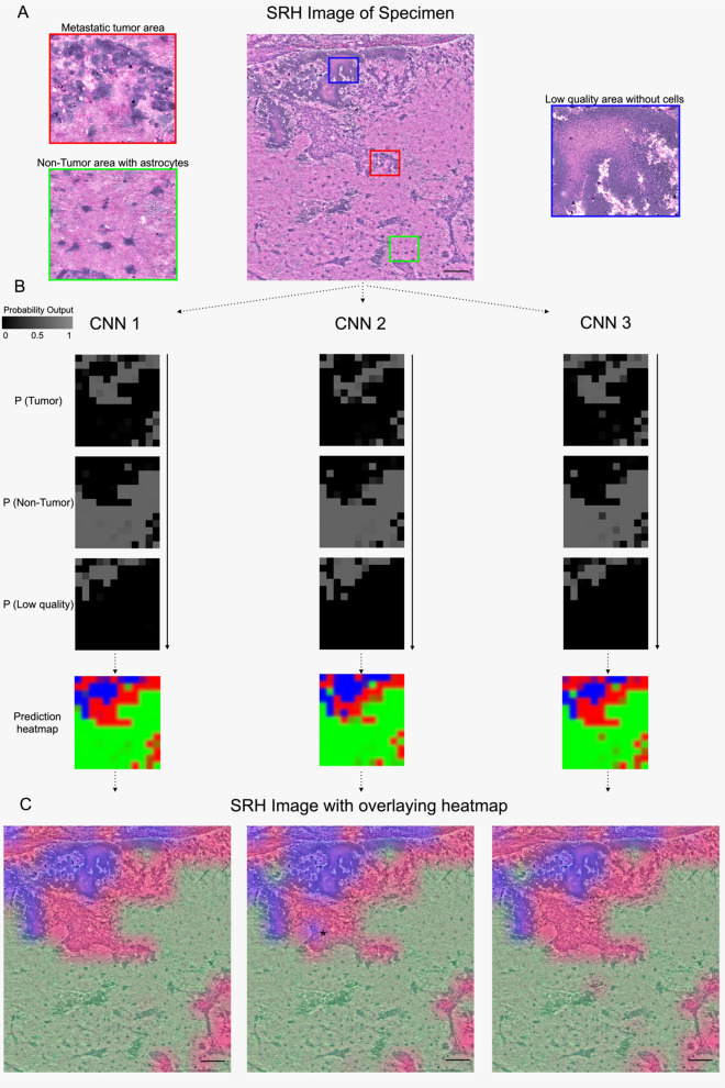
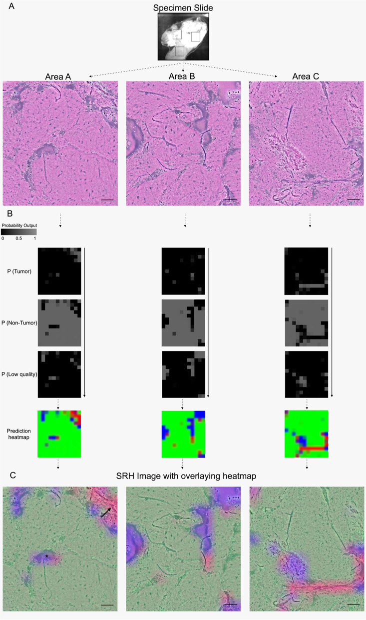
Determining the presence of tumor in biopsies and the decision-making during resections is often dependent on intraoperative rapid frozen-section histopathology. Recently, stimulated Raman scattering microscopy has been introduced to rapidly generate digital hematoxylin-and-eosin-stained-like images (stimulated Raman histology) for intraoperative analysis. To enable intraoperative prediction of tumor presence, we aimed to develop a new deep residual convolutional neural network in an automated pipeline and tested its validity. In a monocentric prospective clinical study with 94 patients undergoing biopsy, brain or spinal tumor resection, Stimulated Raman histology images of intraoperative tissue samples were obtained using a fiber-laser-based stimulated Raman scattering microscope. A residual network was established and trained in ResNetV50 to predict three classes for each image: (1) tumor, (2) non-tumor, and (3) low-quality. The residual network was validated on images obtained in three small random areas within the tissue samples and were blindly independently reviewed by a neuropathologist as ground truth. 402 images derived from 132 tissue samples were analyzed representing the entire spectrum of neurooncological surgery. The automated workflow took in a mean of 240 s per case, and the residual network correctly classified tumor (305/326), non-tumorous tissue (49/67), and low-quality (6/9) images with an inter-rater agreement of 89.6% (κ = 0.671). An excellent internal consistency was found among the random areas with 90.2% (Cα = 0.942) accuracy. In conclusion, the novel stimulated Raman histology-based residual network can reliably detect the microscopic presence of tumor and differentiate from non-tumorous brain tissue in resection and biopsy samples within 4 min and may pave a promising way for an alternative rapid intraoperative histopathological decision-making tool.
Keywords: Artificial intelligence; Brain tumor; Deep learning; Neurosurgery; Stimulated Raman histology; Tissue detection.
© 2022. The Author(s).
Conflict of interest statement
C.F., A.I.M., and F.K. are employees of Invenio Imaging Inc, the manufacturer of the SRH imaging system. C.F., A.I.M., and F.K. did not take part in any data analysis or influenced the report or interpretation of the results. No other conflicts of interest to declare.
Figures


References
-
- Novis DA, Zarbo RJ. Interinstitutional comparison of frozen section turnaround time. A College of American Pathologists Q-Probes study of 32868 frozen sections in 700 hospitals. Arch Pathol Lab Med. 1997;121:559–67. - PubMed
Publication types
MeSH terms
Substances
LinkOut - more resources
Full Text Sources
Medical
