

Three pairs of surrogate redox partners comparison for Class I cytochrome P450 enzyme activity reconstitution
- PMID: 35933448
- PMCID: PMC9357085
- DOI: 10.1038/s42003-022-03764-4
Three pairs of surrogate redox partners comparison for Class I cytochrome P450 enzyme activity reconstitution
Abstract
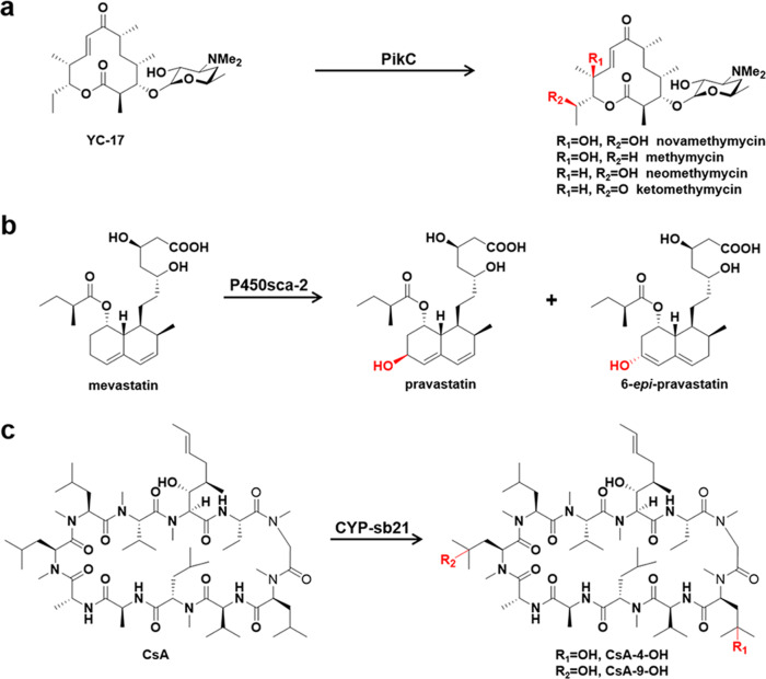
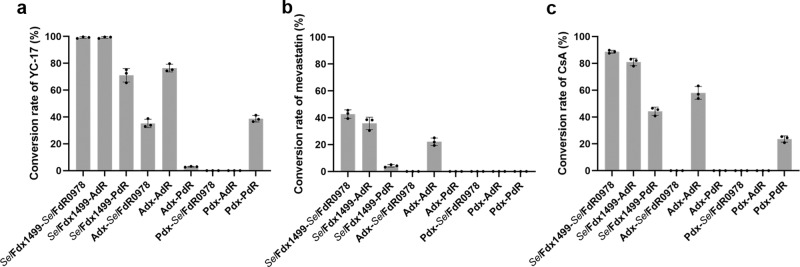
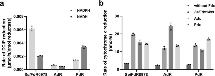
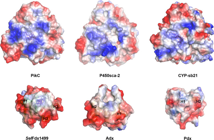
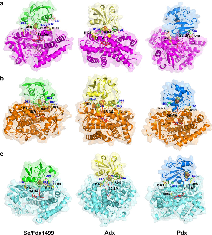
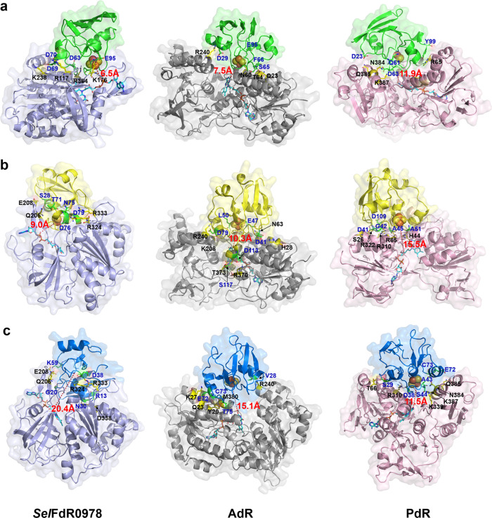
Most P450s require redox partners for the electron transfer during catalysis. However, little information is available on cognate redox partners for P450s, which greatly limits P450 function exploration and practical application. Thus, the stategy of building various hybrid P450 catalytic systems with surrogate redox partner has often adopted to engineer P450 biocatalysts. In this study, we compare three pairs of frequently-used surrogate redox partner SelFdx1499/SelFdR0978, Adx/AdR and Pdx/PdR and in terms of their electron transfer properties. The three selected bacterial Class I P450s include PikC, P450sca-2 and CYP-sb21, which are responsible for production of high-value-added products. Here we show that SelFdx1499/SelFdR0978 is the most promising redox partner compared to Adx/AdR and Pdx/PdR. The results provide insights into the domination for P450-redox partner interactions in modulating the catalytic activity of P450s. This study not only produces a more active biocatalyst but also suggests a general chose for a universal reductase which would facilitate engineering of P450 catalyst.
© 2022. The Author(s).
Conflict of interest statement
The authors declare no competing interests.
Figures
Similar articles
-
A large-scale comparative analysis of affinity, thermodynamics and functional characteristics of interactions of twelve cytochrome P450 isoforms and their redox partners.Biochimie. 2019 Jul;162:156-166. doi: 10.1016/j.biochi.2019.04.020. Epub 2019 Apr 26. Biochimie. 2019. PMID: 31034920
-
Semi-rational engineering of cytochrome P450sca-2 in a hybrid system for enhanced catalytic activity: insights into the important role of electron transfer.Biotechnol Bioeng. 2013 Nov;110(11):2815-25. doi: 10.1002/bit.24960. Epub 2013 Jun 4. Biotechnol Bioeng. 2013. PMID: 23737252
-
Biological diversity of cytochrome P450 redox partner systems.Adv Exp Med Biol. 2015;851:299-317. doi: 10.1007/978-3-319-16009-2_11. Adv Exp Med Biol. 2015. PMID: 26002740 Review.
-
P450BM-3; a tale of two domains--or is it three?Steroids. 1997 Jan;62(1):117-23. doi: 10.1016/s0039-128x(96)00169-9. Steroids. 1997. PMID: 9029725 Review.
-
Engineering of a hybrid biotransformation system for cytochrome P450sca-2 in Escherichia coli.Biotechnol J. 2013 Jul;8(7):785-93. doi: 10.1002/biot.201200097. Epub 2013 Jun 21. Biotechnol J. 2013. PMID: 23744742
Cited by
-
Unnatural activities and mechanistic insights of cytochrome P450 PikC gained from site-specific mutagenesis by non-canonical amino acids.Nat Commun. 2023 Mar 25;14(1):1669. doi: 10.1038/s41467-023-37288-0. Nat Commun. 2023. PMID: 36966128 Free PMC article.
-
Cooperative Substrate Binding Controls Catalysis in Bacterial Cytochrome P450terp (CYP108A1).J Am Chem Soc. 2023 Feb 13:10.1021/jacs.2c12388. doi: 10.1021/jacs.2c12388. Online ahead of print. J Am Chem Soc. 2023. PMID: 36779970 Free PMC article.
-
Pharmaceutical removal from wastewater by introducing cytochrome P450s into microalgae.Microb Biotechnol. 2024 Jun;17(6):e14515. doi: 10.1111/1751-7915.14515. Microb Biotechnol. 2024. PMID: 38925623 Free PMC article. Review.
-
Phylogeny-metabolism dual-directed single-cell genomics for dissecting and mining ecosystem function by FISH-scRACS-seq.Innovation (Camb). 2025 Jan 16;6(3):100759. doi: 10.1016/j.xinn.2024.100759. eCollection 2025 Mar 3. Innovation (Camb). 2025. PMID: 40098675 Free PMC article.
-
Designing cytochrome P450 enzymes for use in cancer gene therapy.Front Bioeng Biotechnol. 2024 May 24;12:1405466. doi: 10.3389/fbioe.2024.1405466. eCollection 2024. Front Bioeng Biotechnol. 2024. PMID: 38860140 Free PMC article. Review.
References
Publication types
MeSH terms
Substances
LinkOut - more resources
Full Text Sources
Molecular Biology Databases
Research Materials
