. 2022 Aug 6;13(1):4583.
doi: 10.1038/s41467-022-32325-w.

Neutrophils restrain sepsis associated coagulopathy via extracellular vesicles carrying superoxide dismutase 2 in a murine model of lipopolysaccharide induced sepsis

Affiliations

Neutrophils restrain sepsis associated coagulopathy via extracellular vesicles carrying superoxide dismutase 2 in a murine model of lipopolysaccharide induced sepsis

Wenjie Bao et al. Nat Commun. .

Abstract

Disseminated intravascular coagulation (DIC) is a complication of sepsis currently lacking effective therapeutic options. Excessive inflammatory responses are emerging triggers of coagulopathy during sepsis, but the interplay between the immune system and coagulation are not fully understood. Here we utilize a murine model of intraperitoneal lipopolysaccharide stimulation and show neutrophils in the circulation mitigate the occurrence of DIC, preventing subsequent septic death. We show circulating neutrophils release extracellular vesicles containing mitochondria, which contain superoxide dismutase 2 upon exposure to lipopolysaccharide. Extracellular superoxide dismutase 2 is necessary to induce neutrophils' antithrombotic function by preventing endothelial reactive oxygen species accumulation and alleviating endothelial dysfunction. Intervening endothelial reactive oxygen species accumulation by antioxidants significantly ameliorates disseminated intravascular coagulation improving survival in this murine model of lipopolysaccharide challenge. These findings reveal an interaction between neutrophils and vascular endothelium which critically regulate coagulation in a model of sepsis and may have potential implications for the management of disseminated intravascular coagulation.


Conflict of interest statement

The authors declare no competing interests.

Figures

Fig. 1
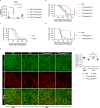
Fig. 1. Transfer of LPS-primed circulating neutrophils protects against sepsis without affecting the initial cytokine storm.
a Schematic diagram of transferring PBS or LPS-primed neutrophils into wild-type mice followed by lethal LPS challenge. b Survival of mice after lethal LPS challenge. Mice were pre-transferred with LPS-primed peripheral blood (PB) neutrophils (PB LPSneutrophils) (n = 19), PBS-primed PB neutrophils (PB PBSneutrophils) (n = 19) or PBS only (n = 19). c Survival of mice after lethal LPS challenge. Mice were pre-transferred with LPS-primed bone marrow (BM) neutrophils (BM LPSneutrophils) (n = 8), PBS-primed BM neutrophils (BM PBSneutrophils) (n = 8) or PBS only (n = 8). d Survival of mice after the cecal ligation and puncture (CLP). Mice were pre-transferred with LPS-primed peripheral blood (PB) neutrophils (n = 22), PBS-primed PB neutrophils (n = 22) or PBS only (n = 22). p = 0.0270. e Survival of mice after lethal Con A challenge. Mice were pre-transferred with LPS-primed PB neutrophils (n = 7), PBS-primed PB neutrophils (n = 7) or PBS only (n = 7). p = 0.0242. f Plasma levels of IL6, TNFα and MCP1 of the indicated recipient mice after lethal LPS (For IL6: 0 h: PBS n = 3, PBSneutrophils n = 3, LPSneutrophils n = 4; 1 h: PBS n = 5, PBSneutrophils n = 8, LPSneutrophils n = 7; 4 h: PBS n = 3, PBSneutrophils n =4, LPSneutrophils n = 3; For TNFα: 0 h: PBS n = 4, PBSneutrophils n = 4, LPSneutrophils n = 4; 1 h: PBS n = 5, PBSneutrophils n = 8, LPSneutrophils n = 7; 4 h: PBS n = 3, PBSneutrophils n = 3, LPSneutrophils n = 3; For MCP1: 0 h: PBS n = 3, PBSneutrophils n = 4, LPSneutrophils n = 3; 1 h: PBS n = 5, PBSneutrophils n = 8, LPSneutrophils n = 7; 4 h: PBS n = 3, PBSneutrophils n = 4, LPSneutrophils n = 3;). g Plasma levels of CXCL2 and CXCL15 of the indicated recipient mice after lethal LPS (For CXCL2: 0 h: PBS n = 5; 1 h: PBS n = 6, PBSneutrophils n = 6, LPSneutrophils n = 6; 4 h: PBS n = 5, PBSneutrophils n = 6, LPSneutrophils n = 5; For CXCL15: 0 h: PBS n = 5; 1 h: PBS n = 4, PBSneutrophils n = 4, LPSneutrophils n = 4; 4 h: PBS n = 4, PBSneutrophils n = 4, LPSneutrophils n = 4;). h Plasma levels of IL6, TNFα and MCP1 of the indicated recipient mice after PBS or lethal Con A (For IL6: 1 h PBS n = 5; 1 h ConA: PBS n = 6, PBSneutrophils n = 6, LPSneutrophils n = 6; For TNFα: 1 h PBS n = 6; 1 h ConA: PBS n = 6, PBSneutrophils n = 6, LPSneutrophils n = 6; For MCP1: 1 h PBS n = 6; 1 h ConA: PBS n = 6, PBSneutrophils n = 6, LPSneutrophils n = 6). Source data are provided as a Source Data file. Data are representative of, or pooled from at least two independent experiments. Data are mean ± SD. Log-rank (Mantel–Cox) test was used for be; Two-tailed unpaired t tests was used for fh. *p  <  0.05, **p  <  0.01, ****p  <  0.0001, N.S. not significant.
Fig. 2
Fig. 2. Transfer of LPS-primed neutrophils mitigate the occurrence of DIC and alleviate endothelial dysfunction.
a Plasma levels of LDH of mice after PBS (n = 3). Plasma levels of LDH of mice 1 h after lethal LPS. Mice were pre-transferred with LPS-primed PB neutrophils (LPSneutrophils) (n = 5), PBS-primed PB neutrophils (PBSneutrophils) (n = 5) or PBS only (n = 5). b Representative imaging of liver functional vessel in the indicated recipient mice 1 h after lethal LPS. FITC-Dextran (green) labeled blood vessels, and DID-labeled red blood cells (red) indicated blood flow. Argatroban, a thrombin inhibitor, was used as the positive control. Scale bar, 100 μm. c Mean ratios of the functional vessel area to the total vessel area in mice 1 h after PBS (n = 5). Mean ratios of the functional vessel area to the total vessel area in mice 1 h after lethal LPS. Mice were pre-transferred with LPS-primed PB neutrophils (n = 5), PBS-primed PB neutrophils (n = 4) or PBS only (n = 6). d Representative images of immunofluorescence staining of fibrin (top panel; scale bar, 200 μm) and H&E staining (bottom panel; scale bar, 50 μm) of liver sections from indicated recipient mice 4 h after PBS or lethal LPS. The white arrows indicate thrombi in liver vessels. e Representative flow plots showing Annexin V binding to hepatic endothelial cells 1 h after PBS or lethal LPS. f Mean percentages of Annexin V+ hepatic endothelial cells in the indicated mice (n = 5 per group). Source data are provided as a Source Data file. Data are representative of, or pooled from at least three independent experiments. Data are mean ± SD. Two-tailed unpaired t tests were used for statistical analyses. *p  <  0.05, **p  <  0.01.
Fig. 3
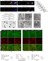
Fig. 3. Sod2 is required for neutrophils to restrain septic DIC.
a Quantitative Real-time PCR analysis of Sod2 mRNA levels in PB or BM neutrophils challenged with PBS (PBSneutrophils) or LPS (LPSneutrophils) (n = 4 per group). b Survival of mice after lethal LPS challenge. Mice were pre-transferred with LPS-primed PB neutrophils treated with (LPSneutrophilsMock) (n = 8), or without CsA (LPSneutrophilsCsA) (n = 7), PBS-primed PB neutrophils treated with (PBSneutrophilsMock) (n = 8), or without CsA (PBSneutrophilsCsA) (n = 7) or PBS only (n = 8). c Survival cure of Sod2+/− mice (n = 20) and WT (n = 20) littermates after lethal LPS. p = 0.0131. d Survival of mice after lethal LPS challenge. Mice were pre-transferred with LPS-primed Sod2+/− PB neutrophils (LPSneutrophilsSod2+/−) (n = 6), LPS-primed WT PB neutrophils (LPSneutrophilsWT) (n = 6) or PBS only (n = 9). e Representative imaging of liver functional vessel in the indicated recipient mice 1 h after lethal LPS. FITC-Dextran (green) labeled blood vessels, and DID-labeled red blood cells (red) indicated blood flow. Scale bar, 100 μm. f Mean ratios of the functional vessel area to the total vessel area in mice 1 h after PBS (n = 5). Mean ratios of the functional vessel area to the total vessel area in mice 1 h after lethal LPS. Mice were pre-transferred with LPS-primed Sod2+/− PB neutrophils (n = 4), LPS-primed PB neutrophils (n = 4) or PBS only (n = 4). Source data are provided as a Source Data file. Data are representative of, or pooled from at least three independent experiments. Data are mean ± SD. Log-rank (Mantel–Cox) test was used for bd; Two-tailed unpaired t tests was used for a and f. *p  <  0.05, **p  <  0.01, ***p  <  0.001, N.S. not significant.
Fig. 4
Fig. 4. Mitochondrion-containing EVs from neutrophils exhibit Sod2-dependent antithrombotic function.
ab Confocal images of Sod2 (Red), Mito-Dendra2 (Green) and Ly6G (Grey) among blood leukocytes from PhAMfloxed × Mrp8-Cre mice. Scale bar in a, 3 μm; Scale bar in b, 2 μm. cd Confocal images of SOD2 (Red), TOM22 (Green) and CD16 (Grey) among blood leukocytes from healthy donors. Scale bar in c, 3 μm; Scale bar in d, 2 μm. e Schematic of sequential centrifugation to isolate EVs from mouse vasculature lavage fluid. f Western blot analysis of Sod2, COX IV and Ly6G protein levels in different pellets (P1-P5) purified from mice 4 h after PBS or LPS. g Representative transmission electron microscope (TEM) images of the isolated EVs from P1 (left) and P2 (right). Scale bar, 2 μm. h Representative TEM images of isolated EVs containing small mitochondria (red arrows). Scale bar, 0.2 μm. i Representative TEM images of a liver section showing a mitochondrion-containing EV (red arrow) within an intravascular neutrophil. Scale bar, 2 μm. The boxed area is enlarged at the bottom panel. Scale bar, 1 μm. j, m Representative imaging of liver functional vessel in the indicated recipient mice 1 h after lethal LPS. FITC-Dextran (green) labeled blood vessels, and DID-labeled red blood cells (red) indicated blood flow. Scale bar, 100 μm. k Mean ratios of the functional vessel area to the total vessel area in mice 1 h after lethal LPS. Mice were pre-transferred with LPS-primed EVs (LPSEVs) (n = 6), PBS-primed EVs (PBSEVs) (n = 7) or PBS only (n = 7). p = 0.0396. l Survival of mice after lethal LPS (20 mg/kg) challenge. Mice were pre-transferred with LPS-primed EVs (n = 21), PBS-primed EVs (n = 21) or PBS only (n = 21). n Mean ratios of the functional vessel area to the total vessel area in mice 1 h after lethal LPS. Mice were pre-transferred with LPS-primed Sod2+/− PB EVs (LPSEVsSod2+/−) (n = 6), LPS-primed EVs (LPSEVsWT) (n = 7) or PBS only (n = 7). Source data are provided as a Source Data file. Data are representative of or pooled from at least three independent experiments. Data are mean ± SD. Log-rank (Mantel–Cox) test was used for i. Two-tailed unpaired t tests were used for statistical analyses in k and m. *p  <  0.05.
Fig. 5
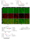
Fig. 5. Neutrophils release mitochondrion-containing EVs during migration and reduce endothelial ROS accumulation.
ab Representative images from intravital imaging of neutrophils (Ly6G-Red, red arrows) within liver vasculature (Dextran-Green) in wild type mice. The white arrow indicates a vesicle released by a migrating neutrophil. Scale bar in a, 30 μm; Scale bar in b, 5 μm. cd Representative images from intravital imaging of neutrophils (Ly6G-Red) within liver vasculature (CD144-Blue) in PhAMfloxed × Mrp8-Cre mice. Mitochondria of neutrophils were labeled by Mito-Dentra2 (Dendra2-Green). The white arrow indicates a mitochondrion-containing vesicle released by a migrating neutrophil. Scale bar in (c), 15 μm; Scale bar in d, 5 μm. e Flow analysis of ROS levels in hepatic endothelial cells 1 h after PBS or lethal LPS in the indicated recipient mice. f M.F.I. of ROS levels in hepatic endothelial cells 1 h after PBS or lethal LPS in the indicated recipient mice (n = 5 per group). Source data are provided as a Source Data file. Data are representative of or pooled from at least three independent experiments. Data are mean ± SD. Two-tailed unpaired t tests were used for statistical analyses. *p  <  0.05, **p  <  0.01, ***p  <  0.001.
Fig. 6
Fig. 6. Transfer of Sod2-rich EVs alleviates the coagulopathy in neutropenic mice upon lethal LPS challenge.
a Survival of α-Ly6G (1A8) (n = 15), or isotype antibody (Rat IgG2a) (n = 15) treated mice after lethal LPS. p = 0.0078. b Survival of DT-treated PMNDTR (ROSA26-iDTRKI × Mrp8-Cre) (n = 15) mice and PMNWT (Mrp8-Cre) (n = 15) littermates after lethal LPS. p = 0.0185. c Representative imaging of liver functional vessel in the neutropenic mice 1 h after PBS or lethal LPS. FITC-Dextran (green) labeled blood vessels, and DID-labeled red blood cells (red) indicated blood flow. Scale bar, 100 μm. d Mean ratios of the functional vessel area to the total vessel area in α-Ly6G (1A8) (n = 4), or isotype antibody (n = 4) treated mice after PBS. Mean ratios of the functional vessel area to the total vessel area in α-Ly6G (1A8) (n = 8), or isotype antibody (n = 6) treated mice after lethal LPS. Mean ratios of the functional vessel area to the total vessel area in α-Ly6G (1A8) treated mice after lethal LPS. Mice were pre-transferred with LPS-primed EVs (LPSEVs) (n = 5). e Western blot analysis of Ly6G protein levels in different pellets (P1-P5) purified from WT or TSPAN9−/− mice 4 h after LPS. f Survival of mice after lethal LPS challenge. Mice were pretransferred with LPS-primed TSPAN9−/− PB neutrophils (LPSneutrophilsTSPAN9−/−) (n = 6), LPS-primed WT PB neutrophils (LPSneutrophilsWT) (n = 10) or PBS only (n = 10). Source data are provided as a Source Data file. Data are representative of, or pooled from at least three independent experiments. Data are mean ± SD. log-rank (Mantel–Cox) test was used for statistical analyses in a, b, f. Two-tailed unpaired t tests were used for statistical analyses in d. *p  <  0.05, **p  <  0.01, N.S. not significant.
Fig. 7
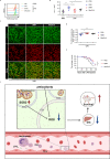
Fig. 7. Antioxidants reduces endothelial ROS, attenuates DIC and improves survival in sepsis.
a Flow analysis of ROS levels in hepatic endothelial cells 1 h after PBS or lethal LPS in the indicated recipient mice. b M.F.I. of ROS levels in hepatic endothelial cells 1 h after PBS or lethal LPS in the indicated mice (n = 5 per group). c Representative imaging of liver functional vessel in the indicated mice 1 h after lethal LPS. FITC-Dextran (green) labeled blood vessels, and DID-labeled red blood cells (red) indicated blood flow. Scale bar, 100 μm. d Mean ratios of the functional vessel area to the total vessel area in mice 1 h after lethal LPS. Mice were injected with mitoQ 10 (5 mg/kg, 30 min before lethal LPS, n = 7), NMN (300 mg/kg, 1 h before lethal LPS, n = 7) or PBS only (n = 8). e Survival of mice after lethal LPS challenge. Mice were injected with mitoQ 10 (5 mg/kg, 30 min before lethal LPS, n = 20), NMN (300 mg/kg, 1 h before lethal LPS, n = 20) or PBS only (n = 20). f A graphical summary showing that circulating neutrophils employ Sod2-rich EVs to diminish intravascular ROS, thereby mitigating DIC and protecting host against sepsis. Alternatively, antioxidants administration displays protective effects similar to the Sod2-rich EVs, highlighting the potentials of antioxidants in managing sepsis. Source data are provided as a Source Data file. Data are representative of, or pooled from at least three independent experiments. Data are mean ± SD. log-rank (Mantel–Cox) test was used for statistical analyses in e. Two-tailed unpaired t tests were used for statistical analyses in b and d. *p  <  0.05, **p  <  0.01.

Similar articles

Cited by

References

    1. Singer M, et al. The Third International Consensus Definitions for Sepsis and Septic Shock (Sepsis-3) JAMA. 2016;315:801–810. doi: 10.1001/jama.2016.0287. - DOI - PMC - PubMed
    1. Giustozzi, M. et al. Coagulopathy and sepsis: Pathophysiology, clinical manifestations and treatment. Blood Rev., 100864, 10.1016/j.blre.2021.100864 (2021). - PubMed
    1. Tang D, Wang H, Billiar TR, Kroemer G, Kang R. Emerging mechanisms of immunocoagulation in sepsis and septic shock. Trends Immunol. 2021;42:508–522. doi: 10.1016/j.it.2021.04.001. - DOI - PMC - PubMed
    1. Mayadas TN, Cullere X, Lowell CA. The multifaceted functions of neutrophils. Annu Rev. Pathol. 2014;9:181–218. doi: 10.1146/annurev-pathol-020712-164023. - DOI - PMC - PubMed
    1. Wang J. Neutrophils in tissue injury and repair. Cell Tissue Res. 2018;371:531–539. doi: 10.1007/s00441-017-2785-7. - DOI - PMC - PubMed

Publication types