. 2022 Oct;11(20):e2200964.
doi: 10.1002/adhm.202200964. Epub 2022 Aug 18.

Living Biomaterials to Engineer Hematopoietic Stem Cell Niches

Affiliations

Living Biomaterials to Engineer Hematopoietic Stem Cell Niches

Michaela Petaroudi et al. Adv Healthc Mater. 2022 Oct.

Abstract

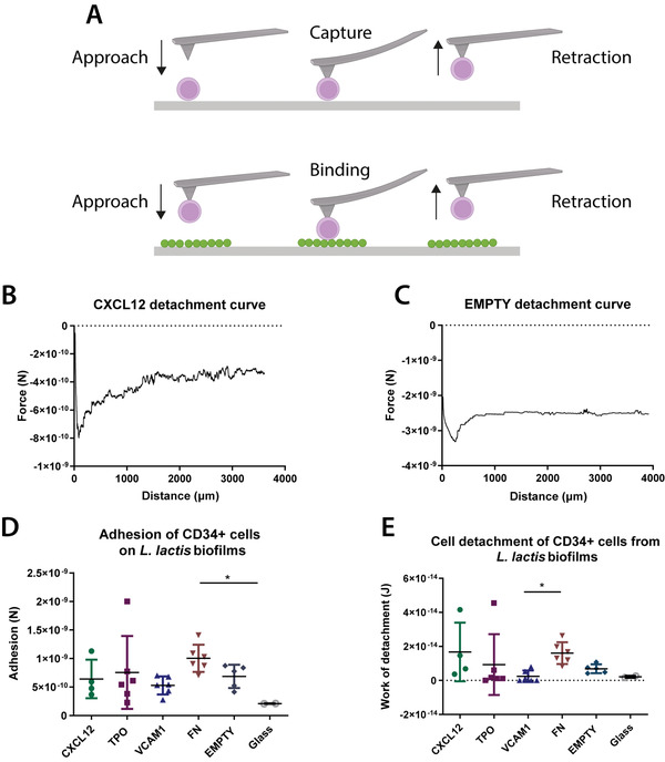
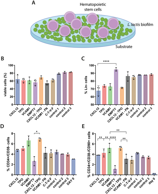
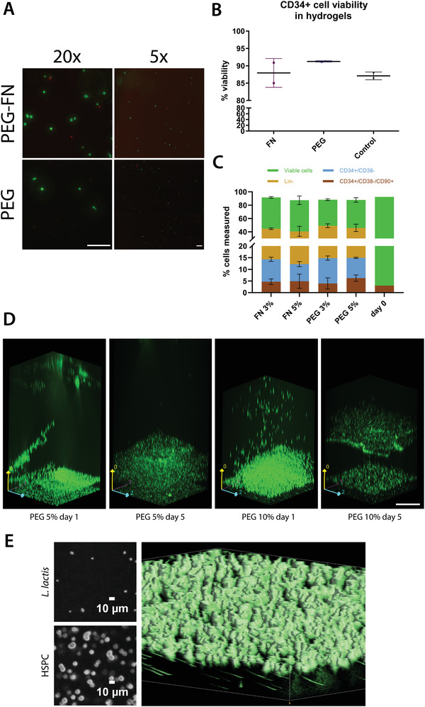
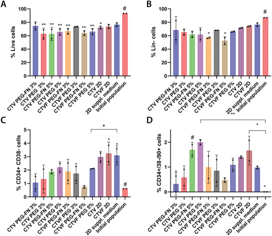
Living biointerfaces are a new class of biomaterials combining living cells and polymeric matrices that can act as biologically active and instructive materials that host and provide signals to surrounding cells. Here, living biomaterials based on Lactococcus lactis to control hematopoietic stem cells in 2D surfaces and 3D hydrogels are introduced. L. lactis is modified to express C-X-C motif chemokine ligand 12 (CXCL12), thrombopoietin (TPO), vascular cell adhesion protein 1 (VCAM1), and the 7th-10th type III domains of human plasma fibronectin (FN III7-10 ), in an attempt to mimic ex vivo the conditions of the human bone marrow. These results suggest that living biomaterials that incorporate bacteria expressing recombinant CXCL12, TPO, VCAM1, and FN in both 2D systems direct hematopoietic stem and progenitor cells (HSPCs)-bacteria interaction, and in 3D using hydrogels functionalized with full-length human plasma fibronectin allow for a notable expansion of the CD34+ /CD38- /CD90+ HSPC population compared to the initial population. These results provide a strong evidence based on data that suggest the possibility of using living materials based on genetically engineered bacteria for the ex-vivo expansion of HSPC with eventual practical clinical applications in HSPCs transplantation for hematological disorders.

Keywords: cell engineering; genetic engineering; hematopoiesis; living materials; microenvironment engineering; stem cells; synthetic biology.


Conflict of interest statement

The authors declare no conflict of interest.

Figures

Figure 1
Figure 1
Adhesion strength between CD34+ HSPC cells and L. lactis biofilms. A) A diagrammatic sketch of the HSPC capture on poly‐d‐lysine‐coated silicon nitride AFM cantilevers and the following adhesion force measurement on L. lactis biofilms for 15 s. B,C) Sample atomic force microscopy (AFM) curves from representative samples show the force of adhesion of CD34+ HSPC cells to CXCL12 and EMPTY biofilms. D) The force of adhesion and E) work of detachment of the stem cells to L. lactis biofilms expressing CXCL12, TPO, VCAM1, and FN, as well as EMPTY biofilms and glass coverslips (controls) were measured using AFM. There is a significant increase of adhesion force of cells on the FN‐expressing biofilm compared to D) the negative control (EMPTY) and E) VCAM1. This result agrees with previously published results in the literature. The work of detachment was calculated as the area under the curve from z = 0 to the detachment point, shown as the area between the curve and the dotted line. Data analysis in (D) and (E) was performed using a nonparametric Kruskal–Wallis test with Dunn's post hoc multiple comparison test, α = 0.05 (*p < 0.05). Data are presented as mean ± SD, n ≥ 2.
Figure 2
Figure 2
CD34+ cell populations as assessed by flow cytometry after 5 days of culture on top of L. lactis biofilms, as depicted in (A). Graph (B) represents stem cell viability, suggesting that it remains unaffected by the presence of the biofilms and is comparable to the traditional HSPC expansion methods (control 1 to 3, described later in this caption). The co‐cultures with the biofilms can also be associated with C) the maintenance of a lineage‐negative phenotype and D) the traditionally recognized CD34+/38 cell phenotype. E) Finally, the engrafting CD34+/CD38/CD90+ population of HSPCs is also maintained in the conditions where a biofilm is present, at similar levels to the positive controls. Interestingly, this is not the case in the EMPTY condition, where the biofilm produces no recombinant proteins, and where both the CD34+/CD38 and CD90+ cell populations are significantly lower than all other conditions. In all cases, except for the EMPTY condition, the stem cell populations of interest have shown increased expansion compared to the initially seeded population (shown as Day 0). The data in B are presented as mean ± SD and was analyzed using a one‐way ANOVA with Tukey post hoc test (n ≥ 2, α = 0.05, significance values *p < 0.05, **p < 0.01, ****p < 0.0001, compared to the reference condition). Data in (C)–(E) were analyzed using a nonparametric Kruskal–Wallis test, with a Dunn's post hoc multiple comparison test, n ≥ 2, α = 0.05, significance values *p < 0.05, **p < 0.01, ****p < 0.0001. Explanation of the control conditions: Control 1: CD34+ cells cultured on Sigmacote‐coated coverslips in the absence of bacteria in IMDM, 20% BIT, 10% L‐glutamine, 10 ng mL–1 soluble SCF/FLT3L, and 5 ng mL–1 TPO. Control 2: same as control 1, with SR1 at 1 × 10−6 m. Control 3: same as control 2, but no Sigmacote‐coated glass coverslip was used, the cells were cultured directly on the polystyrene surface of the multiwell plate.
Figure 3
Figure 3
CD34+ cell viability in hydrogels. A) Representative images of the live/dead assay performed to assess CD34+ cell viability. The live cells are shown in green, and the dead cells are depicted in red. Scale bar is 200 µm for both images. B) CD34+ cell viability was recorded above ≈85% in all hydrogels, including nonfunctionalized PEG and PEG‐FN hydrogels. The results were compared to a 2D control, where the stem cells were cultured in the traditional HSPC maintenance media, IMDM plus the cytokine cocktail, in the absence of a hydrogel. No statistically significant differences were observed between the cell viability in the hydrogels and the 2D control, using a Kruskal–Wallis nonparametric test with Dunn's post hoc multiple comparison test (n ≥ 2, α = 0.05). Data are presented as mean ± SD. C) CD34+ cell viability, the lineage‐negative CD34+ cell population, the naïve HSPC population, and the engrafting HSPC population as measured by flow cytometry after a 5 day incubation in different PEG‐based hydrogels. All hydrogels used in this experiment were based on PEG, with the addition of FN. The hydrogels were engineered for different Young's modulus, based on the percentage of the polymer content (3% and 5% w/v), ranging from 2 kPa (3% w/v polymer) to 5 kPa (5% w/v polymer). The results were compared to the initially seeded populations (day 0). No statistically significant differences were observed after data analysis using a two‐way ANOVA with Tukey's multiple comparison post hoc test (n = 3, α = 0.05, presented as mean ± SD). The results are the aggregate of three independent cell experiments. D) Confocal Ζ‐stack images showing an EMPTY L. lactis biofilm and a hydrogel containing encapsulated CD34+ cells incubated on top of the bacteria. Both the biofilm and stem cells express GFP and are depicted in green for visual purposes. In the PEG 10%, day 5 image, there is a slight outgrowth of bacteria on top of the biofilm. Images were taken in a Zeiss spinning disk confocal microscope. Scale bar is 200 µm. E) HSPCs are shown embedded in the hydrogel while L. lactis cells are located under the hydrogel, with a smaller diameter. The two small images in the left part of the panel show two slices of the z‐stack, where the L. lactis cells show an approximate diameter of 1.5 µm while the HSPC cells have an approximate diameter of 10 µm. The 3D volume reconstruction of the gel, the bigger image of the panel, shows the HSPCs on top with a bigger diameter while L. lactis cells, in the bottom, are visibly smaller. The elongated shape of the L. lactis cells is due to a small drift of the hydrogel in the stage while imaging the hydrogel.
Figure 4
Figure 4
CD34+ viability and cell populations as assessed by flow cytometry after 5 days of culture inside different hydrogels, and in the presence of L. lactis biofilms expressing different recombinant cytokines. This experiment features PEG and PEG‐FN hydrogels of different stiffnesses (3% and 5% w/v of polymer in each case, corresponding to a G’ of 2 and 5 kPa, respectively), and L. lactis biofilms expressing different cytokines, including CXCL12 (C), TPO (T), VCAM1 (V), and FN (F). After 5 days of culture, we performed flow cytometric analysis of the populations, recording A) CD34+ cell viability, B) the lineage‐negative cell population, as well as C) the HSPC content, and D) engrafting HSPCs population. All conditions were compared to the initially seeded population and a 2D control, where the stem cells were cultured in expansion media with added cytokines, without the presence of bacteria (2D cytokines). The data were analyzed using a two‐way ANOVA with Tukey's post hoc test (*p < 0.05, **p < 0.01) and the statistical differences are shown compared to the reference condition (#). Results are the aggregate of two independent experiments combining the data from three different donors.

Similar articles

Cited by

References

    1. Henrich T. J., Lelièvre J.‐D., Curr. Opin. HIV AIDS 2018, 13, 381. - PubMed
    1. Hogan L. E., Hanhauser E., Hobbs K. S., Palmer C. D., Robles Y., Jost S., Lacasce A. S., Abramson J., Hamdan A., Marty F. M., Kuritzkes D. R., Henrich T. J., PLoS One 2018, 13, e0197298. - PMC - PubMed
    1. Roβmanith T., Schröder B., Bug G., Müller P., Klenner T., Knaus R., Hoelzer D., Ottmann O. G., Stem Cells 2001, 19, 313. - PubMed
    1. Branco A., Bucar S., Moura‐Sampaio J., Lilaia C., Cabral J. M. S., Fernandes‐Platzgummer A., Lobato Da Silva C., Front. Bioeng. Biotechnol. 2020, 8, 1154. - PMC - PubMed
    1. Porter R. L., Georger M. A., Bromberg O., Mcgrath K. E., Frisch B. J., Becker M. W., Calvi L. M., Stem Cells 2013, 31, 372. - PMC - PubMed

Publication types