. 2022 Oct;237(10):3927-3943.
doi: 10.1002/jcp.30849. Epub 2022 Aug 7.

PTH1R translocation to primary cilia in mechanically-stimulated ostecytes prevents osteoclast formation via regulation of CXCL5 and IL-6 secretion

Affiliations

PTH1R translocation to primary cilia in mechanically-stimulated ostecytes prevents osteoclast formation via regulation of CXCL5 and IL-6 secretion

Irene Tirado-Cabrera et al. J Cell Physiol. 2022 Oct.

Abstract

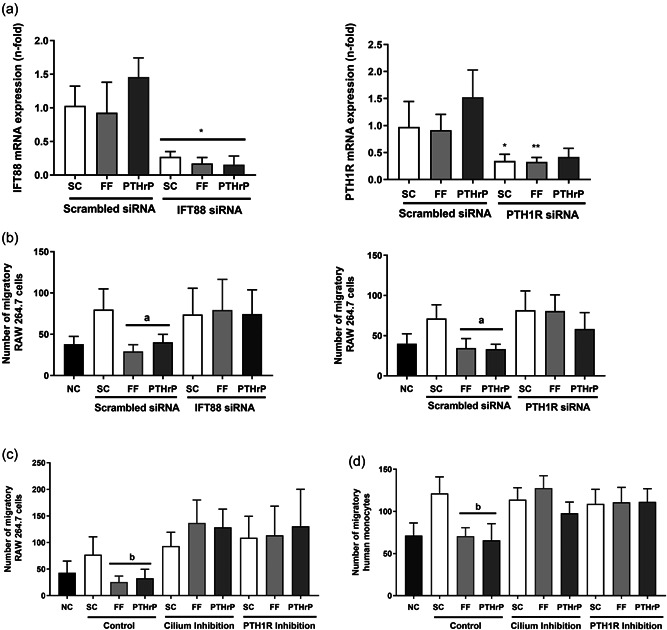
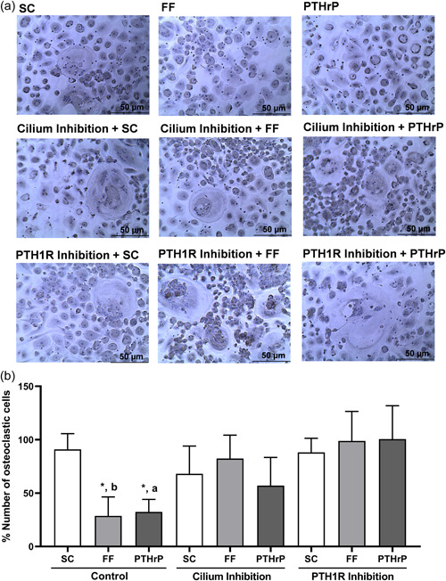
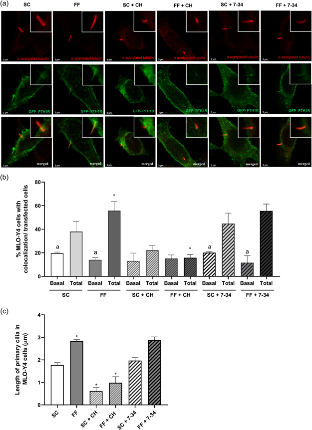
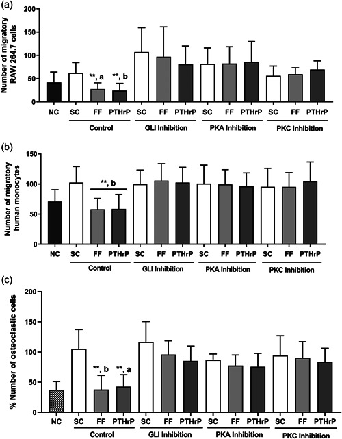
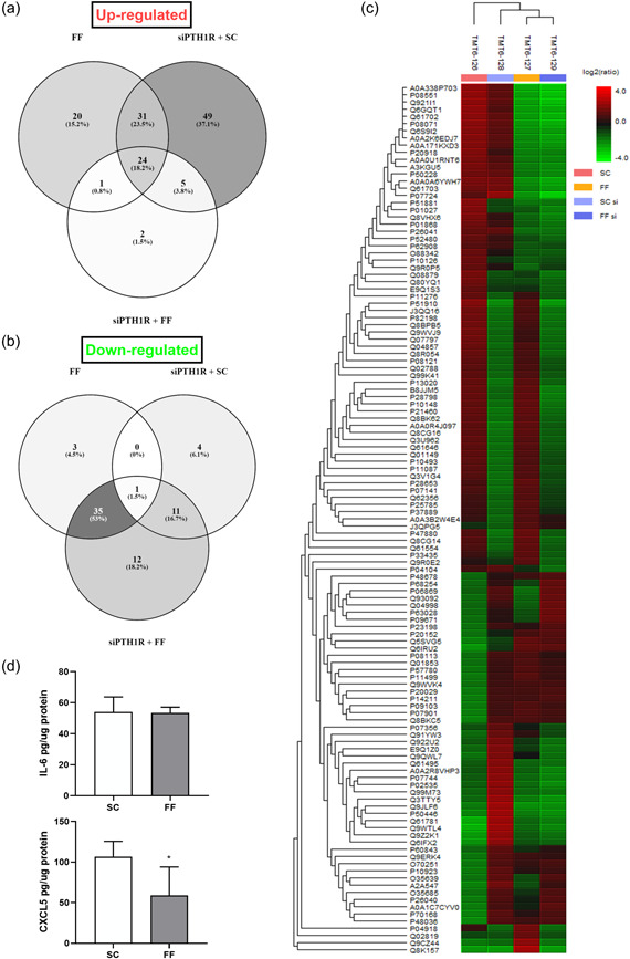
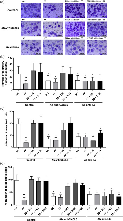
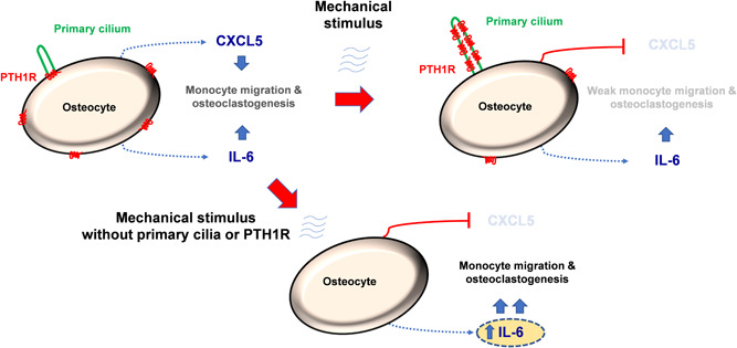
Osteocytes respond to mechanical forces controlling osteoblast and osteoclast function. Mechanical stimulation decreases osteocyte apoptosis and promotes bone formation. Primary cilia have been described as potential mechanosensors in bone cells. Certain osteogenic responses induced by fluid flow (FF) in vitro are decreased by primary cilia inhibition in MLO-Y4 osteocytes. The parathyroid hormone (PTH) receptor type 1 (PTH1R) modulates osteoblast, osteoclast, and osteocyte effects upon activation by PTH or PTH-related protein (PTHrP) in osteoblastic cells. Moreover, some actions of PTH1R seem to be triggered directly by mechanical stimulation. We hypothesize that PTH1R forms a signaling complex in the primary cilium that is essential for mechanotransduction in osteocytes and affects osteocyte-osteoclast communication. MLO-Y4 osteocytes were stimulated by FF or PTHrP (1-37). PTH1R and primary cilia signaling were abrogated using PTH1R or primary cilia specific siRNAs or inhibitors, respectively. Conditioned media obtained from mechanically- or PTHrP-stimulated MLO-Y4 cells inhibited the migration of preosteoclastic cells and osteoclast differentiation. Redistribution of PTH1R along the entire cilium was observed in mechanically stimulated MLO-Y4 osteocytic cells. Preincubation of MLO-Y4 cells with the Gli-1 antagonist, the adenylate cyclase inhibitor (SQ22536), or with the phospholipase C inhibitor (U73122), affected the migration of osteoclast precursors and osteoclastogenesis. Proteomic analysis and neutralizing experiments showed that FF and PTH1R activation control osteoclast function through the modulation of C-X-C Motif Chemokine Ligand 5 (CXCL5) and interleukin-6 (IL-6) secretion in osteocytes. These novel findings indicate that both primary cilium and PTH1R are necessary in osteocytes for proper communication with osteoclasts and show that mechanical stimulation inhibits osteoclast recruitment and differentiation through CXCL5, while PTH1R activation regulate these processes via IL-6.

Keywords: PTH1R; mechanotransduction; osteoclasts; osteocytes; primary cilia.


Figures

Figure 1
Figure 1
CM from mechanically‐stimulated MLO‐Y4 cells inhibit the migration of monocytes by a mechanism dependent on the primary cilium and PTH1R. MLO‐Y4 osteocytic cells were transfected with three IFT88, three PTH1R siRNAs, or with a scrambled siRNA for 24 h followed by serum‐deprivation for 24 h. The efficiency of IFT88 and PTH1R silencing was tested by real time PCR (a). Alternatively, osteocyte cells were serum‐deprived for 24 h and treated with 1 mM aqueous chloral hydrate or with 100 nM PTHrP (7‐34) for 1 h. Cells were subsequently stimulated with shear stress (10 dynes/cm2) or with 100 nM PTHrP (1‐37) for 10 min. CM was collected after 18 h (b−d). To evaluate the number of migratory cells, RAW 264.7 (b and c) or human monocytic cells from buffy coat (d) were cultivated in transwell cell culture chamber inserts with an 8 µm pore size. In the lower compartment 20% of each MLO‐Y4 cell‐conditioned medium was added. After 6 h, cells were fixed, stained with crystal violet, and counted with an inverted optical microscope. The number of monocytic cells evaluated with ImageJ software are represented. Results are the mean ± SD of triplicates. *p < 0.05 versus corresponding scrambled siRNA; **p < 0.01 versus corresponding scrambled siRNA; a p < 0.01 versus SC or corresponding IFT88 siRNA/PTH1R siRNA; b p < 0.01 versus SC or corresponding cilium or PTH1R inhibition. CM, conditioned media; FF; fluid flow; IFT88, intraflagellar transport 88 protein; mRNA, messenger RNA; NC, negative control; PTHrP, PTH‐related protein; PTH1R, PTH 1 receptor; SC, static control; SD, standard deviation; siRNA, small interfering RNA.
Figure 2
Figure 2
Secretome from mechanically‐stimulated MLO‐Y4 cells reduces the differentiation of human monocytes into osteoclasts by a mechanism dependent on the primary cilium and PTH1R. MLO‐Y4 cells were serum‐deprived for 24 h, treated with 1 mM aqueous chloral hydrate or with 100 nM PTHrP (7‐34) for 1 h and stimulated with shear stress (10 dynes/cm2) or with 100 nM PTHrP (1‐37) for 10 min. CM was collected after 18 h. To evaluate the differentiation of monocytic cells into osteoclasts, human monocytes were treated with 20 ng/ml M‐CSF and 20 ng/ml RANKL plus the corresponding 20% MLO‐Y4 cell‐conditioned mediums. Then, cells were fixed, permeabilized, and stained with hematoxylin. The differentiation of human monocytes into osteoclasts was determined by evaluation of the morphology, observing the formation of giant cells (≥50µm) with at least three or more nuclei. Representative images of each condition are shown (a). The percentage of cells with three or more nuclei evaluated with ImageJ software is represented (b). Results are the mean ± SD of triplicates. *p < 0.05 versus SC; **p < 0.01 versus SC; a p < 0.05 versus corresponding PTH1R inhibition; b p < 0.01 versus corresponding cilium or PTH1R inhibition. CM, conditioned media; FF; fluid flow; M‐CSF, macrophage colony‐stimulating factor; PTHrP, PTH‐related protein; PTH1R, PTH 1 receptor; RANKL, receptor activator for nuclear factor κ B ligand; RANKL and (+) α‐MEM medium; SC, static control; SD, standard deviation.
Figure 3
Figure 3
PTH1R colocalizes with primary cilium in mechanically‐stimulated MLO‐Y4 cells, being distributed throughout the whole cilium. MLO‐Y4 cells were transfected with 1 μg of a (GFP)‐PTH1R construct using lipofectamine 3000 for 4 h at 37°C. Cells were subsequently serum‐deprived for 6 h, treated with 1 mM aqueous chloral hydrate or with 100 nM PTHrP (3‐37)  for 1 h and stimulated with shear stress (10 dynes/cm2) for 10 min. To evaluate the colocalization of PTH1R with primary cilia, cells were fixed, permeabilized, blocked, and incubated overnight at 4°C with mouse α‐acetylated tubulin antibody. Then, cells were incubated for 1 h with Alexa fluor 546‐conjugated anti‐mouse IgG secondary antibodies. Representative images of each condition are shown (a). The percentage of cells with PTH1R colocalization just at the base (basal) and throughout the whole (total) primary cilium was analyzed in each condition in cells transfected with a GFP‐PTH1R plasmid (b). Evaluation of the length of primary cilia in MLO‐Y4 cells using ImageJ software are represented (c). Results are the mean ± SD of triplicates. *p < 0.05 versus SC or corresponding control; a p < 0.05 versus corresponding total conditions. 7‐34, PTHrP (7‐34); CH, chloral hydrate; FF; fluid flow; (GFP)‐PTH1R, green fluorescent protein‐PTH1R; IG, immunoglobulin G; SC, static control; SD, standard deviation.
Figure 4
Figure 4
Osteocytic hegdehog, PKA, and PKC signaling pathways are involved in MLO Y4 CM‐dependent regulation of monocyte migration and osteoclastogenesis. MLO‐Y4 cells were serum‐deprived for 24 h and treated with 10 μM GANT61, 100 μM of the adenylate cyclase inhibitor SQ22536, or with 1 μM of the phospholipase C inhibitor U73122 for 1 h. Cells were subsequently stimulated with shear stress (10 dynes/cm2) or with 100 nM PTHrP (1‐37) for 10 min. CM was collected after 18 h. To evaluate the number of migratory cells, RAW 264.7 (a) or human monocytic cells from buffy coat (b) were cultured in transwell cell culture chamber inserts with an 8 µm pore size, and in the lower compartment with 20% of each MLO‐Y4 cell‐conditioned medium. After 6 h, cells were fixed, stained with crystal violet, and counted with an inverted optical microscope. The number of monocytic cells evaluated with ImageJ software are represented (a and b). To evaluate the differentiation of monocytes into osteoclasts, human monocytes were treated with 20 ng/ml M‐CSF and 20 ng/ml RANKL, plus the corresponding 20% MLO‐Y4 cell‐conditioned mediums. Cells were fixed, permeabilized, and stained with hematoxylin. The percentage of cells with three or more nuclei evaluated with ImageJ software are represented (c). Results are the mean ± SD of triplicates. **p < 0.01 versus SC; a p < 0.05 versus corresponding GLI, PKA, or PKC inhibition; b p < 0.01 versus corresponding GLI, PKA, or PKC inhibition. CM, conditioned media; FF; fluid flow; GANT61, Gli‐1‐Antagonist 61; M‐CSF, macrophage colony‐stimulating factor; NC, negative control (−) RANKL and (+) α‐MEM medium; PKA, protein kinase A; PKC, protein kinase; PTHrP, PTH‐related protein; RANKL, receptor activator for nuclear factor κ B ligand; SC, static control; SD, standard deviation.
Figure 5
Figure 5
Proteomic analysis of mechanically‐stimulated MLO‐Y4 cells, +/‐ PTH1R silencing. MLO‐Y4 osteocytic cells were transfected with three PTH1R siRNAs or with a scrambled siRNA for 24 h. Cells were subsequently serum‐deprived for 24 h and stimulated with shear stress (10 dynes/cm2) for 10 min. CM were collected after 18 h, lyophilized, and analyzed by tandem mass tag mass spectrometry. Venn diagrams show the profile of secreted proteins that increase (a) or decrease (b) comparing different experimental conditions. (c) A heatmap showing protein secretion differences after proteomic analysis of osteocytes is pictured. The color and intensity of the boxes is used to represent changes (not absolute values) of protein secretion. In the picture above, red represents increased secretion and green represents decreased secretion. Black represents unchanged secretion. (d) IL‐6 and CXCL5 in the MLO‐Y4‐CM were analyzed by ELISA as described in Section 2. *p < 0.05 versus SC. CM, conditioned media; FF; fluid flow; IL‐6, interleukin‐6; PTH1R, PTH 1 receptor; SC, static control; siPTH1R, PTH1R silencing.
Figure 6
Figure 6
Mechanical stimulation inhibits the recruitment and differentiation of osteoclasts through a mechanism dependent on CXCL5 and IL‐6. MLO‐Y4 cells were serum‐deprived for 24 h and treated with 1 mM aqueous chloral hydrate, 100 nM PTHrP (7‐34), 10μM of the Gli inhibitor GANT61, 100 μM of the adenylate cyclase inhibitor SQ22536 or with 1 μM of the phospholipase C inhibitor U73122, for 1 h. Cells were subsequently stimulated with shear stress (10 dynes/cm2) for 10 min. CM were collected after 18 h and 2 µg/ml of neutralizing antibodies anti‐mCXCL5 or 1 µg/ml of anti‐mIL‐6 were added. To evaluate the number of migratory cells, human monocytes from buffy coat (a and b) were cultured in transwell cell culture chamber inserts with an 8 µm pore size. In the lower compartment 20% of each MLO‐Y4 cell‐conditioned medium was added plus the corresponding neutralizing antibody. After 6 h, cells were fixed, stained with crystal violet, and counted with an inverted optical microscope. Representative images of each condition are shown (a). The number of monocytic cells evaluated with ImageJ software are represented (b). To evaluate the differentiation of monocytes into osteoclasts, human monocytes were treated with 20 ng/ml M‐CSF and 20 ng/ml RANKL, plus the corresponding treatments: 20% of each MLO‐Y4 cell‐conditioned medium and the neutralizing antibody. Cells were fixed, permeabilized, and stained with hematoxylin (c and d). The percentage of cells with three or more nuclei evaluated with ImageJ software is represented (c and d). Results are the mean ± SD of triplicates. *p < 0.05 versus SC; **p < 0.01 versus SC. 7‐34, PTHrP (7‐34); Ab, antibody; CH, chloral hydrate; CM, conditioned media; CXCL5, C‐X‐C motif chemokine 5; FF; fluid flow; GANT61, Gli‐1‐Antagonist 61; IL‐6, interleukin‐6; M‐CSF, macrophage colony‐stimulating factor; PKA, protein kinase A; PKC, protein kinase C; RANKL, receptor activator for nuclear factor κ B ligand; SC, static control; SD, standard deviation.
Figure 7
Figure 7
Proposed mechanism for regulation of osteoclast recruitment and differentiation by primary cilium and PTH1R in osteocytes. The presence of both functional primary cilium and PTH1R in osteocytes are necessary for correct communication with osteoclasts. Mechanical stimulation inhibits the recruitment and differentiation of osteoclasts through CXCL5, while PTH1R activation regulate osteoclasts through IL‐6. CXCL5, C‐X‐C Motif Chemokine Ligand 5; IL‐6, interleukin‐6; PTH1R, parathyroid hormone receptor type 1.

Similar articles

Cited by

References

    1. Alonso, R. , Pisa, D. , Marina, A. I. , Morato, E. , Rábano, A. , Rodal, I. , & Carrasco, L. (2015). Evidence for fungal infection in cerebrospinal fluid and brain tissue from patients with amyotrophic lateral sclerosis. International Journal of Biological Sciences, 11(5), 546–558. - PMC - PubMed
    1. Ardura, J. A. , Portal‐Núñez, S. , Alonso, V. , Bravo, B. , & Gortazar, A. R. (2019). Handling parathormone receptor type 1 in skeletal diseases: Realities and expectations of abaloparatide. Trends in Endocrinology and Metabolism, 30(10), 756–766. - PubMed
    1. Baron, R. , & Kneissel, M. (2013). WNT signaling in bone homeostasis and disease: From human mutations to treatments. Nature Medicine, 19(2), 179–192. 10.1038/nm.3074 - DOI - PubMed
    1. Bellido, T. (2014). Osteocyte‐driven bone remodeling. Calcified Tissue International, 94(1), 25–34. - PMC - PubMed
    1. Chen, X. , Macica, C. M. , Ng, K. W. , & Broadus, A. E. (2005). Stretch‐induced PTH—Related protein gene expression in osteoblasts. Journal of Bone and Mineral Research, 20(8), 1454–1461. - PubMed

Publication types

MeSH terms